Nuclear Scintigraphy, CT and MRI
1/37
There's no tags or description
Looks like no tags are added yet.
Name | Mastery | Learn | Test | Matching | Spaced | Call with Kai |
|---|
No analytics yet
Send a link to your students to track their progress
38 Terms
Nuclear Scintigraphy
an imaging modality that utilizes a gamma camera to track radioactive uptake in structures or organs of interest
Atomic Number
Number of protons in a nucleus
Atomic Mass
Sum of the protons and the neutrons in a nucleus
Radioactive
Meaning that the atom is emitting ionizing radiation or particles
Isotope
Same element, but contains a different number of neutrons
Half-Life
The time taken for the radioactivity of a specified isotope to fall to half its original value
Radiopharmacy
Atomic number: Number of protons in a nucleus
Atomic Mass: Sum of the protons and the neutrons in a nucleus
Radioactive: Meaning that the atom is emitting ionizing radiation or particles
Isotope: Same element, but contains a different number of neutrons
Half-life: The time taken for the radioactivity of a specified isotope to fall to half its original value
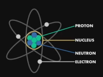
Atomic Structure
Proton +
Neutron: no charge
Electron -
Nucleus: Made up of positively charged protons and neutrons that contain no charge
Radioactive decay
Disruptions of the atom
Nucleus beings to shake
Release of different forms of energy
Alpha emission
Beta emission
Gamm radiation
Alpha emission
loss of 2 neutrons and 2 protons
Beta emission
loss of 1 electron
Gamma Radiation
Pure wave of immense photon energy
Have the shortest wavelength and contain the most energy of any wave on the electromagnetic spectrum
In space, these rays are created by events such as supernovas and black holes
On earth they are created by radioactive decay
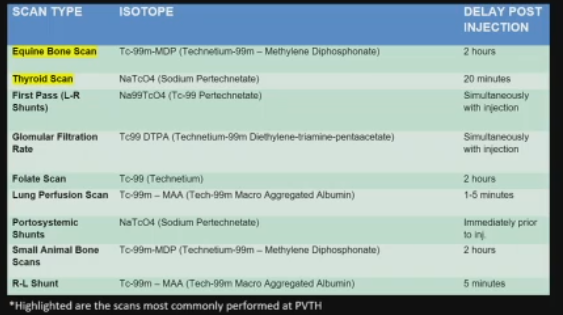
Technetium 99m
Most widely used radioisotope in nuclear medicine
Emits gamma radiation
Short half-life - 6 hours
MDP
Methylene Diphosphonate
Tracer
Seeks out areas of high osseous turnover
Making Technetium 99m
Molybdenum is placed within a generator
The atom is disrupted
It begins to decay
Once it decays into Tc 99 it can be eluted and drawn into a syringe
The syringe is then ready to be injected into the patient
This is a very time sensitive process because the isotope will continue to decay over time. It must be properly calibrated to the time that the isotope will be injected into the patient
Various types of nuclear scans
MAIN ONES
Equine Bone scan
Cat Thyroid scan
Day PRIOR to equine bone scan
Request received
Isotope dose is calculated and ordered to be calibrated in coordination with injection time
Prep camera with colbalt flood
The horse is what is radioactive not any of the imaging equipment
Box is prepared to be taken to stall - leg wraps, foot wrap, radiation signs etc.
Day OF Equine bone scan
Isotope processed
Camera peaked to the isotope
Horses legs/feet wrapped
Catheter placed - horse injected
Wait 2 hours
Give sedation - walk to nuclear room
Put on blocks - start at feet and move up
Bathroom break for horse
Scan caudal half of horse
Horse geigered - hand held tool to check the radiation amount they are reading at
Room surveyed for contamination
Day AFTER equine bone scan
Horse will be geigered again
If it is under 120 mR/hr it will be released to go home or have further work up/diagnostics
Horse moved to new stall
TWO DAYS after equine bone scan
Stall is geigered
Must read background (0.02 mR/hr)
Signs removed from stall
Trash removed from stall
REM contacted for trash pick up
3 phases of nuclear imaging
Vascular phase
Soft tissue Phase
Bone Phase
Vascular phase
Image is acquired simultaneously with injection of the isotope
Traces the isotope as it moved through the circulatory system
Indications for use
Suspect thrombosis
Decreased perfusion
Not commonly performed
Soft tissue phase
Image is acquired 5 minutes post injection of isotope
Detects soft tissue inflammation
Trauma - ex. Kicked by another horse
Saddle soreness
Muscle injury
Used more commonly than the vascular phase
Bone Phase
Scan begins 2-4 hours post injection of isotope
Technetium 99m - MDP seek areas of High osseous turnover
Bone injuries - stress fractures
Osteoarthritis
Neoplasia
To obtain good nuclear images
Requires 2-3 staff members
The horse must remain completely still - typically very sedated
When imaging the distal limb, lead shielding is placed between the legs
Camera face is positioned around the horse
Effectiveness of lead shielding for personnel remains controversial
At Purdue we do not have to where lead shielding
Whole body scan average: 3-4 hours
Half body scans average: 1-2 hours
Time length for whole body nuclear scan
3-4 hours
Time length for half body nuclear scan
1-2 hours
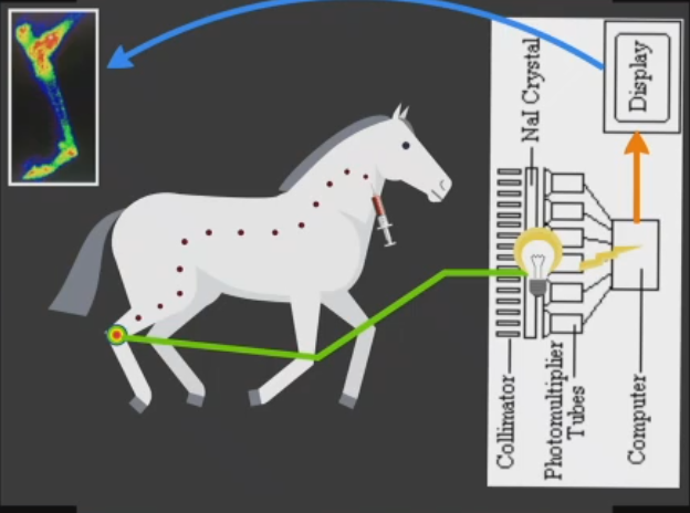
How the equine nuclear image is obtained
Isotope emits gamma rays from area of uptake
Rays pass through the collimator on camera
Rays interact with sodium iodide crystals within camera
Reaction causes rays to be scintillated into photon light energy
Photomultiplier tube converts the light waves into electrical pulses
If the pulses fall within the photo peak window they are transcribed into a digital image
How nuclear images are interpreted
Areas of high uptake will appear bright red or "hot"
Areas of low uptake will appear blue or "cold"
Uptake does not directly relate to pain!
Nuclear image - Red areas
Area of HIGH uptake
Signs of active skeletal growth
Especially useful in localizing bone injuries
Nuclear image - Blue areas
Areas of LOW uptake
Can represent a normal region or an area lacking perfusion
If entire distal limb is blue you probably have a perfusion issue
Information Nuclear scans provide
Nuclear scans are sensitive but not specific
Ex. Could potentially pick up on a stress fracture that would not be able to be seen on radiographs
However, cannot tell you that the problem is a stress fracture
Nuclear scans act as a diagnostic compass
They localize of problem, but don't provide a diagnosis
What happens after a nuclear scan
Nuclear scans must be used in conjunction with other diagnostics
Reassess the lameness
Take radiographs of localized area
Potential Cross sectional imaging
CT is much more specific
Ultrasound if useful
Cat Thyroid Scan
Performed if there is suspicion that the cat is hyperthyroid
Isotope: Sodium Pertechnetate
Cat injected - wait 20 minutes
5 scans - ventral, dorsal, both laterals and pinhole
Looking for uneven distribution of uptake in the thyroid
Isotope for Cat thyroid scan
Sodium Pertechnetate
ADVANTAGES of Nuclear imaging
Ability to scan the entire body
Provides valuable information about soft tissue, bone and vasculature
Does no require general anesthesia (for horses)
Provides valuable information in the diagnostic process
DISADVANTAGES of Nuclear imaging
Not specific enough for diagnosis
Uptake does not necessarily mean the source of lameness
Time consuming
Effectiveness of PPE is controversial
Patient remains radioactive post scanning
Summary of Nuclear Scintigraphy
Good diagnostic tool in terms of localization
Diagnostic compass
Must be used in conjunction with other diagnostics
Relatively low radiation exposure to patient
Detection of bone abnormalities
Does not always uncover the source of lameness