Unit 5 - Muscles of Abdomen & Pelvis
1/7
There's no tags or description
Looks like no tags are added yet.
Name | Mastery | Learn | Test | Matching | Spaced | Call with Kai |
|---|
No analytics yet
Send a link to your students to track their progress
8 Terms
Muscles of abdominal wall
Deep → superficial
Transverse Abdominus m.
Internal Oblique m.
Rectus Abdominus m.
External Oblique m.
(All innervated by ventral rami of T7-L2)

Rectus sheath
Aponeuroses of the 3 anterolateral abdominal wall muscles, forms the anterior & posterior rectus sheet (encloses rectus abdominis muscle)
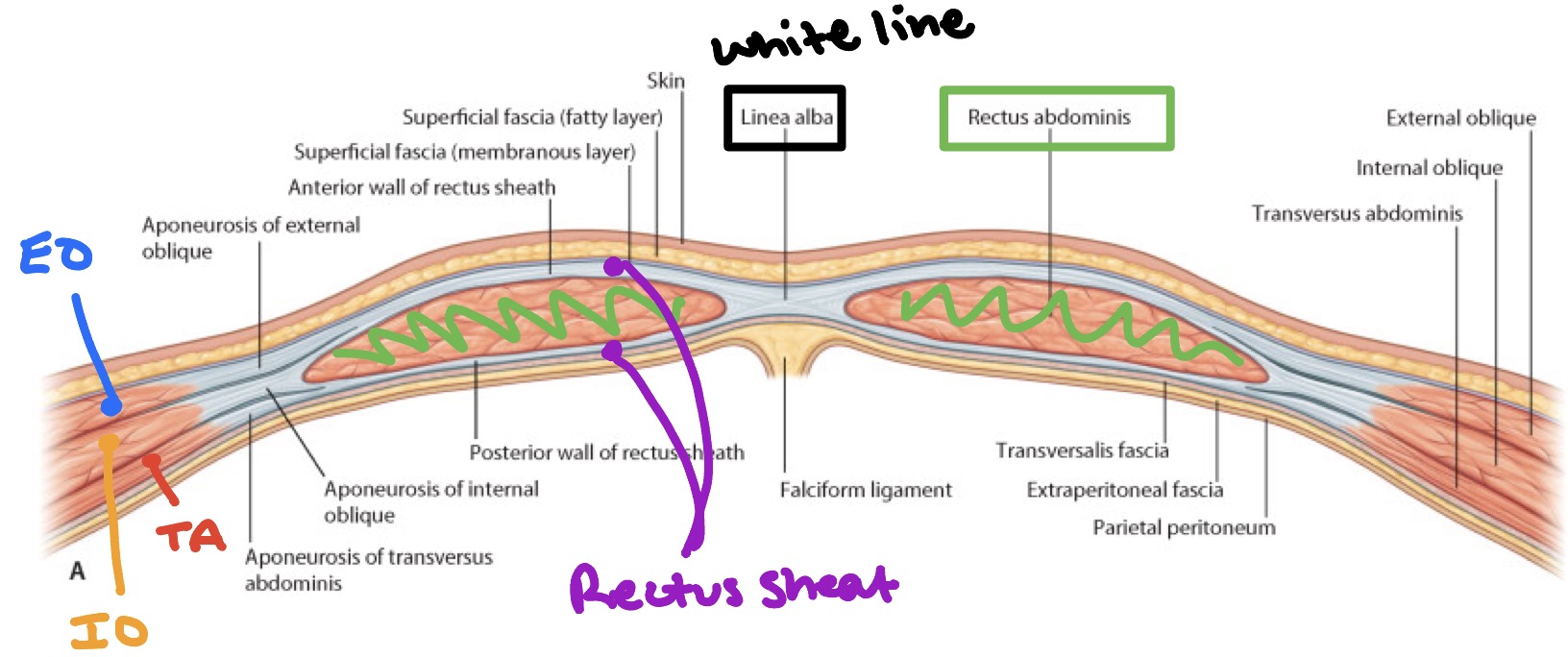
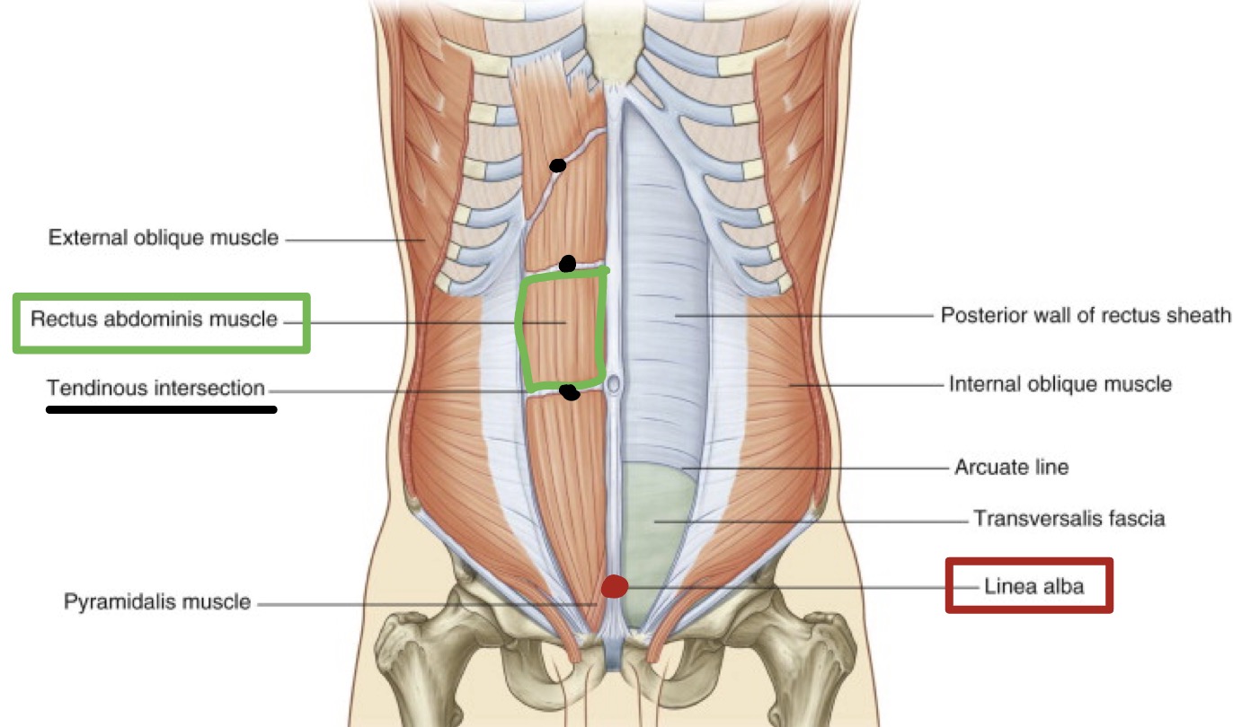
Rectus abdominis muscle
Paired muscle, separated at midline by linea alba
Along its course, it is intersected by 3 or 4 tendinous intersections
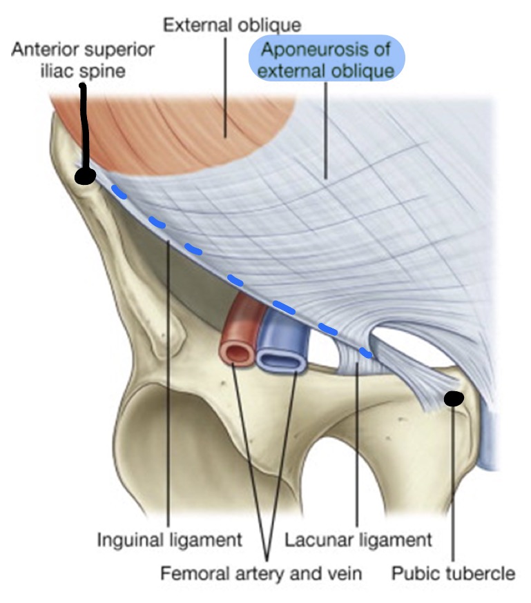
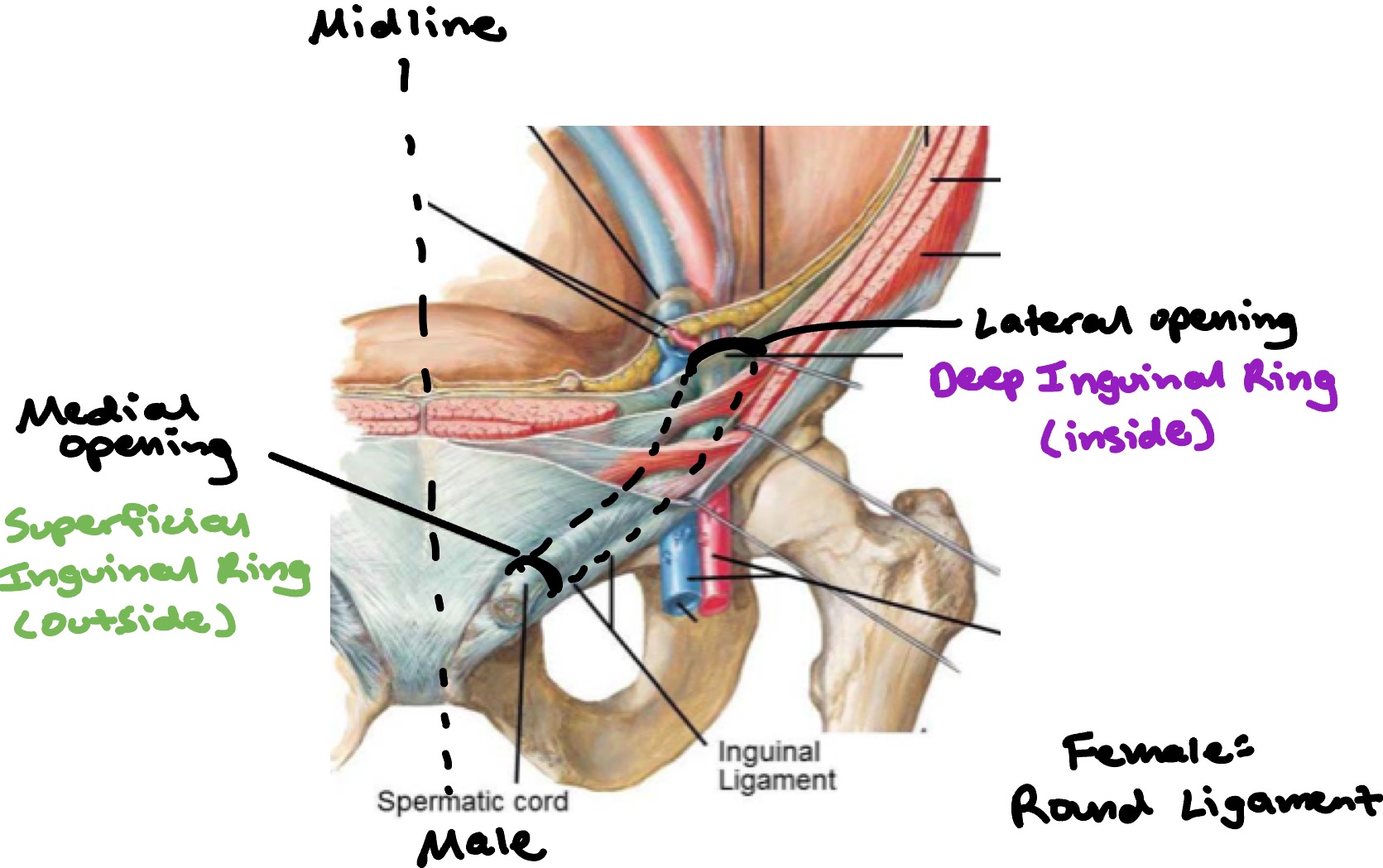
Inguinal region
Area of junction b/w anterior & abdominal wall & thigh
Inferior free border of external oblique aponeurosis folds back on itself to form the inguinal ligament, which runs from anterior superior iliac spine to public tubercle
Inguinal region - parts
Inguinal canal is slit-like passage that extends in a downward & medial direction
Begins at deep inguinal ring & continues for 4 cm, ending at superficial inguinal ring
Contains spermatic cord (testes) & round ligament (uterus)
Inguinal region - visual
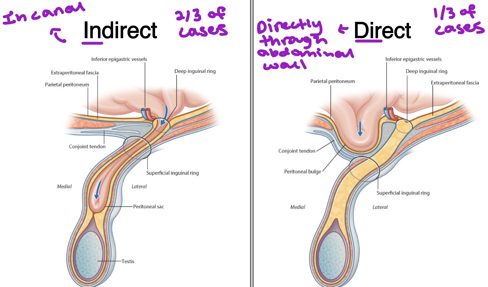
Inguinal Hernias
Indirect = in canal → 2/3 of cases
Direct = directly through abdominal wall → 1/3 of cases
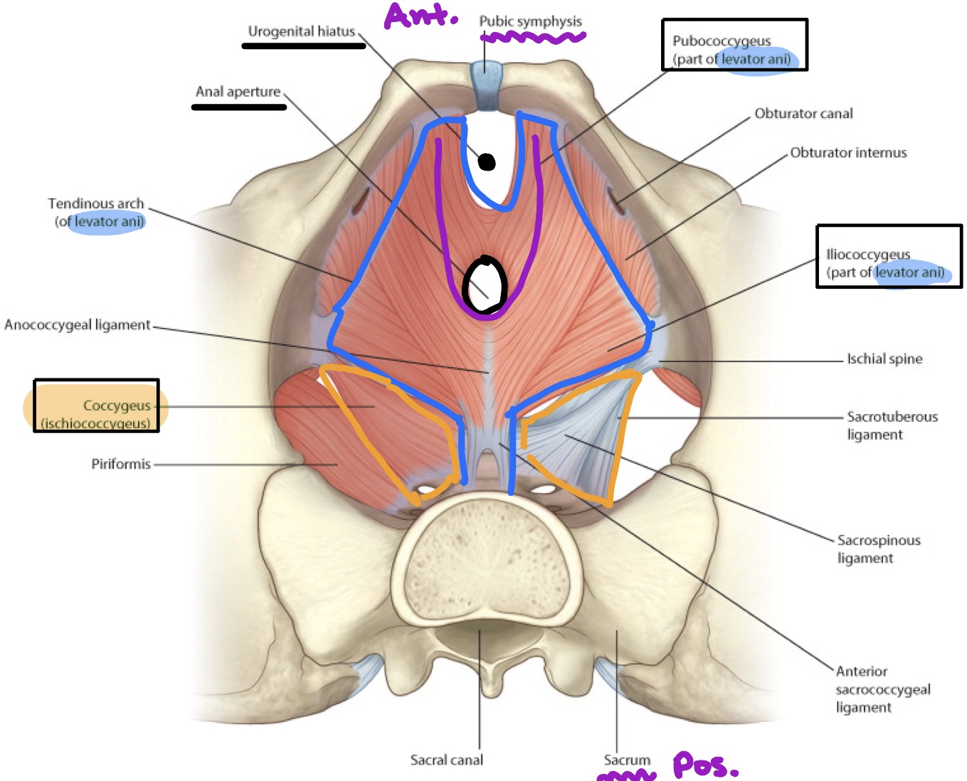
Muscles of Pelvis
Funnel shaped pelvis floor or pelvis diaphragm is composed of 2 paired muscles
Levator ani muscles & coccygeus muscles
Pelvis diaphragm closes the majority of the inferior pelvis aperture
Pierced by rectum & urethra
This diaphragm supports & raises the pelvis floor & assists in the support of the abdominopelvic viscera