Pachychoroid Disease - Posterior Segment & Ocular Disease Spring 2026
1/33
There's no tags or description
Looks like no tags are added yet.
Name | Mastery | Learn | Test | Matching | Spaced | Call with Kai |
|---|
No analytics yet
Send a link to your students to track their progress
34 Terms
-Enhanced-depth imaging (EDI-OCT)
-Swept Source OCT (SS-OCT)
What technology would you use to diagnose pachychoroidal disease?
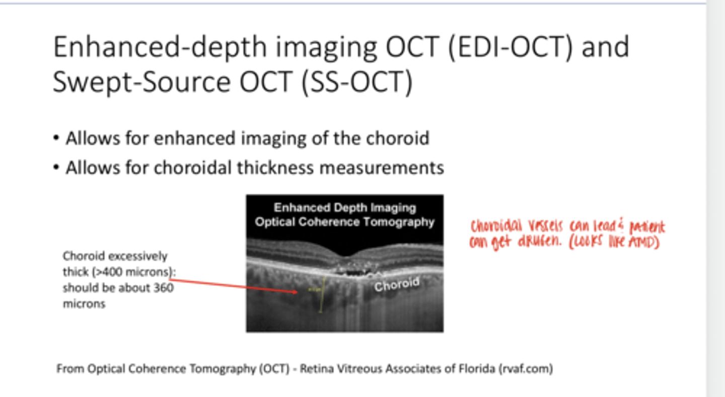
-enhanced imaging of the choroid
-choroidal thickness measurements
What do Enhanced-depth imaging (EDI-OCT) & Swept Source OCT (SS-OCT) allow for?

Focal or diffuse choroidal thickening (>400 microns) d/t dilation of choroidal vessels in Haller's layer (more external)
What happens to the choroid in pachychoroidal disease?
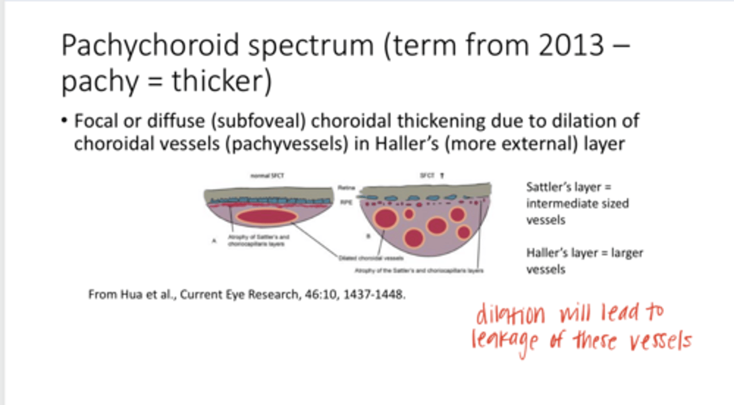
leakage of these vessels
Dilation of the vessels in Haller's layer of the choroid can lead to what?
thinning of the choriocapillaris and Sattler's layer
Dilation of the vessels in Haller's layer of the choroid can lead to thinning of what other layer?

RPE abnormalities and sometimes photoreceptor abnormalities & atrophy
What can pachychoroidal disease be associated with?
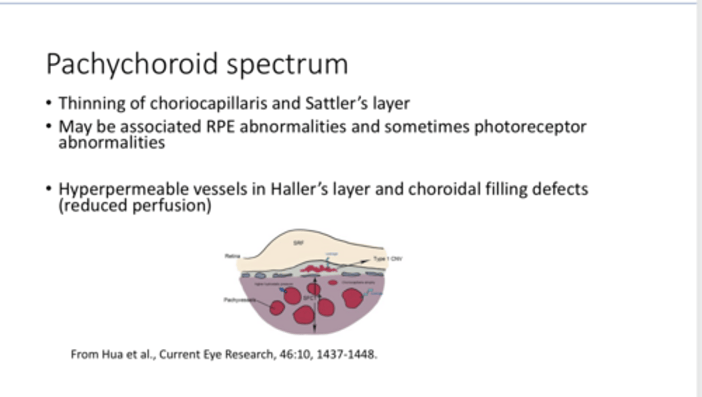
hyper
In pachychoroidal disease, the vessels in Haller's layer will be (hyper/hypo) permeable

Yes
Are choroidal filling defects (reduced perfusion) associated with pachychoroidal disease?

-retinal changes that seem like AMD (serous fluid, serous detachments, drusenoid-like detachments, hemorrhages, hard exudate)
What does pachychoroidal disease lead to?
younger
Patients with pachychoroidal disease may be ____ than a typical AMD patient
true
True or False:
A patient with pachychoroidal disease will probably NOT have large drusen?
-Photodynamic therapy (PDT)
-Combo PDT and Anti-VEGF meds
Pachychoroidal disease sometimes responds well to _______
-Central serous chorioretinopathy (CSC)
-Pachychoroid Pigment Epitheliopathy (PPE)
-Pachychoroid Neovasculopathy (PVN)
-Polypoidal Choroidal Vasculopathy/Aneurysmal Type 1 Neovascularization (PCV/AT1)
-Focal Choroidal Excavation (FCE)
-Peripapillary Pachychoroid Syndrome (PPS)
What are the different kinds of pachychoroidal disease?
Central Serous Chorioretinopathy on OCT (Pic)
Central Serous Chorioretinopathy on OCT (Pic)
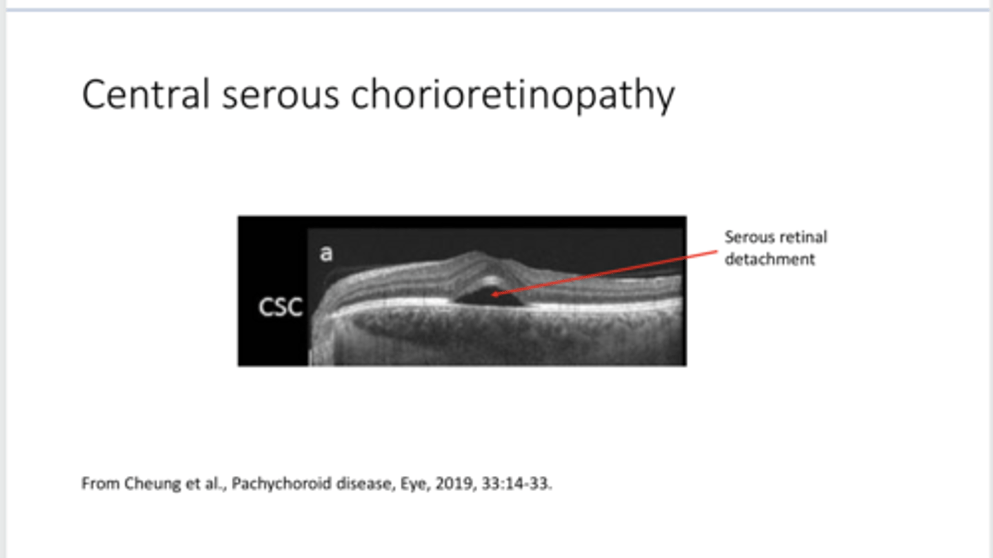
Serous retinal detachment d/t leakage of vessels in the choroid
What is shown in this pic?

Central serous chorioretinopathy with Large Pachyvessels (Pic)
Central serous chorioretinopathy with Large Pachyvessels (Pic)
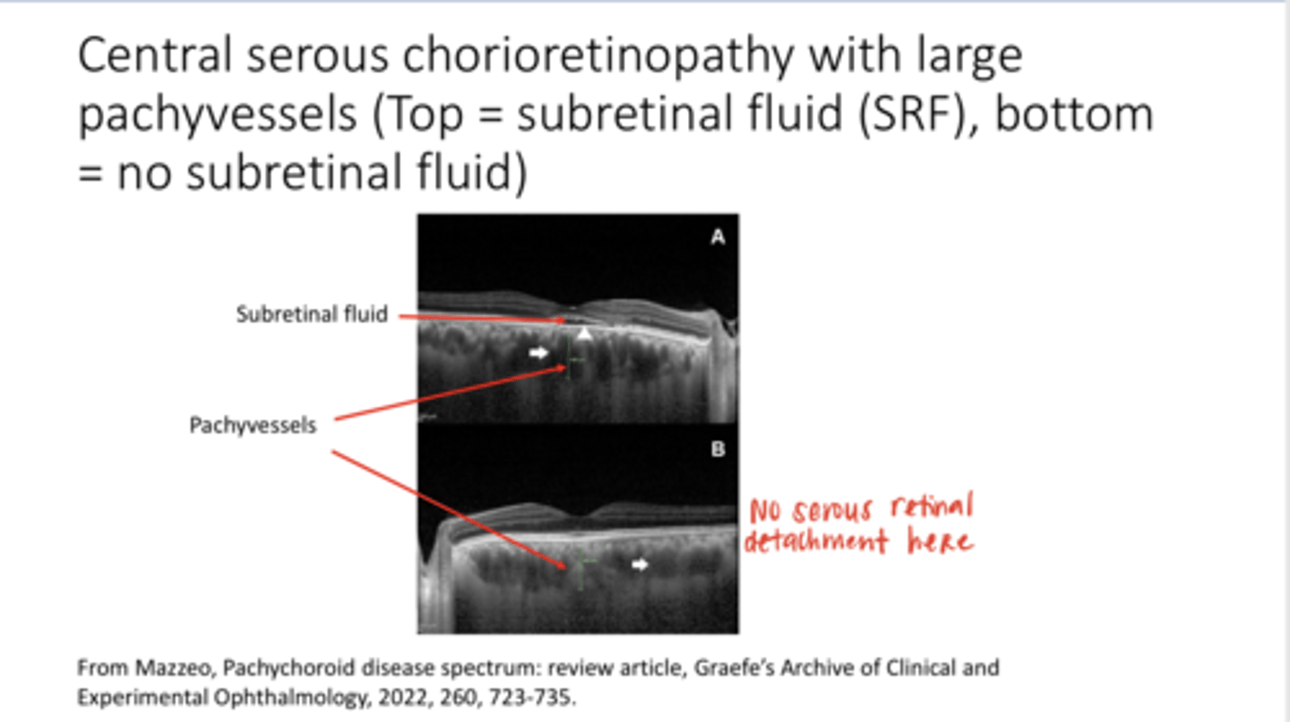
Yes? They will have the problem in both eyes but may not progress to a retinal detachment in both
Is central serous bilateral?

Central Serous Chorioretinopathy and Focal Choroidal Excavation (Pic)
Central Serous Chorioretinopathy and Focal Choroidal Excavation (Pic)
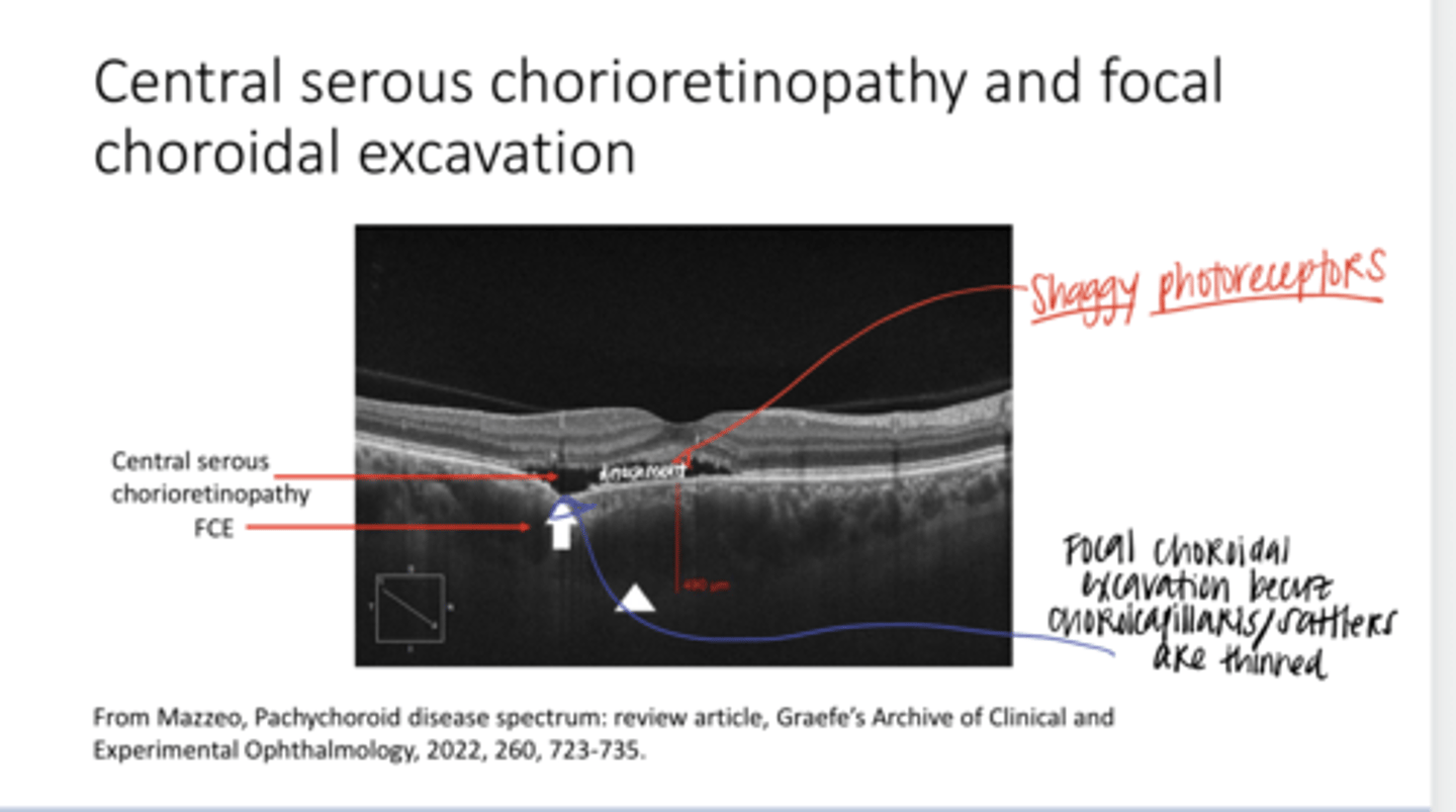
thinned choriocapillaris and Sattler's layer of the choroid
Why does a focal choroidal excavation present?

Dunno
Does FCE only occur in pachychoroidal disease?

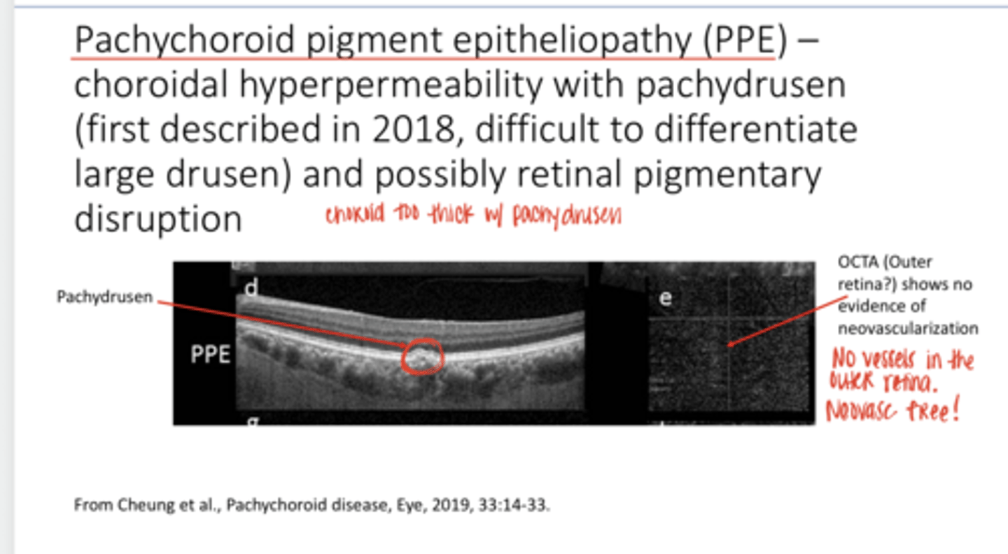
Pachychoroidal Pigment Epitheliopathy (PPE) -- Choroidal Hyperpermeability with Pachydrusen and Possibly Retinal Pigmentary Disruption (Pic)
Pachychoroidal Pigment Epitheliopathy (PPE) -- Choroidal Hyperpermeability with Pachydrusen and Possibly Retinal Pigmentary Disruption (Pic)

No -- OCT will show no evidence of neovascularization
On OCT-A will there be vessels in the outer retina of a patient with Pachychoroidal Pigment Epithelialopathy?
Pachychoroidal Neovasculopathy (PVN) -- Choroidal Hyperpermeability and T1 Neovascularization (Pic)
Pachychoroidal Neovasculopathy (PVN) -- Choroidal Hyperpermeability and T1 Neovascularization (Pic)
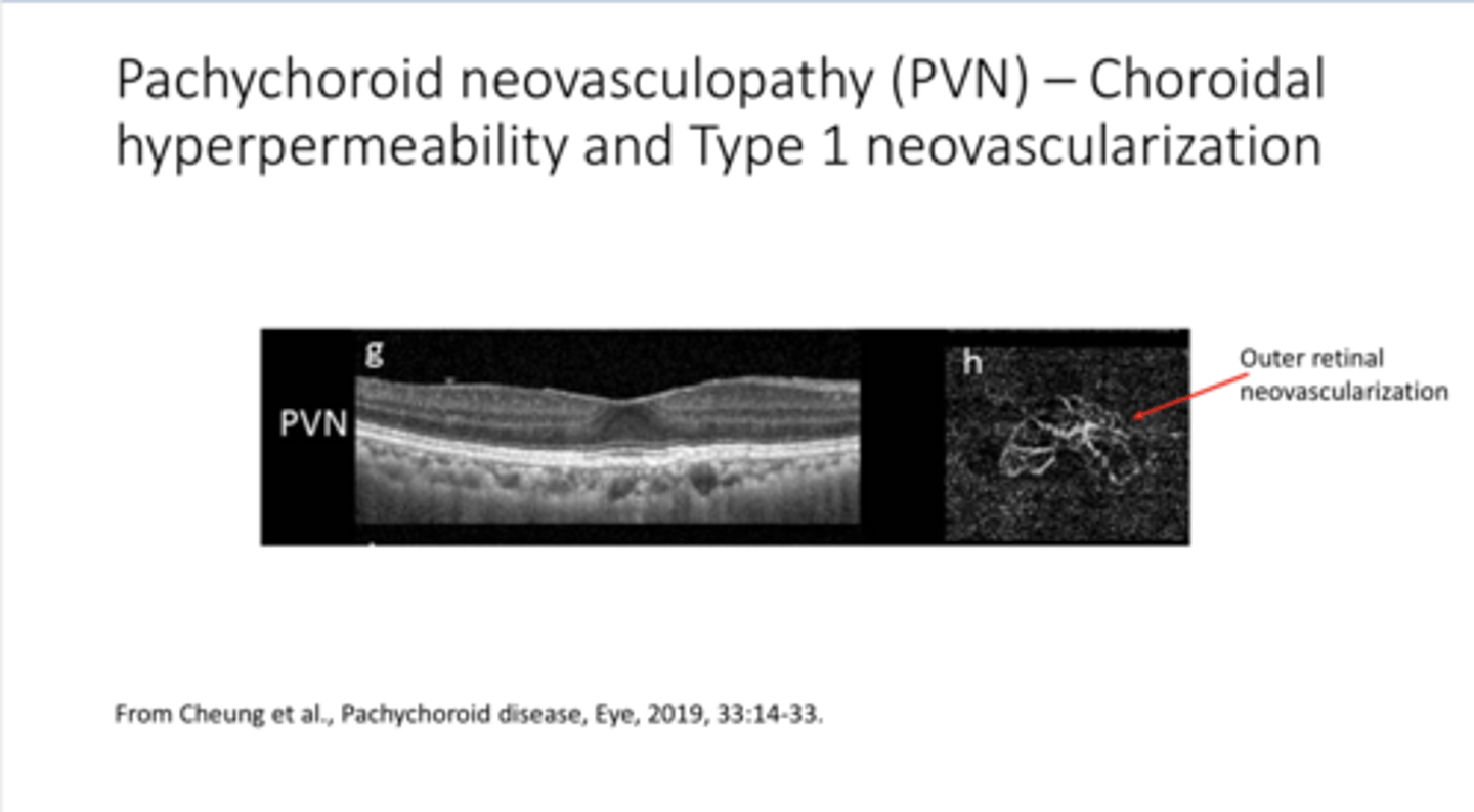
Neovasc
What is present in the outer retina of Pachychoroidal Neovasculopathy (PVN)?

Prob
Will the neovasc in Pachychoroidal Neovasculopathy (PVN) leak?

Choroidal Neovasc Membrane located below the RPE
Type 1 ("Occult") Choroidal Neovasc
Choroidal Neovasc Membrane that grows through the RPE into the subretinal space
Type 2 ("Classic") Choroidal Neovasc
Retinal angiomatous proliferation; retinal neovascularization that moves posteriorly
Type 3 Choroidal Neovasc
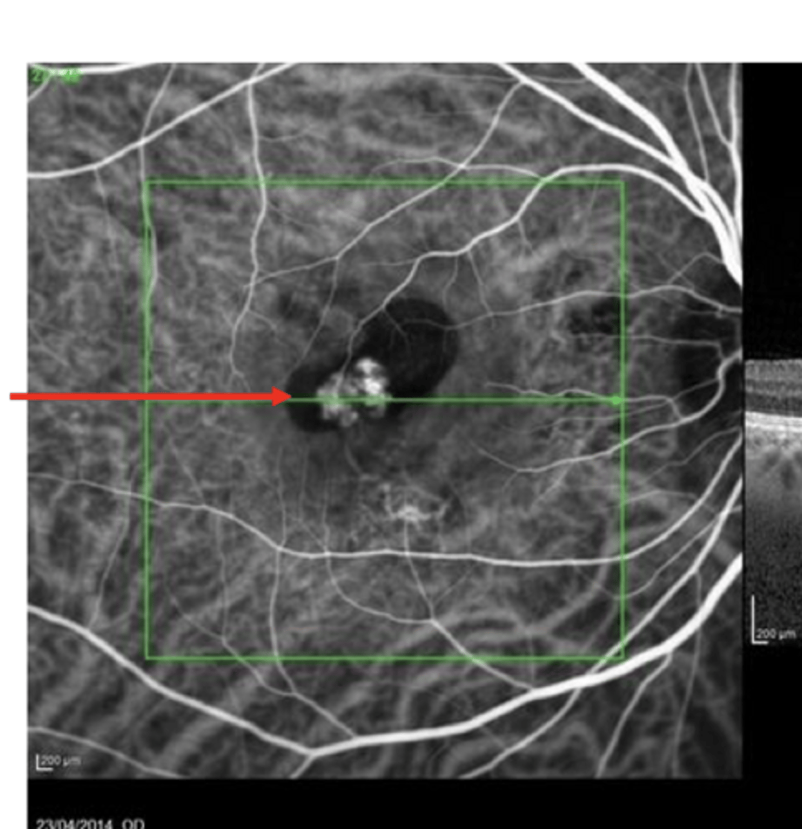
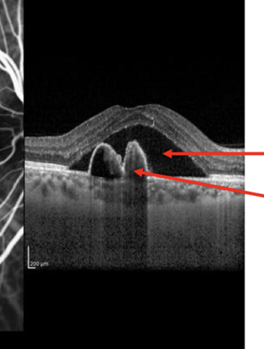
Polypoidal Choroidal Vasculopathy/Aneurysmal Type 1 Neovascularization (PCV/AT1)
Polypoidal Choroidal Vasculopathy/Aneurysmal Type 1 Neovascularization (PCV/AT1)
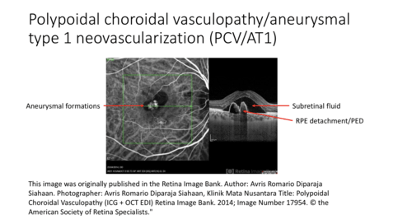
Aneurysms of the choroidal vessels
What are these bright bulbs on OCT-1?
-Serous Detachment
-RPE Detachment
-In the presence of choroidal aneurysms in the same place
What is present in this pic?
Yes
Does Polypoidal Choroidal Vasculopathy/Aneurysmal Type 1 Neovascularization (PCV/AT1) usually have neovasc associated with it?

PDT w/ possible Anti-VEGF meds
What is Polypoidal Choroidal Vasculopathy/Aneurysmal Type 1 Neovascularization (PCV/AT1) treated with?

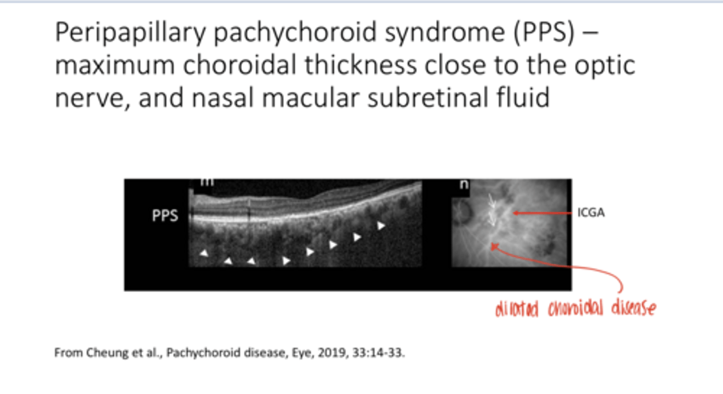
Peripapillary Pachychoroid Syndrome (PPS) -- Maximum Choroidal Thickness Close to the Optic Nerve and Nasal Macular Subretinal Fluid (Pic)
Peripapillary Pachychoroid Syndrome (PPS) -- Maximum Choroidal Thickness Close to the Optic Nerve and Nasal Macular Subretinal Fluid (Pic)