L8 Limb Development of Muscoskeletal components of the limb
1/119
There's no tags or description
Looks like no tags are added yet.
Name | Mastery | Learn | Test | Matching | Spaced | Call with Kai |
|---|
No analytics yet
Send a link to your students to track their progress
120 Terms
The limbs
Muscles with tissue junctions and tendons
Bones with joints
BONE DEVELOPMENT
What are the two types of bone formation in the limbs
Endochondral ossification (formation of cartilage template that is replaced by bone)
AND
Intramembranous ossification (bones forms directly from mesenchymal cells)
What forms by intramembranous ossification
Mainly flat bones
How do long bones of the skeleton form
By endochondral ossification where mesenchymal cells first condense and differentiate into chondrocytes to create a cartilage model that is later replaced by bone.
Endochondral ossification diagram, slide 6
What initiates chondrocyte differentiation during endochondral ossification
Expression of SOX9 initiates condensation of mesenchymal cells and Indian hedgehog (Ihh) regulated their differentiation into chondrocytes
What are the two types of bone formation in the limbs?
The two types are endochondral ossification and intramembranous ossification
How do long bones of the limbs form?
Long bones form by endochondral ossification where a cartilage template is replaced by bone
What initiates condensation of mesenchymal cells during bone development?
SOX9 initiates mesenchymal condensation and chondrocyte differentiation
What regulates chondrocyte differentiation during endochondral ossification?
Indian hedgehog (Ihh) regulates chondrocyte differentiation
What are the main stages of endochondral ossification?,
Endochondral ossification begins with SOX9-driven condensation of mesenchymal cells which differentiate into chondrocytes under control of Ihh. Chondrocytes produce collagen type II and form a cartilage template. These cells become hypertrophic, express collagen type X and the matrix becomes calcified
How does ossification begin in the cartilage model?
Ossification begins when blood vessels invade the centre of the cartilage model forming the primary ossification centre in the diaphysis. Pre-osteoblasts differentiate into osteoblasts and begin producing collagen type I to form mineralised bone
How does the long bone continue to grow after the primary ossification centre forms?
Ossification spreads from the diaphysis toward the epiphyses. A bone collar forms around the diaphysis which thickens to form a cortical bone layer. Proliferating chondrocytes remain organised in the growth plate allowing longitudinal growth (bone lengthens)
what do osteoclasts do during enchondrol ossification
brought in via the vasculature. They function to digest bone tissue as part of ongoing growth and remodelling throughout life
What is the function of RUNX2 in bone development?
RUNX2 is required for osteoblast differentiation and bone mineralisation. it allows cells to become osteoblasrts and is necessary for bone mineralisation
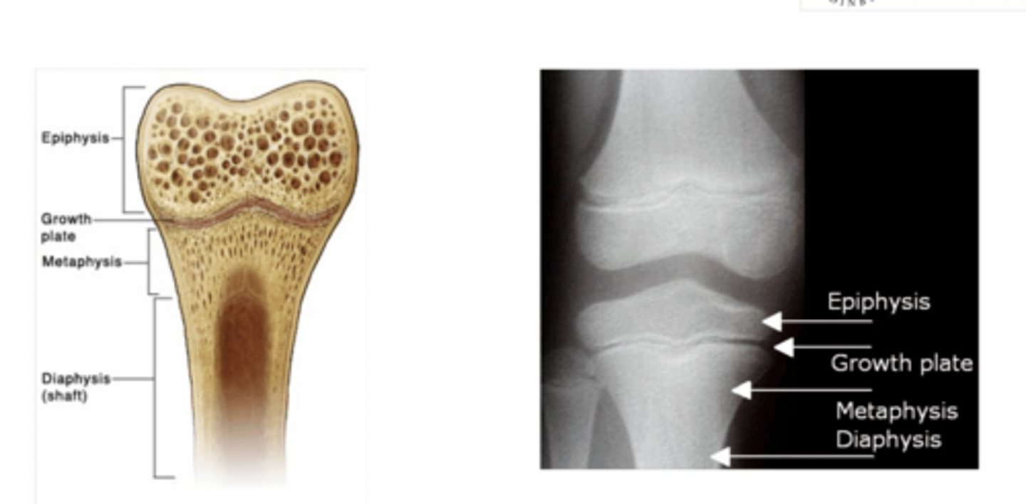
Where does the primary ossification centre form?
It forms in the diaphysis of long bones and is the first site where cartilage is replaced by bone
Where do secondary ossification centres develop?
They develop in the epiphyses after birth
What is the function of the growth plate?
The growth plate allows longitudinal bone growth through organised chondrocyte proliferation.
At what age do growth plates usually fuse?
Growth plates usually fuse around 20 years of age
What happens at birth and after in endochondral ossification?
At birth the diaphysis is ossified but the epiphyses remain cartilaginous. Secondary ossification centres form in the epiphyses after birth and growth continues at the epiphyseal plate until it fuses around 20 years of age
Which molecules regulate chondrocyte differentiation and growth plate activity?
Indian hedgehog, PTHrP and FGF18 regulate chondrocyte proliferation and differentiation within the growth plate
Why is knowledge of ossification centres clinically important?
It helps determine skeletal age and distinguish fractures from growth plates. Assists in forensic age estimation
Primary ossification centres are present at birth
visible on x-ray from birth
- metacarpal diaphyses: 9 weeks in utero
- phalangeal diaphyses: 9-12 weeks in utero
primary ossification centres developing after birth
Ossification of the carpal bones occurs in a predictable sequence,starting with the capitate and ending with the pisiform.
At birth, there is no calcification in the carpal bones. Although there isgreat individual variabilit
diagram of bone epiphysis, growth plate, metaphysis, diaphysis
MUSCLE DEVELOPMENT
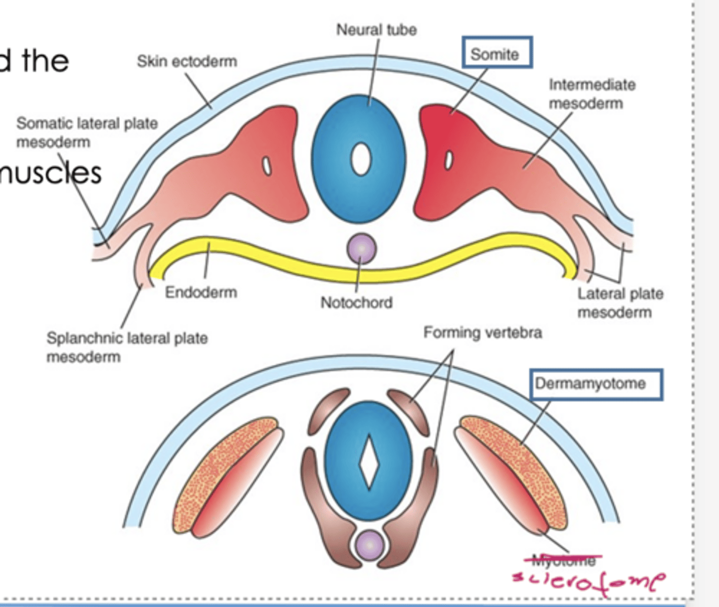
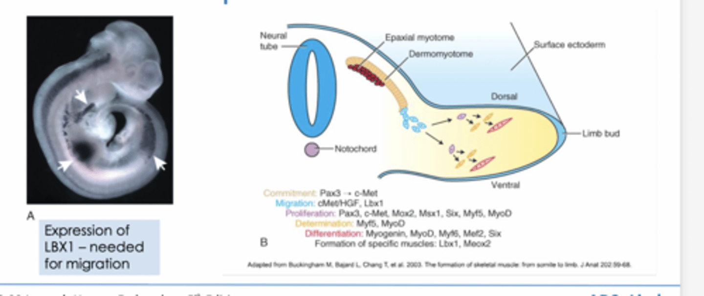
From which embryological structure do limb skeletal muscles arise?
Limb skeletal muscles arise from the somites of the paraxial mesoderm. Form blocks in a regulated pattern
Cells of the somite are subdivided into the sclertotome and the dermamytome
What does the dermomyotome give rise to?
It gives rise to skeletal muscle and dermis
myo = muscle, derma = dermis
Figure of muscle development
When do muscle precursor cells migrate into the limb bud?
Migration begins during the 5th week of development
How do muscle precursor cells reach the limb bud
Limb musculator muscle group mpves ventrallt into the limb buds. Muscle precursor cells delaminate from the dermomyotome and migrate into the limb bud during the 5th week of development, a process that requires expression of the transcription factor LBX1
How is limb musculature initially organised within the limb bud?
Once inside the limb bud, migrating myogenic cells form two condensations called the dorsal and ventral muscle masses, which will later split into specific muscle groups
Dorsal – extensors and supinators (UL)extensors and abductors (LL)
Ventral - flexors and pronators (UL) flexors andadductors (LL)
What does the dorsal muscle mass form?
It forms extensors and supinators in the upper limb and extensors and abductors in the lower limb
What does the ventral muscle mass form?
It forms flexors and pronators in the upper limb and flexors and adductors in the lower limb
Which transcription factor is required for migration of muscle precursors?
LBX1 is required for migration
ventral and dorsal muscle mass (upper limb)
ventral: anterior compartment of arm and forearm
dorsal: posterior compartment of muscles of arm and forearm
ventral and dorsal muscle mass (lower limb)
ventral: medial compartment muscles of thigh, posterior compartment of muscles of thigh, posterior compartments of leg
dorsal: anterior compartment muslces of thigh and leg
How does limb rotation affect final muscle orientation?
The upper limb rotates laterally and the lower limb rotates medially during development, which explains why extensors are posterior in the upper limb but anterior in the lower limb and why the great toe lies medially in anatomical position
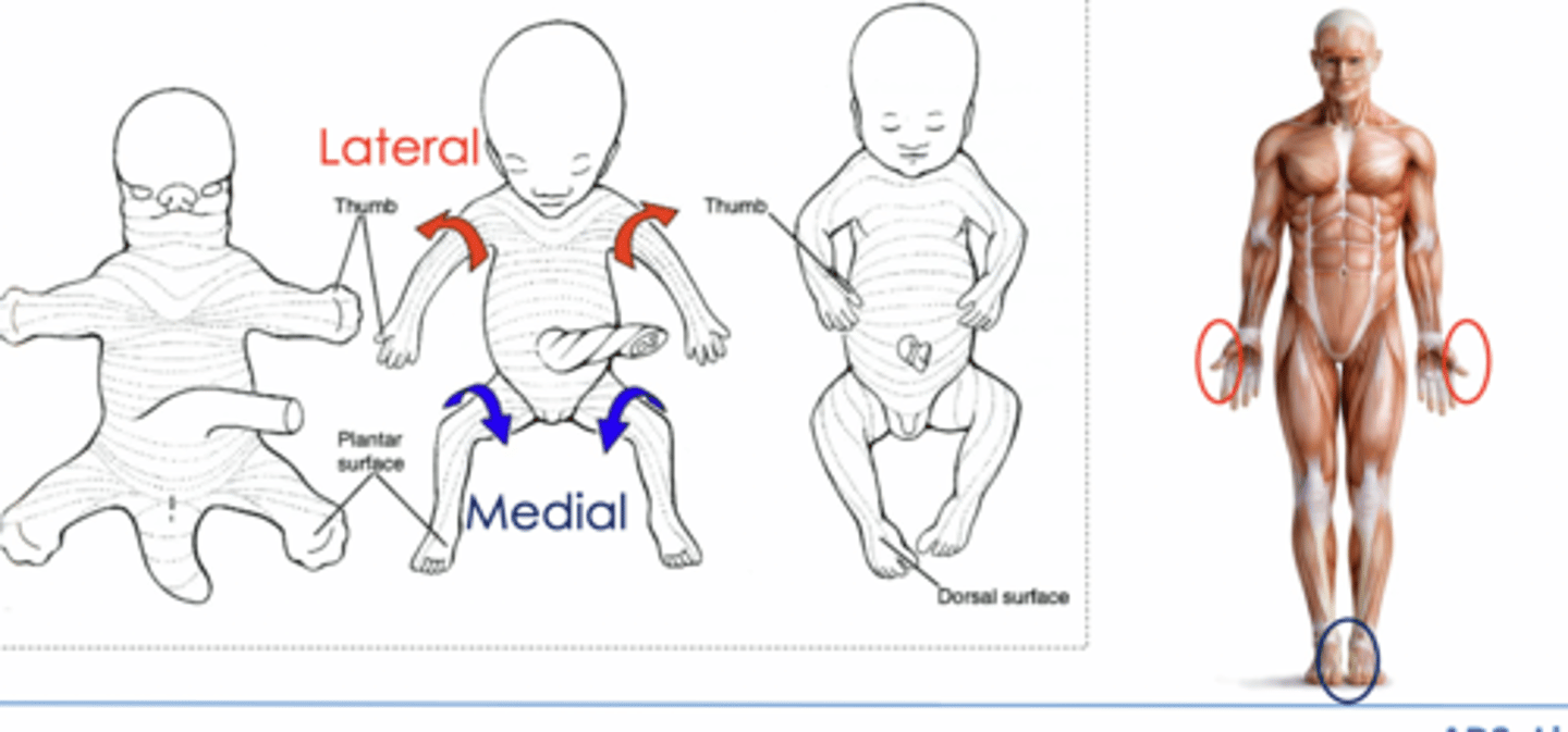
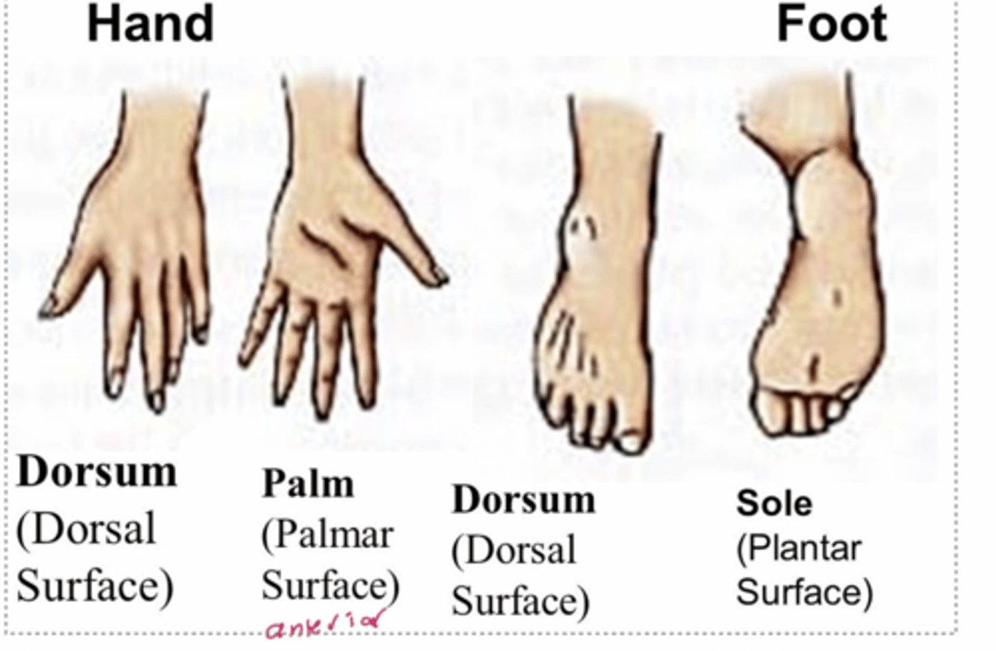
specific surfaces of the hands and feet used in anatomical
Dorsal of the foot is the top - this is because of the rotation which has occurred
Dorsiflexion and plantarflexion
muscle development process
1. primary myogenesis
2. secondary myogenesis
3. postnatal muscle growth
What is primary myogenesis?
Primary myogenesis is the embryonic stage where myoblasts proliferate and differentiation to form myocytes then fuse to form multinucleated muscle fibres (myotubes)
What is secondary myogenesis?
Secondary myogenesis is the fetal stage where most muscle fibres are formed as primary muscle fibres fuse
What cells are responsible for postnatal muscle growth?
Satellite cells (quiescent cells in muscles) are responsible for postnatal muscle growth and repair. Respond to exercise/damage and form myocytes
molecular regulators of limb muscle development
lbx1 AND others
TENDON DEVELOPMENT
major structural component of tendon
Collagen type I, found in many other body tissues. Until recently lack of suitable markers avaialble for studying tendon develpment, now we have Scx
From which embryological tissue do limb tendons arise?
Limb tendons arise from the lateral plate mesoderm
What is Scleraxis (Scx)?
Scx is an early marker of tendon development that regulates collagen type I expression
what does Scx do
Regulates type I collagen in mouse tendons
BUT not thought out as the master regulator since Scx mutant mice still have tendons that attach muscle to bone so likely works with othermarkers
Loss of Scx activity will severely disrupt force-anchoring tendons (such as those in the limb)but only affect other tendons moderately.
other genes involved in tendon development
Tenomodulin (Tnmd) – marker for differentiated tendon cells
Mohawk (Mkx) – can reduce collagen typeIa expression
Early growth response 1 (Egr1) – KO shows reduced collagen fibrilorganisation and mechanical weakness in tendons
where do tendons originate from
Tendons of different body regions originate from different embryological origins.
- Limb tendonsoriginate from the lateral platemesoderm (differsfrom skeletalmuscle)
Do limb tendons require muscle for full maturation?
Yes, limb tendons require interaction with muscle to fully mature but this varies with anatomical location
- e.g. Limb tendons will initially form without muscle, but their maturation depends onmuscle-interaction
- Axial tendons will not form if axial muscle isabsent
JOINT DEVELOPMENT
joint development diagram
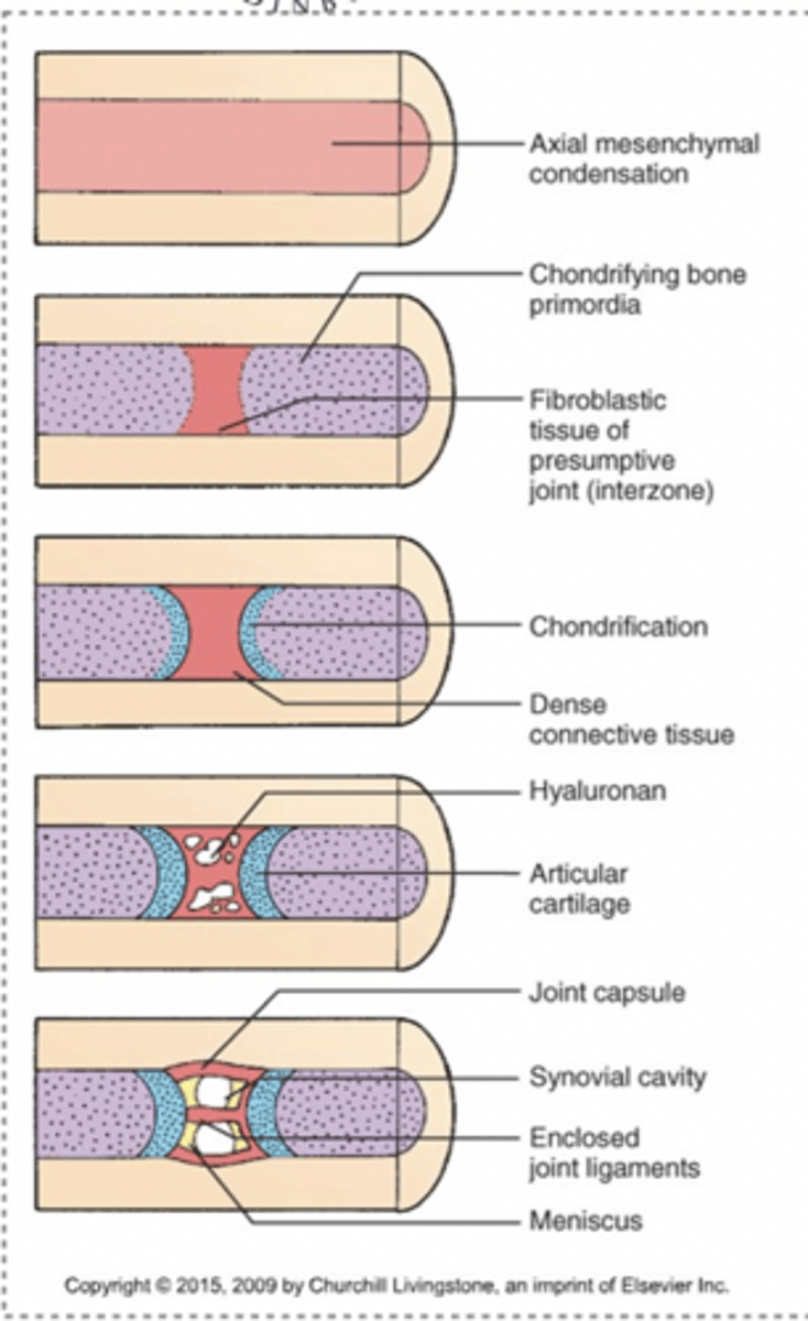
synovial joint formation process
1. Interzone forms (area between future bondes becomes fibrous tissue)
2. Articular cartilage forms on proximal and distal bone ends
3. Connective tossie condenses to form synovial tissue lining, ligaments, menisci, etc.
4. Cavitation creates a fluid filled joint cavity
What is the interzone in joint development?
The interzone is fibroblastic tissue that marks the future site of a synovial joint
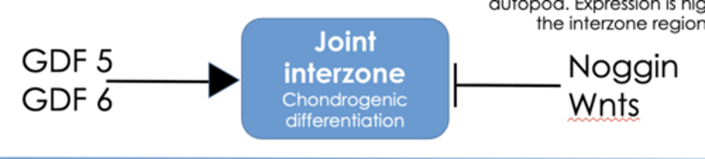
Which growth factor regulates interzone formation?
GDF5 regulates interzone and joint development
What is cavitation in joint development?
Cavitation is the formation of the synovial joint cavity
molecular regulation of joint development control
- Growth differentiation factors (GDF) 5/6
- Noggin
- Wnts
TISSUE JUNCTIONS
two interfaces of the tissue junctions
bone-tendon interface and muscle-tendon interface
What is the enthesis? (bone tendon interfaces)
The enthesis is the mechanical graded interface between tendon and bone. It transfers force from compliant tendon to stuff bone
What is the structural organisation of the enthesis from tendon to bone?
The enthesis transitions from tendon to unmineralised fibrocartilage to mineralised fibrocartilage and finally to bone, creating a gradual change in stiffness
Plug of Fibrocartilage, has a lot of water so is a shock absorber

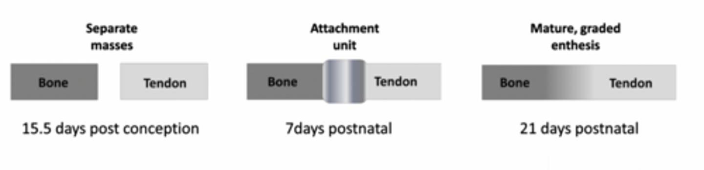
How does the enthesis develop during embryonic and postnatal life?
Bone and tendon initially develop as separate tissues in the embryo and a direct attachment forms after birth, with a mature graded enthesis developing postnatally in response to mechanical loading.
Mature enthesis not observed until 21 days postnatally.
what happens to enthesis if you paralyse a limb
Evidence that if you paralyse a limb you don't get the mature graded enthesis, important that normal forces are at play
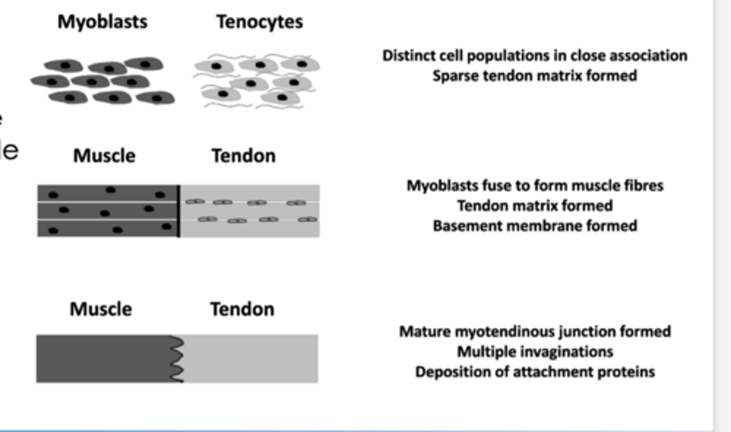
What is the myotendinous junction?
It is the specialised interface between muscle and tendon that increases surface area for force transmission
An important transition site between the contractile muscle tissue and the strong, elastic tendon
How is the myotendinous junction structurally adapted for force transmission?
The myotendinous junction increases surface area through structural folding, allowing stronger attachment and efficient force transfer
Two different tissue masses
(myoblasts and the coyotes), these will fuse together but there is still separation between each other
What is the significance of muscle-tendon interaction during development?
Muscle presence maintains expression of tendon markers such as Scleraxis and is necessary for proper organisation and mechanical strength of the tendon
- seen in varies species; mouse, zebrafish, chick
- In drosophila, tendon cells do not need muscle to form but needinteraction with muscle cells to fully differentiate and mature.
MECHANOBIOLOGY IN LIMB BIOLOGY
What is mechanobiology in limb development?
Mechanobiology is the study of how mechanical forces such as tension, compression and muscle contraction influence bone, tendon, ligament and joint development
Why is muscle contraction important in limb development?
Muscle contraction provides mechanical forces that influence bone development growth, joint formation and tissue maturation
- without it we would not have fully formed tissues
mechanobiology in. limb development Bone development and growth
growth generated strain Wolpert proposed that radial stress from the perichondrium restricts circumferential growth and promotes longitudinal bone growth, although the exact mechanisms are still not fully understood
Muscular loading Directional strain on bone from musclecontraction. Paralysis results in skeletal malformations . Generally accepted view that musclecontraction influences bone development andremodelling
mechanobiology in limb development: Enthesis Maturation
At birth the enthesis is a simple attachment but mechanical input from muscle contraction is required for it to mature into a graded and well-organised interface
mechanobiology in limb development: Joint Formation
Muscle contraction helps drive joint cavitation and shapes developing articular surfaces, and absence of muscle activity can prevent proper joint cavity formation
What happens to joint development in muscle-less or paralysed embryos?,
In the absence of muscle contraction, joint cavitation fails to occur and cartilage markers such as Sox9 and Col2a1 remain highly expressed
Why is mechanobiology clinically important
Understanding mechanobiology helps explain skeletal deformities caused by paralysis or immobilisation and guides rehabilitation, tissue engineering and regenerative medicine
What happens if muscle contraction is absent during development?
Absence of muscle contraction leads to skeletal malformations and failure of proper joint formation
What is phocomelia?
Phocomelia is a congenital condition where long bones are absent or severely shortened
What is polydactyly?
Polydactyly is duplication of digits
What is syndactyly?
Syndactyly is failure of separation of digits
WORKSHOP QUESTIONS
Which limb bone forms by endochondral ossification?
The femur forms by endochondral ossification
Which bone forms by intramembranous ossification?
The occipital bone forms by intramembranous ossification
Which signalling molecule controls differentiation of mesenchymal cells into chondrocytes?
Indian hedgehog (Ihh) controls chondrocyte differentiation
What is the function of RUNX2 in bone development?
RUNX2 is required for osteogenic differentiation and bone mineralisation
Which stain colours mineralised bone red?
Alizarin red stains mineralised tissue red
Around what age do the last growth plates fuse in humans?
Growth plates fuse at around 20 years of age
At what age does the secondary ossification centre appear in the patella?
The patella secondary ossification centre appears around 3 to 5 years of age
Why is knowledge of primary and secondary ossification centres important?
It helps identify normal growth patterns and distinguish growth plates from fractures
Give one clinical use of knowing ossification centre timing?
It is used for skeletal age estimation in radiology and forensics
From which embryological structure do limb muscles originate?
Limb muscles originate from somites
Into which two parts do somites subdivide?
Somites subdivide into the sclerotome and dermomyotome
What else does the dermomyotome form besides skeletal muscle?
The dermomyotome also forms the dermis of the skin
During which week do muscle precursors migrate into the limb bud?
Muscle precursors migrate during the 5th week of development
Which muscle mass gives rise to the anterior compartment of the upper limb?
The ventral muscle mass forms the anterior compartment of the upper limb
How does limb rotation explain final lower limb orientation?
Medial rotation of the lower limb causes the great toe to lie medially in anatomical position
Which transcription factor is required for migration of muscle precursors?
LBX1 is required for migration
Do all muscles develop at the same time or via the same signals?
No, different muscles develop at different times and use different signals