An overview of Dental Anatomy
1/109
There's no tags or description
Looks like no tags are added yet.
Name | Mastery | Learn | Test | Matching | Spaced | Call with Kai |
|---|
No analytics yet
Send a link to your students to track their progress
110 Terms
Anterior
Forward, towards front of mouth and midline
Apical
Towards root, apex of tooth
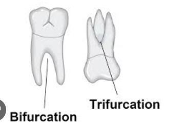
Bifurcated
Single tooth with 2 roots
Buccal
Surface facing cheeks
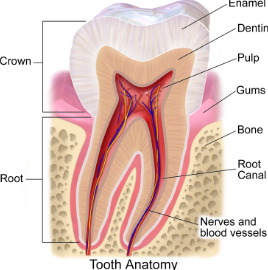
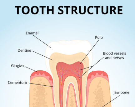
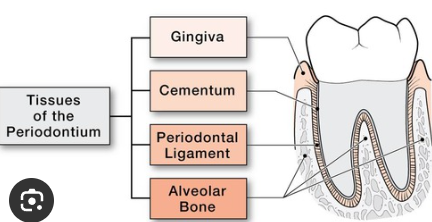
Cementum
Tissue covering root of tooth
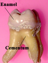
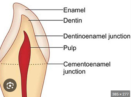
Cementoenamel junction (CEJ)
Line where enamel and cementum meet
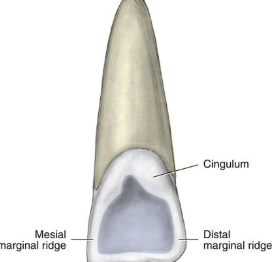
Cingulum
Small bump near cervcial third of anterior tooth on lingual side
Crown
Portion of tooth above gumline — starting at cementoenamel junction


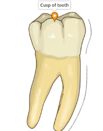
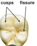
Cusp
Top portion of tooth, tip or point
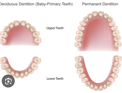
Deciduous teeth
Primary teeth (first set of teeth)
Dentin
Tissue of the tooth between pulp and enamel and cementum; majority of the tooth
Dentition
Set of teeth
(could be primary, mixed, or permanent dentition)
Dentoenamel junction (DEJ)
Where enamel meets dentin
Distal
Surface of the tooth away from midline of face
Dorsal
Top surface
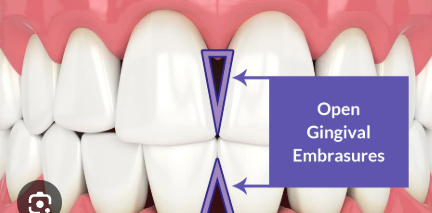
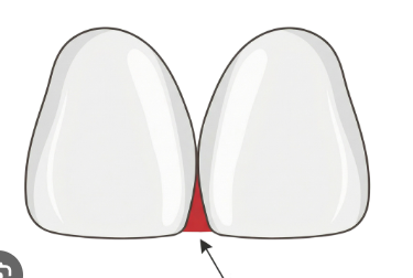
Embrasure
Space between teeth — begins at contact area and widens down towards gingiva
Filled by interdental papillae
Enamel
Tissue covering crown of the tooth; hardest substance in the body
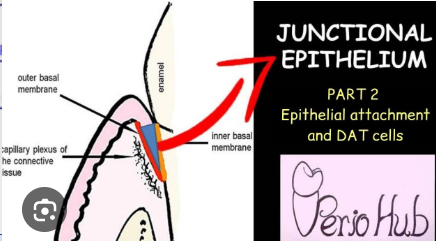
Epithelial attachment
Base of sulcus where epithelial tissue is attached to tooth
Facial
Towards lips or cheeks
Buccal — cheeks
Labial — lips
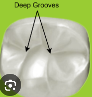
Fissure
Natural groove in tooth
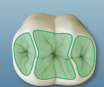
Fossa
Shallow depression in tooth
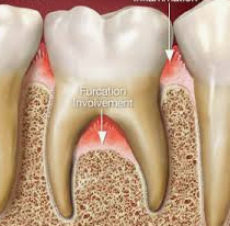
Furcation
Multi—rooted teeth, area where roots divide
Gingiva
Tissue that surrounds the teeth, aka gums
Groove
Depression in tooth that is narrow and linear
Incisal
Towards the cutting edge of the anterior teeth — opposite of apex of tooth
Interdental
Space between 2 adjacent teeth
Interproximal
Surface between the 2 adjacent teeth
Keratinized
Firm stippled texture — like tissue of attached gingiva
Lateral
Sides
Lingual
Towards tongue
Mandibular
Lower jaw
Maxillary
Upper jaw
Mesial
Surface of the tooth facing towards the midline of the face
Midline
Imaginary line that divides the body into equal right and left halves
Mucogingival junction
Area where mucosa meets attached gingiva
Occlusal
Chewing surface of posterior (back) teeth
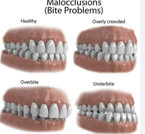
Occlusion
term to define how upper and lower teeth meet when closing the jaws
Palate
Roof of mouth— hard and soft palate
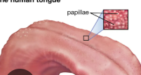
Papillae
Small projection of tissue — often nipple like
Periapical
Around apex of tooth
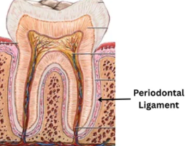
Periodontal ligaments
Thin fibers connecting tooth to bone
Peridontium
Surrounding supporting structures of the teeth
Posterior
Towards back of mouth, behind
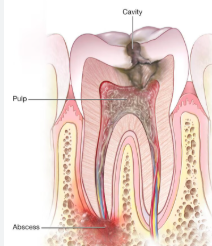
Pulp
Most iner part of tooth, contains blood vessels and nerves
Quadrant
Mouth divided into 4 quadrants
2 maxillary
2 mandibular
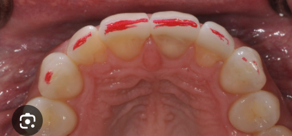
Ridge
Rounded, elevated, linear area on enamel of tooth
There are 5 types of ridges on teeth
Marginal
Triangular
Oblique
Transverse
Cingula
Succedaneous
Permanent teeth that replace the 20 primary teeth
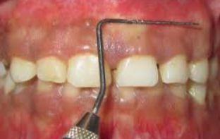
Sulcus
Formed by free gingiva (non attached) lying nxt to tooth
Trough area, also known as gingiva crevice
Ventral
Towards bottom surface; underside of tooth is — ventral surface
______________________________________________________
Bone structures of the face
Everything below this point
Ethmoid
Midline cranial bone
Frontal
Bone that forms the forehead
Lacrimal
Paired bones that form the wall of the orbit
Mandible
Bone that forms the lower jaw
Maxilla
Bone that forms the upper jaw
Nasal
Paired bones that form the bridge of the nose
Occipital
Bone that forms the posterior portion of the head
Parietal
Paired bones at the top of the skull. They articulate with each other and other bones in the skull
Sphenoid
Midline bone that has several processes (bone areas or plates) associated with it
Temporal
Paired bones that form the lateral walls of the skull and articulate with the mandible at the temporomandibular joint
Zygomatic
Paired facial bones that form the cheeks
___________________________________________
Facial landmarks
everything below this point
Ala
Wing of the nose
Inner canthus of the eye
The inner corner of the eye
Labial commissures
Corners of the mouth
Labiomental groove
Area that separates the lower lip and the chin
Naris
Nostril
Nasal septum
Vertical separation of the nasal cavity
Nasiolabial sulcus
Groove that runs upward from the commissures of the mouth and the ala of the nose
Nasion
Midline junction between the eyes just below the eyebrows
Outer canthus of the eye
The outer corner of the eye
Philtrum
Vertical groove or depression in the midline above the upper lip
Tragus
Triangle flap of cartilage that is at the external opening of the ear
Tubercle of the lip
Midline of the upper lip that has a small tissue projection
Vermillion border
Where the skin and lips meet
Vermillion zone
The reddish portion of the lips
___________________________________________
Landmarks of the oral cavity
Tissues and structures below this point
Fauces
Passageway from oral cavity to pharynx
Frenum
Raised folds of tissue that extend from the alveolar and the buccal and labial mucosa
Gingiva
Mucosal tissue surrounding portions of the maxillary and mandibular teeth and bone
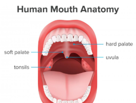
Hard palate
Anterior portion of the palate which is formed by the processes of the maxilla
Incisive papilla
A tissue projection that covers the incisive foramen on the anterior of the hard palate, just behind the maxillary central incisors
Maxillary tuberosity
A bulge of bone posterior to the most posterior maxillary molar
Maxillary/Mandibular tori
Normal bony enlargements that can occur either on the maxilla or mandible
Mucosa
Mucous membrane lines the oral cavity. It can be highly keratinized (such as what covers the hard palate), or lightly keratinized (such as what covers the floor of the mouth and the alveolar processes) or thinly keratinized (such as what covers the cheeks and inner surfaces of the lips
Palatine rugae
Firm ridges of tissues on the hard palate
Parotid papilla
Slight fold of tissue that covers the opening to the parotid gland on the buccal mucosa adjacent to maxillary first molars
Pillars of Fauces
Two arches of muscle tissue that defines the fauces
Soft palate
Posterior portion of the palate. This is non-bony and is comprised of soft tissue
Sublingual folds
Small folds of tissue in the floor of the mouth that cover the openings to the smaller ducts of the sublingual salivary gland
Submandibular gland
Located near the inferior border of the mandible in the submandibular fossa
Tonsils
Lymphoid tissue located in the oral pharynx
Uvula
A non-bony, muscular projection that hangs from the midline at the posterior of the soft palate
Vestibule
Space between the maxillary or mandibular teeth, gingiva, cheeks and lips
Wharton’s duct
Salivary duct opening on either side of the lingual frenum on the ventral surface of the tongue
____________________________________________________________
Tongue landmarks
everything beyond this point
Apex of the tongue
The tip of the tongue
Circumvallate papillae
Two v-shaped rows of larger, flat, cup shaped papillae on the posterior dorsum of the tongue. Each contains taste buds
Dorsal surface
The top surface of the tongue
Filiform papillae
Fine, small, cone shaped papillae covering most of the dorsum of the tongue. They are responsible for giving the tongue its texture and are responsible for the sensation of touch