Lecture 2 - Bacteria I
1/101
There's no tags or description
Looks like no tags are added yet.
Name | Mastery | Learn | Test | Matching | Spaced | Call with Kai |
|---|
No analytics yet
Send a link to your students to track their progress
102 Terms
Bacterial and archaea structure and function (recap)
- Prokaryotes differ from eukaryotes in the size and simplicity (most prokaryotes lack a internal membrane systems, including nucleus)
Prokaryotes are divided into two taxa
Bacteria and Archaea
Shape, Arrangement, and Size
SHAPE
- Cocci and rods most common
- various others
ARRANGEMENT
- determined by plane of division
- determined by separation (after division) or not
SIZE
- varies
Shape - Cocci (s., coccus)
Spheres
Shape: Cocci - diplococci (s., diplococcus)
Pairs
Shape: Cocci - Streptococci
Chains
Shape: Cocci - Staphylocci
Grape-like clusters
Shape: Cocci - Tetrads
4 cocci in a square gemoetry
Shape: Cocci - Sarcinae
Cubic configuration of 8 cocci
Shape & Arrangement: Bacilli (s., bacillus)
Rods
Shape & Arrangement: Coccobacilli
Very short rods
Shape & Arrangement: Vibrios
Resemble rods, comma shaped
Shape & Arrangement: Spirilla (s., spirillum)
rigid helices
Shape & Arrangement: Spireochetes
Flexible helices
Size
- Smallest - 0.3 micrometers (Myocoplasma)
- Average rod: 1.1 - 1.5 x 2-6 micrometers (E.coli)
- Very large: 200-700 micrometers
Size: Oscillatoria Red blood cell
7000 nanometers
Size: E. Coli
1300 x 4000 nanometers
Size: Strepococcus
800-1000 nanometers
Size: poxvirus
230 x 320 nanometers
Size: Influenza virus
85 nanometers
Size: T2 E.coli bacteriophage
65 x 95 nanometers
Size: Tobacco mosaic virus
15 x 300 nanometers
Size: Poliomyelitis virus
27 nanometers
1 micrometer = ? nanometers
1000 nanometers
Cell organization in Prokaryotes
External structures
Cell envelope
Cytoplasm
Components of a prokaryotic cells
- Nucleoid
- Ribosome
- Cytoplasmic Membrane
- Cell wall
- Capsule
- Flagella
- Fimbriae
Prokaryotes: Bacterial cell envelope
Plasma membrane, Cell wall, Layers outside the cell wall (Sometimes)
Prokaryotes: Bacterial Plasma membrane
absolute requirements for all living organisms
Plasma Membrane Functions
- Encompasses the cytoplasm
- Selectively permeable barrier
Interacts with external environment:
- Receptors for detection of and response to chemicals in surroundings
- transport systems
- Metabolic processes (ATP)
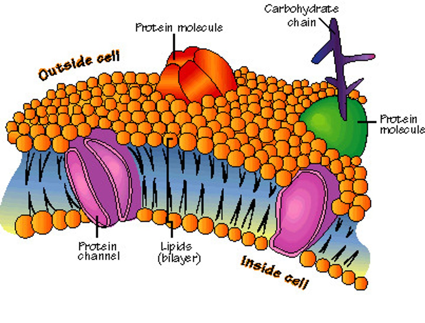
Fluid Mosaic Model of Membrane structure
Lipid bilayers with floating proteins:
- Amphipathic lipids
= Polar ends (hydrophilic - interact with water)
= Non-polar tails (hydrophobic - insoluble in water)
Membrane proteins
Fluid Mosaic Model
Structural model of the plasma membrane where molecules are free to move sideways within a lipid bilayer.
Membrane Proteins: Perpheral
- loosely connected to membrane
- easily removed
Membrane proteins: Integral
- Amphipathic = embedded within membrane
- Carry out important functions
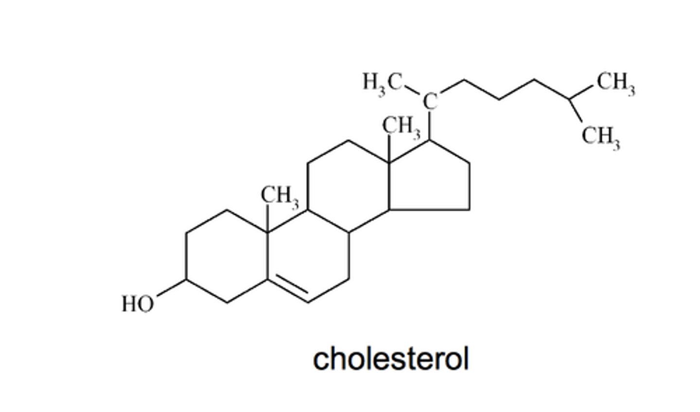
Membrane Steroids
Cholesterol (a steroid) is found in eucaryotes
Membrane Hopanoids
A bacteriohopanetetrol (a hopanoid) is found in bacteria
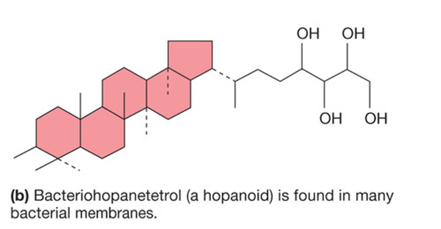
Bacterial Lipids
- Bacterial saturation levels of membrane lipids reflect the environmental conditions such as temperature
- Membranes lack sterols but do contain sterol-like molecules, instead contain hopanoids (Stabilize membrane)
Bacterial cell wall
peptidoglycan (only bacteria) - rigid structure hat lies just outside the cell membrane
Bacterial Cell Wall - gram positive
- Stain purple; THICK peptidoglycan
Bacterial Cell Wall - gram negative
- Stain pink or red; THIN peptidoglycan and outer membrane
Cell Wall Functions
- Maintains the shape of the bacterium (almost all bacteria have one)
- Helps protect cells from osmotic lysis
- Helps protect from toxic materials
- May contribute to pathogenicity (ability to cause disease)
peptidoglycan structure
- Meshlike polymer of identical subunits forming long strands
Peptidoglycan alternating sugars
2 ALTERNATING SUGARS:
- N-acetylglucosamine (NAG)
- N-acetylmuramic acid
Alternating D- (only bacteria) and L- (other protein structures) amino acids
4 most common amino acids
= L-alanine
= D-glutamic acid
= Meso-diaminopimelic acid
= D-alanine
Strands are corossed link
- Peptidoglycan strands have a helical shape
- Peptidoglycan chains are crosslinked by peptides for strength
- Nature of cross linking also provides flexibility to an extent
Chemical structure of cross-linking peptidoglycan components of cell wall
- Peptide chain
- N-acetyl glucosamine
- Pentapeptide bridge
- N-acetyl muramic acid
Gram-positive cell walls
- Composed primarily of peptidoglycan (lots)
- May also contain large amounts of TEICHOIC ACIDS (negatively charged)
- Some gram-positive bacteria have layer of proteins on surface of peptidoglycan
Teichnoic Acid
- Help maintain cell envelop
- Protect from environmental substances
- May bind to host cells
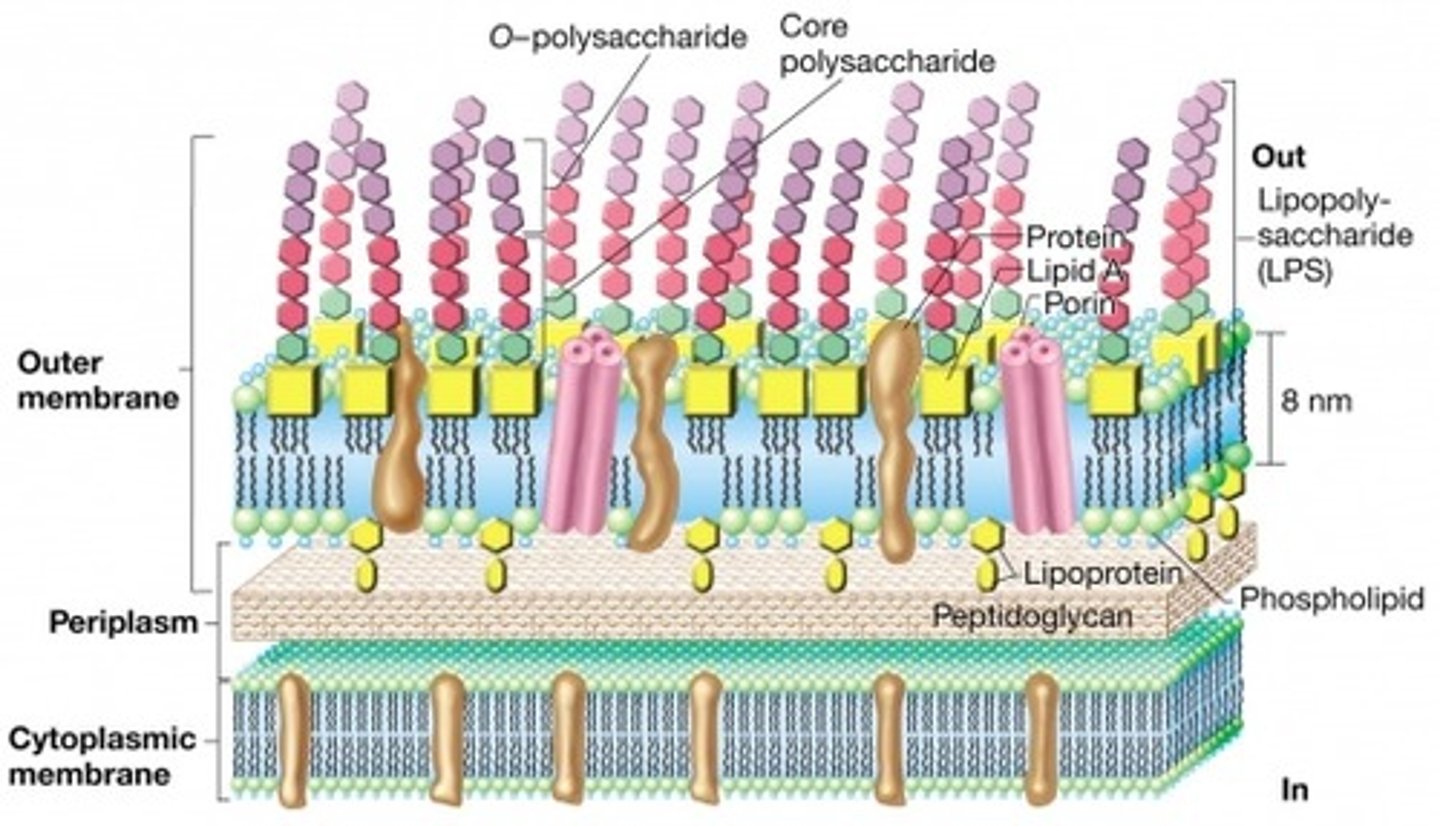
Gram-negative cell walls (has outer membrane)
- More complex than gram-positive
- Consists of a thin layer of peptidoglycan surrounded by an outer membrane
- Outer membrane composed of lipids, lipoproteins, and lipopolysaccharide (LPS)
- No teichoic acid
Gram-negative outer membrane
- Outer membrane lies outside the thin peptidoglycan layer
- Braun's lipoproteins connect outer membrane to peptidoglycan
- Other adhesion sites reported
Gram-negative cell walls
- Peptidoglycan is ~5-10% of cell wall weight
- Have a periplasmic space between the outer membrane and the inner membrane
Periplasmic Space
The space between the inner and outer cell membranes in Gram-negative bacteria.
- May constitute 20-40% of cell volume
- Many enzymes present in periplasm
= hydrolytic enzymes, transports proteins, and other proteins
Structural Features of Gram-positive and gram-negative cell walls
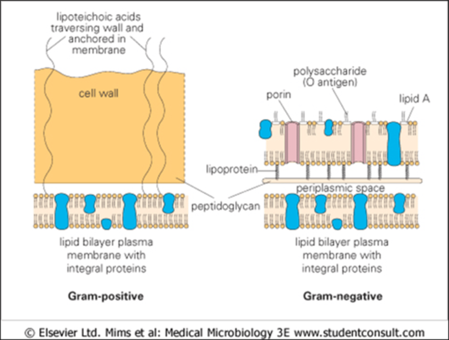
Lipopolysaccharides (LPSs) in gram (-) negative outer membranes
CONSISTS OF THREE PARTS:
- Lipid A (embedded in the outer membrane)
- Core polysaccharide (extends out from the cell)
- O side chain or O antigen (extends out from the cell) - immunogenic
Gram negative bacterial cell wall
Importance of LPS
- Contributes to negative charge on cell surface
- Helps stabilize outer membrane structure
- May contribute to attachment to surfaces and biofilm formation
- Creates a permeability barrier
- Protection from host defenses (O antigen)
- Can act as an endotoxin (lipid A)
LPS: can act as a endotoxin (lipid A)
"endotoxin" response elicited by gram (-) bacteria only...dont confuse with exotoxin shock or the broader term septic shock
Gram-negative outer membrane permeability
more permeable than plasma membrane due to presence of porin proteins and transporter proteins
- Porin proteins form channels through which small molecules
- (600-700 daltons) can pass
Osmosis
Movement of solvent (such as water) through a semipermeable membrane) such as the plasma membrane in a living cell) into a solution of higher solute (such as sugar or salt) concentration that tends to equalize the concentrations of the solute on the two sides of that semipermeable membrane.
osmosis example
Osmotic Protection - Hypotonic Environments
Solute concentration outside the cell is less than the inside the cell.
Water moves into cell and cell swells - can burst
Osmotic Protection - Hypertonic Environment
Solute concentration outside the cell is higher than inside, water leaves the cell.
Plasmolysis occurs - cell shrinks
Plasmolysis
Cell shrinks
Evidence of protection nature of the cell wall - Lysozyme
breaks down the bond between N-acetyl glucosamine and N-acetylmuramic acid
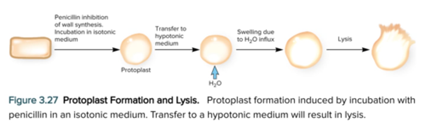
Evidence of protection nature of the cell wall - Penicillin
Inhibits peptidoglycan synthesis
Cell wall: Lysozyme & Penicillin
If cells are treated with either of the above they will lyse if they are in a hypotonic solution
Cells may survive loss of cell wall ONLY in isotonic conditions
- Protoplasts
- Spheroplasts
These forms are "man-made" by manipulation of bacteria using cell wall-damaging agents (lysozyme or penicillin)
Protoplasts
May survive loss of cell wall in only an isotonic environment:
Protoplasts (cell wall-less form derived from gram+ bacteria)
Spheroplasts
May survive loss of cell wall in only an isotonic environment:
Spheroplasts (cell wall-less form derived from gram- bacteria)
Cells survive loss of cell wall only isotonic environments
- Another name- Lawless (L) forms (derived from bacterial when treated with penicillin and are susceptible to lysis in hypotonic solutions; can revert back to normal form)
Mycoplasma (exception)
- Does not produce a cell wall EVER
- Plasma membrane more resistant to osmotic pressure and can survive in hypotonic environment - born this way
Lysis of a protoplast
Components outside of the cell wall
Extracellular material secreted by bacteria in form of:
- Slime layer
or
- Capsule
Slime layer
- Are diffuse, unorganized and easily removed
- Made of exopolysaccharides, glycoproteins, and glycolipids
- Slime may protect the bacteria from harsh environments and aid in adherence
Capsule
- Capsule is an amorphous gelatinous layer surrounding the entire bacterium
- Composed of polysaccharide or sometimes protein (e.g. Bacillus anthracis)
- The sugar component of the capsule varies in different bacterial species and also determines serological type within a species
Bacterial and archaeal cytoplasmic structures
- Cytoskeleton
- Intracytoplasmic membranes
- Inclusions
- Ribosomes
- Nucleoid and plasmids
Protoplast and cytoplasm
- Protoplast is plasma membrane and everything within
- Cytoplasm = material bounded by the plasmid membrane
Inclusions
- Granules of organic or inorganic material that are stockpiled by the cell for future use
- Some are enclosed by a single-layered membrane
= membranes vary in composition
= some made of proteins; others contain lipids
= may be referred to as microcompartments
Storage inclusions
- storage of nutrients, metabolic end products, energy, building blocks
- glycogen storage
- carbon storage (poly-B-hydroxybutyrate (PHB))
- phosphate - polyphosphate (volutin)
- amino acids - cyanophycin granules
Inclusions: Gas Vacuoles
- found in aquatic, photosynthetic bacteria and archaea
- provides buoyancy in gas vesicles
Inclusions: Magnetosomes
- found in aquatic bacteria
- magnetic particles for orientation in earth's magnetic field
- cytoskeletal protein MamK (helps form magnetosome chain)
Ribosomes
COMPLEX STRUCTURES:
- Consisting of protein and RNA
- sites of protein synthesis
ENTIRE RIBOSOME:
- bacterial and archaea ribosome = 70S
- eukaryotic (80S)
- S = svedburg unit
The nucleoid
- Irregular shaped region in bacteria and archaea
- Usually not membrane bound
- Location of chromosome and associated proteins
- Usually 1 (a closed circular, double-stranded DNA molecule
- Supercoiling and nucleoid associating proteins (NAP) probably aid in folding (different proteins differ from histones
Plasmids (extra)
- Extrachromosomal DNA
= found in bacteria, archaea, some fungi
= usually small, closed circular DNA molecules
- Exist and replicate independently of chromosome
= episomes - may integrate into the chromosome
- Contain few non-essential genes (non-core function)
= confer a selective advantage-additive features to host (e.g., drug resistance) - are helpful but NOT required for core survival under normal circumstances
Plasmids (continued)
- May exist in many copies in cell
- Inherited stably during cell division
- curing is the loss of a plasmid
- classification of plasmids based on mode of existence, spread, and function
External structures
- Extend beyond the cell envelop in bacteria and archaea
FUNCTION:
- protection, attachment to surfaces, horizontal gene transfer, cell movement
- pili, fimbriae, and flagella
Fimbriae
- short, thin, hairlike, proteinaceous appendages (up to 1000/cell)
- mediate attachment to surfaces
- some (type IV fimbriae) required for motility or DNA uptake
Sex Pili
- similar to fimbriae except longer, thicker, and less numerous (1-10/cell)
- genes for formation found on plasmids
- required for conjugation
Flagella
- threadlike, locomotor appendages extending outward from the plasma membrane and cell wall
FUNCTIONS:
- motility and swarming behaviour
- attachment to surfaces (may function as a virulence factor)
Bacteria Flagella
- thin, rigid protein structures that cannot be observed with bright-field microscope unless specially stained
- ultrastructure composed of three parts
- pattern of flagellation varies
Patterns of flagella distribution
- monotrichous
- polar flagellum
- amphitrichous
- lophotrichous
- peritrichous
Monotrichous
one flagellum
Polar flagellum
flagellum at one end of the cell
amphitrichous
one flagellum at each end of the cell
lophotrichous
cluster of flagella at one or both ends
Peritrichous
spread over entire surface of cell
three parts of a flagella
- Filament
- Hook
- Basal body
Flagella - Filament
- extends from cell surface to the tip
- hollow, rigid cylinder
- composed of the protein flagellin
- some bacteria have a sheath around filament
Flagella - Hook
- links filament to basal body
Flagella - Basal Body
- series of rings that drive flagellar motor
Endospore Structure (concentrated cell)
- Spore surrounded by a thin covering called exosporium
- Thick layers of protein form the spore coat
- Cortex, beneath the coat, thick peptidoglycan
- Core has nucleoid and ribosomes