HEAD AND NECK CUMULATIVE
 Call Kai
 Learn
Learn Practice Test
 Spaced Repetition
 Match
Match Flashcards
 Knowt Play
1/595

There's no tags or description
Looks like no tags are added yet.
Name | Mastery | Learn | Test | Matching | Spaced | Call with Kai |
|---|
No analytics yet
Send a link to your students to track their progress
596 Terms
What are the two main parts of the skull?
The calvaria (upper part) and cranial floor (base of the skull).
What is the significance of infant fontanels?
Infant fontanels allow for skull flexibility during birth and later fuse to form adult sutures.
How many cranial bones are there, and what are they?
There are 8 cranial bones: frontal, parietal (2), temporal (2), occipital, sphenoid, and ethmoid.
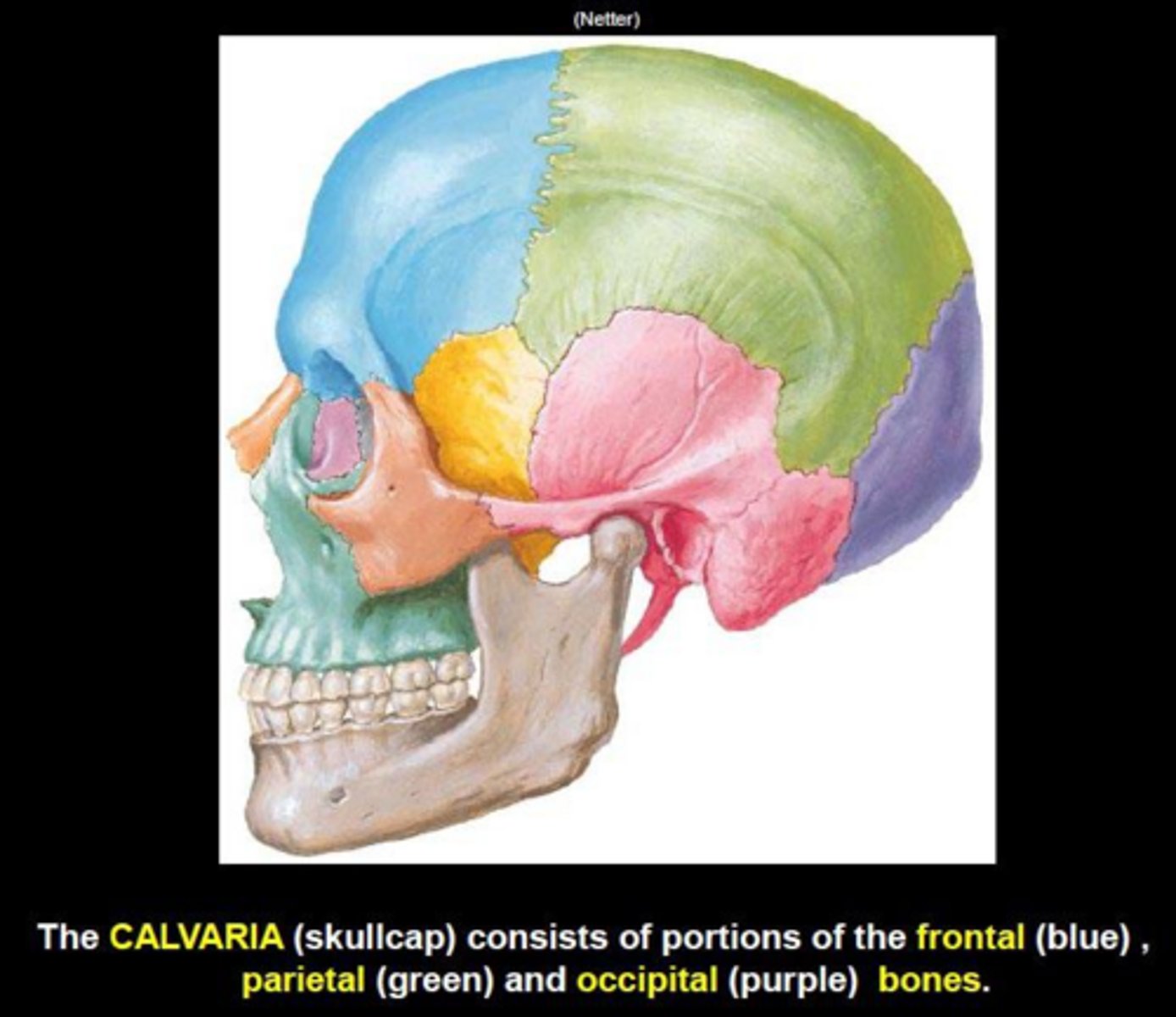
How many facial bones are there, and what are they?
There are 14 facial bones: maxilla (2), mandible, zygomatic (2), nasal (2), palatine (2), lacrimal (2), inferior nasal concha (2), and vomer.
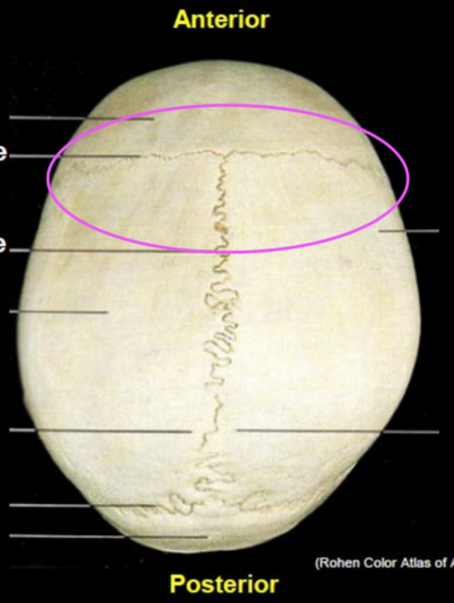
Coronal suture
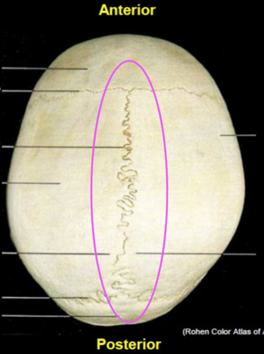
Sagittal suture
Lambdoid suture
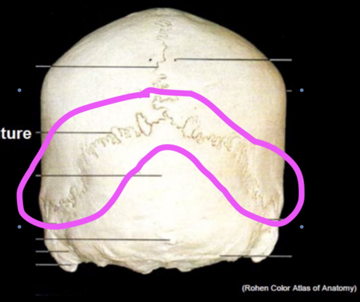
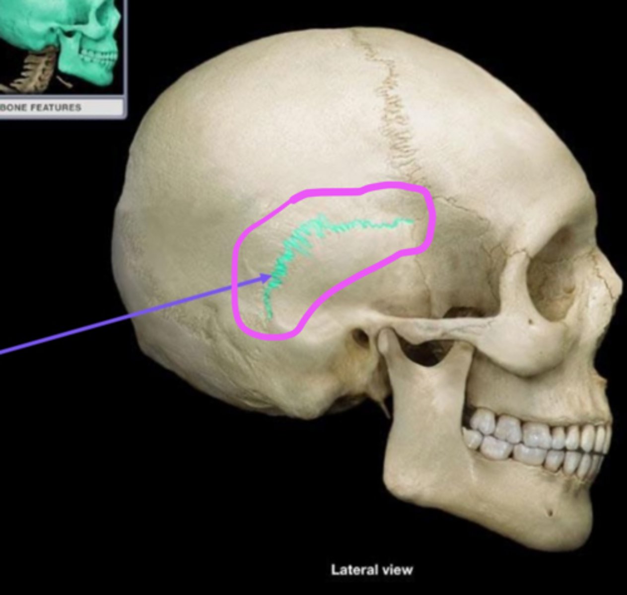
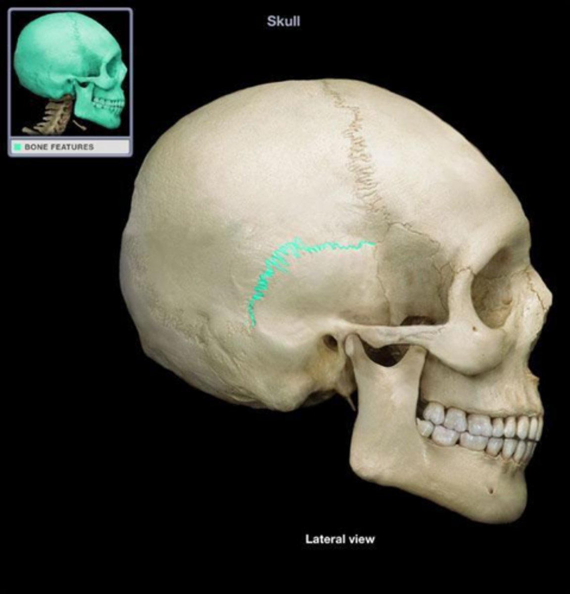
Squamous Suture
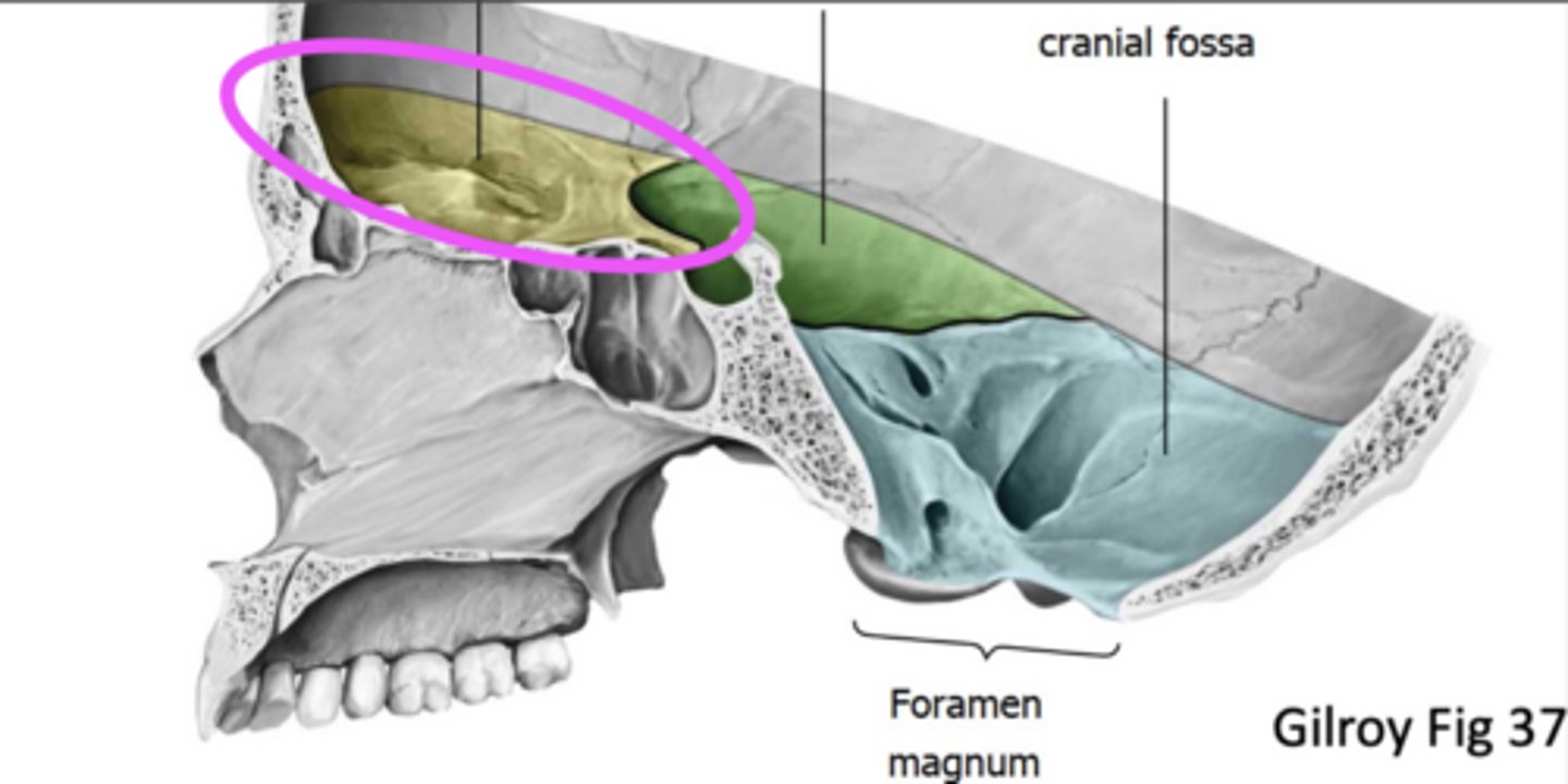
Anterior cranial fossa
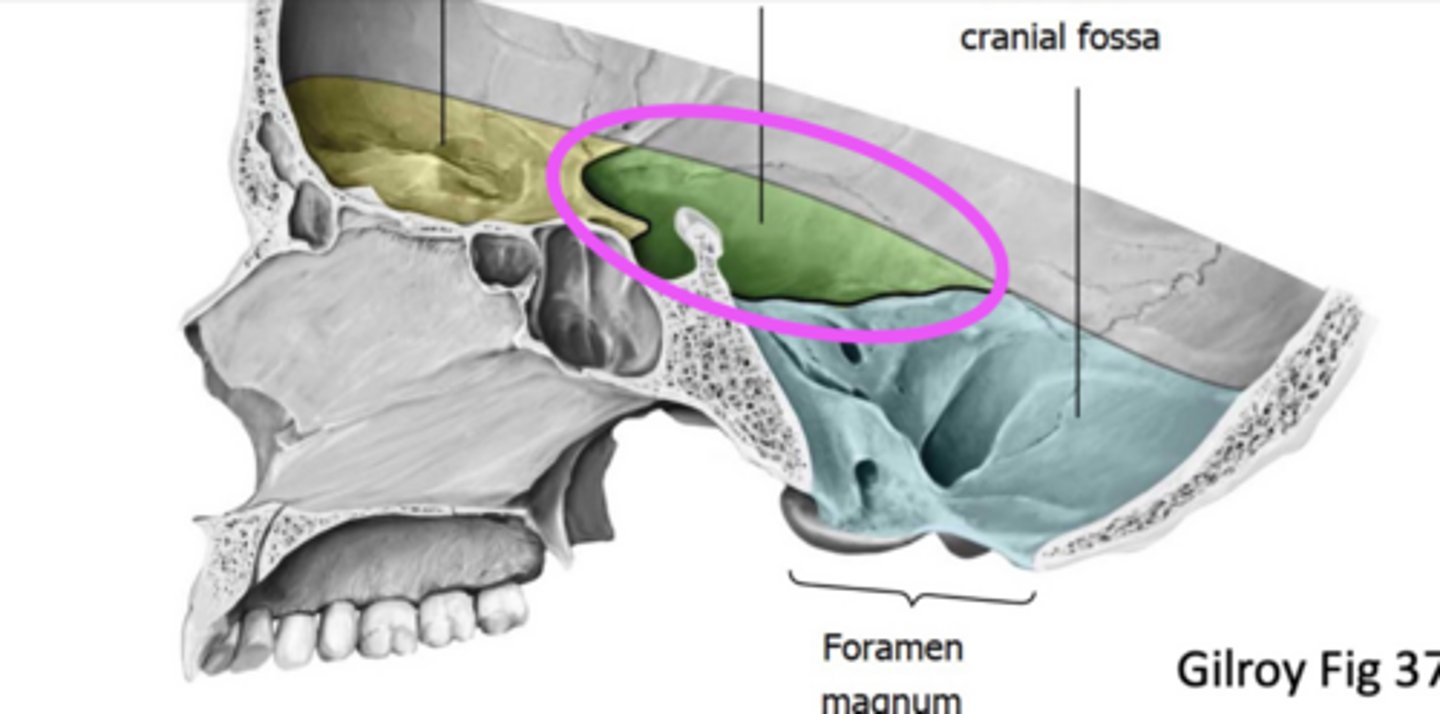
Middle cranial fossa
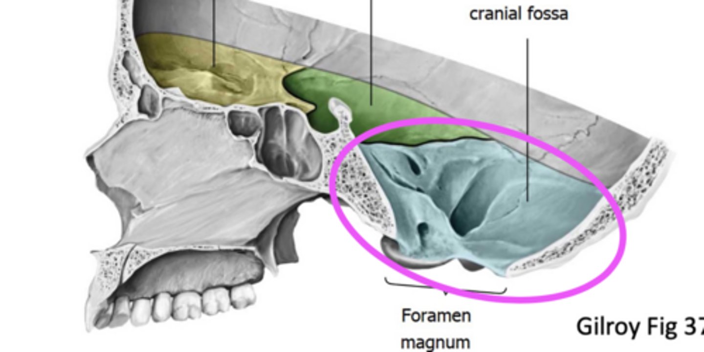
Posterior cranial fossa
The glabella on the frontal lobe is the ------ area between the eyes.
smooth
What are the three regions of the cranial floor?
The anterior, middle, and posterior cranial floor.
What is the role of foramina in the skull?
Foramina allow the passage of significant nerves and blood vessels.
What are cranial nerves and why are they important?
Cranial nerves are responsible for various functions, and damage to any nerve can lead to specific functional deficits.
What are the actions of facial expression muscles?
Facial expression muscles control movements such as smiling, frowning, and other expressions.
What are the main components of the skull?
The skull consists of the calvaria (skull cap), cranial cavity, two orbits, two ears, two nasal cavities, and an oral cavity.
What is the calvaria?
The calvaria is the skull cap formed from many fused bony plates that protect the upper cranium.
What are infant fontanels?
Infant fontanels are soft tissues in infants that later fuse to form solid sutures in adults.
How many cranial bones are there?
There are 8 cranial bones.
How many facial bones are there?
There are 14 facial bones.
What is the significance of the cranial fossae?
Cranial fossae are depressions in the skull that accommodate the brain and its structures.
What is the function of the cribriform plate of the ethmoid bone?
The cribriform plate allows olfactory nerves to penetrate the skull into the nasal area.
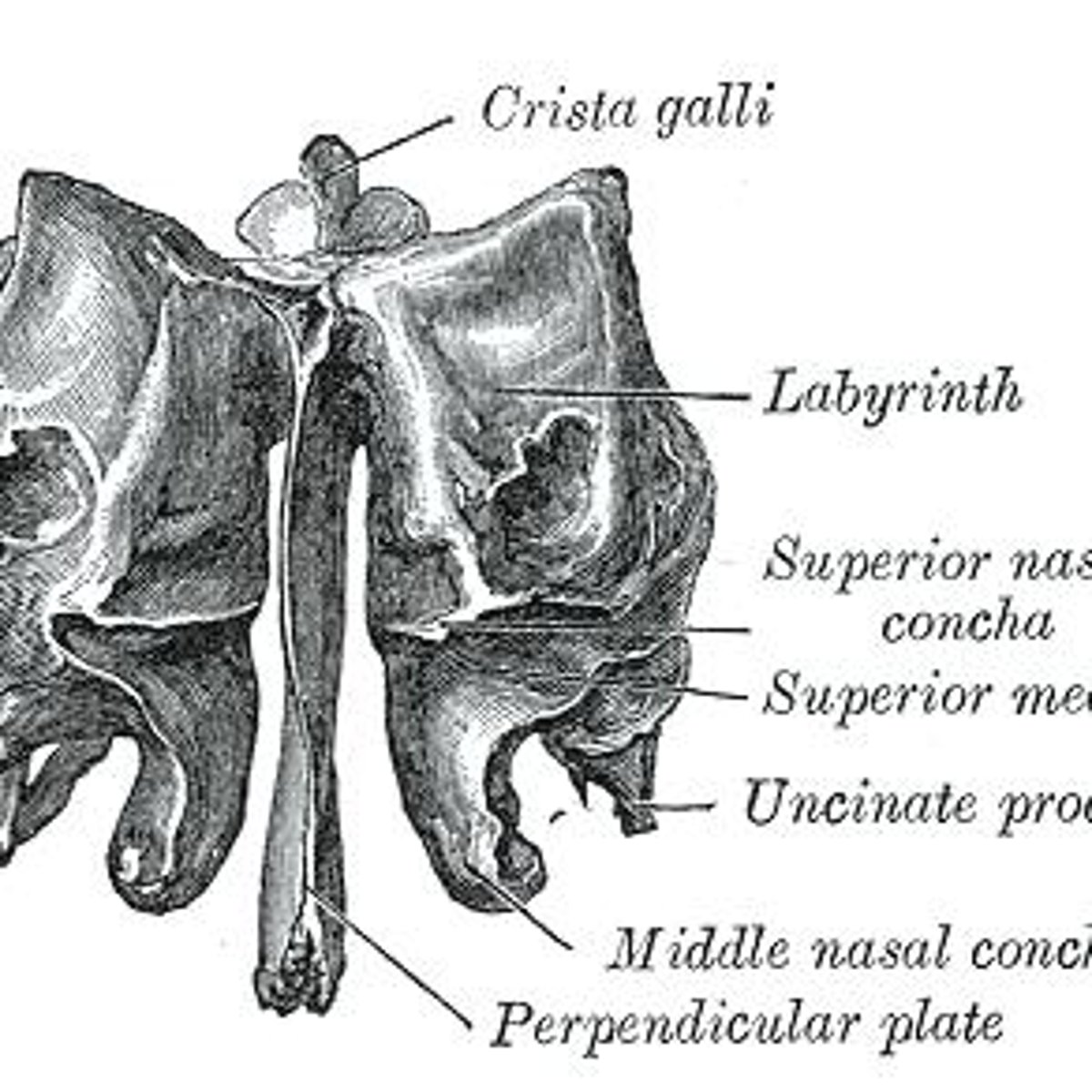
What is the crista galli?
The crista galli is a vertical projection of the ethmoid bone providing attachment for the dura mater.
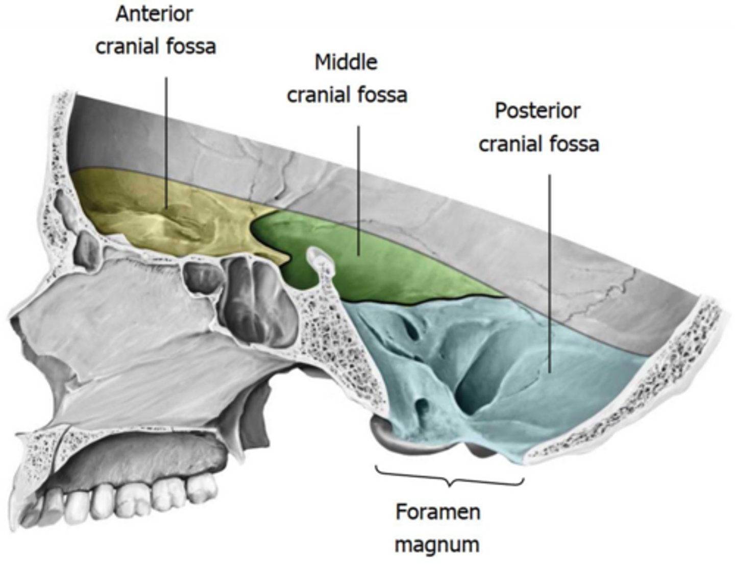
What structures are found in the middle cranial fossa?
The middle cranial fossa contains the temporal lobes, cavernous sinus, internal carotid artery, and pituitary gland.
What is the sella turcica?
The sella turcica is a saddle-shaped region in the sphenoid bone that houses the pituitary gland.
What are the foramina of the sphenoid bone?
Important foramina include the optic canal, superior orbital fissure, foramen rotundum, and foramen ovale.
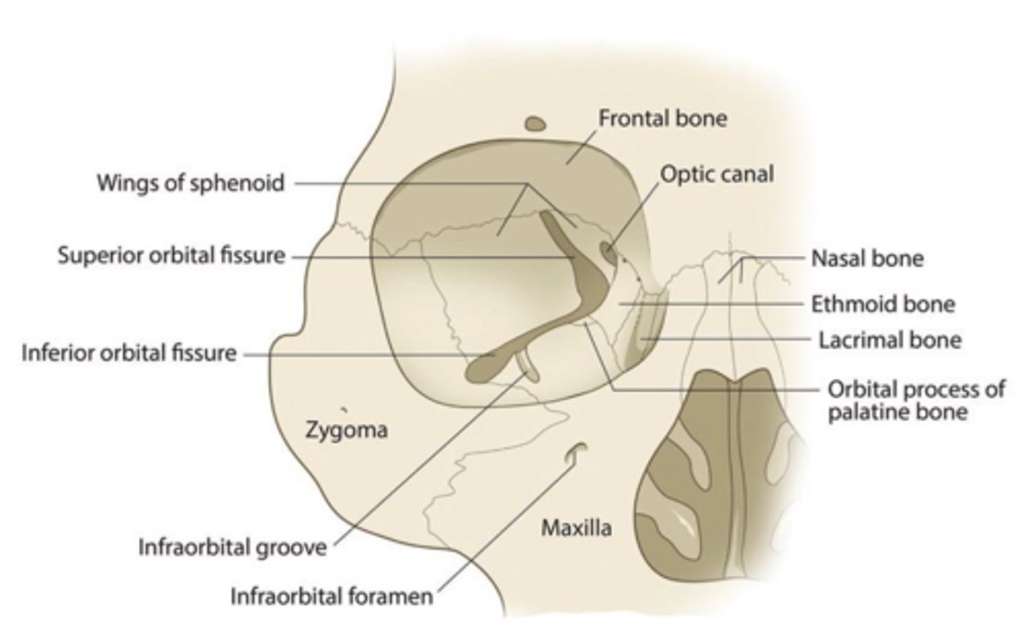
What is the role of the internal acoustic meatus?
The internal acoustic meatus transmits the facial nerve (CN VII) and vestibulocochlear nerve (CN VIII).
Which cranial nerve exits through the jugular foramen?
The glossopharyngeal nerve (CN IX), vagus nerve (CN X), and accessory nerve (CN XI) exit through the jugular foramen.
What is the function of the temporal bone?
The temporal bone houses structures related to hearing and balance and contains the internal acoustic meatus.
What is the significance of the occipital bone?
The occipital bone forms the posterior cranial fossa and contains important foramina for nerve and blood vessel passage.
What is the function of the facial expression muscles?
Facial expression muscles are responsible for the movements of the face, enabling expressions.
What is the role of the middle meningeal artery?
The middle meningeal artery supplies the meninges and is located in the foramen spinosum.
What is the function of the hypoglossal canal?
The hypoglossal canal allows the hypoglossal nerve (CN XII) to exit the skull.
What are the contents of the posterior cranial fossa?
The posterior cranial fossa contains the cerebellum, brainstem, and vertebrobasilar arteries.
What is the function of the olfactory bulb?
The olfactory bulb processes sensory information related to smell and is located in the anterior cranial fossa.
What is the significance of the foramen magnum?
The foramen magnum allows the spinal cord to connect with the brain and is a passage for vertebral arteries.
What is the role of the styloid process?
The styloid process serves as the origin for several important pharyngeal and extrinsic tongue muscles.
What structures pass through the carotid canal?
The internal carotid artery passes through the carotid canal.
What is the function of the superior orbital fissure?
The superior orbital fissure allows passage for several cranial nerves and the ophthalmic vein.
What is the function of the maxillary branch of the trigeminal nerve?
The maxillary branch (V2) exits through the foramen rotundum and provides sensory innervation to the midface.
What is the significance of the parietal foramen?
The parietal foramen allows passage for the emissary vein, which drains into the superior sagittal sinus.
What is the stylopharyngeus muscle?
A muscle involved in the swallowing process.
What is the stylomastoid foramen?
A tiny opening in the temporal bone through which cranial nerve VII (Facial nerve) leaves the cranium.
What are the occipital condyles?
Rounded projections lateral to the foramen magnum that articulate with the first cervical vertebrae (atlas).
What are the fused facial bones?
Maxillary (2), Nasal (2), Palatine (2).
What bone forms the upper alveoli teeth sockets?
Maxillae.
What is the infraorbital foramen?
An opening under the orbit carrying the Maxillary nerve V2 (infraorbital nerve).
What is the palatine bone?
The posterior portion of the hard palate.
What do the nasal bones form?
The bridge of the nose.
What is the zygomatic bone?
The bone that forms the majority of the cheek area and part of the lateral orbit.
What is the vomer bone?
A blade-shaped bone making up the inferior portion of the nasal cavity.
What are the inferior nasal conchae?
Thin curved bones along the lateral portions of the nasal cavity that increase surface area and slow down air.
What is the mental foramen?
An opening in the mandible through which the mental nerve passes to innervate the lower jaw, lips, and anterior mandibular teeth.
What is the coronoid process?
A site of muscle attachment (temporalis muscle) on the mandible.
What is the mandibular condyle?
The articulation point of the mandible with the mandibular fossa of the temporal bone.
What is the epidural space?
A potential space that can become an epidural hematoma.
What is the dura mater?
The outermost layer of the meninges that adheres tightly to the skull.
What is the subdural space?
A potential space that can become a subdural hematoma.
What is the arachnoid mater?
A layer of the meninges that lines the dura and is not adhered to it.
What is the subarachnoid space?
The true space that contains circulating CSF and cerebral arteries and veins.
What is the falx cerebri?
A crescent-shaped downward projection of meningeal dura mater that passes between the two cerebral hemispheres.
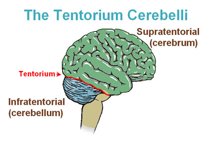
What does the tentorium cerebelli do?
Covers and separates the cerebellum from the posterior parts of the cerebral hemispheres.
What is the confluence of sinuses?
The meeting point of the superior sagittal sinus, straight sinus, and occipital sinus.
What is the cavernous sinus?
A bilateral sinus located on either side of the body of the sphenoid bone, draining various veins.
What is an epidural hematoma?
A type of intracranial hemorrhage often due to trauma, characterized by arterial bleeding between the dura and skull.
What is a subdural hematoma?
A type of intracranial hemorrhage that can occur due to trauma or spontaneously, characterized by venous bleeding.
What is a subarachnoid hemorrhage?
An arterial bleed that occurs between the arachnoid mater and pia mater, often due to trauma or a ruptured aneurysm.
What is xanthochromia?
A pale yellow-colored appearance of CSF, often seen in cases of subarachnoid hemorrhage.
What is the classic presentation of a subarachnoid hemorrhage?
Often described as the 'worst headache of my life.'
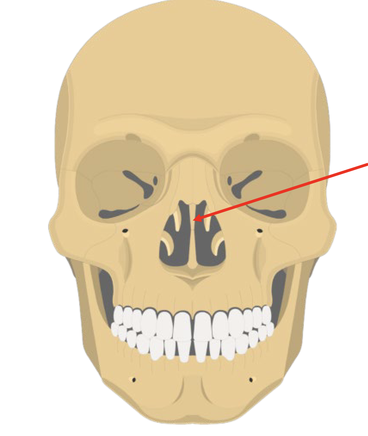
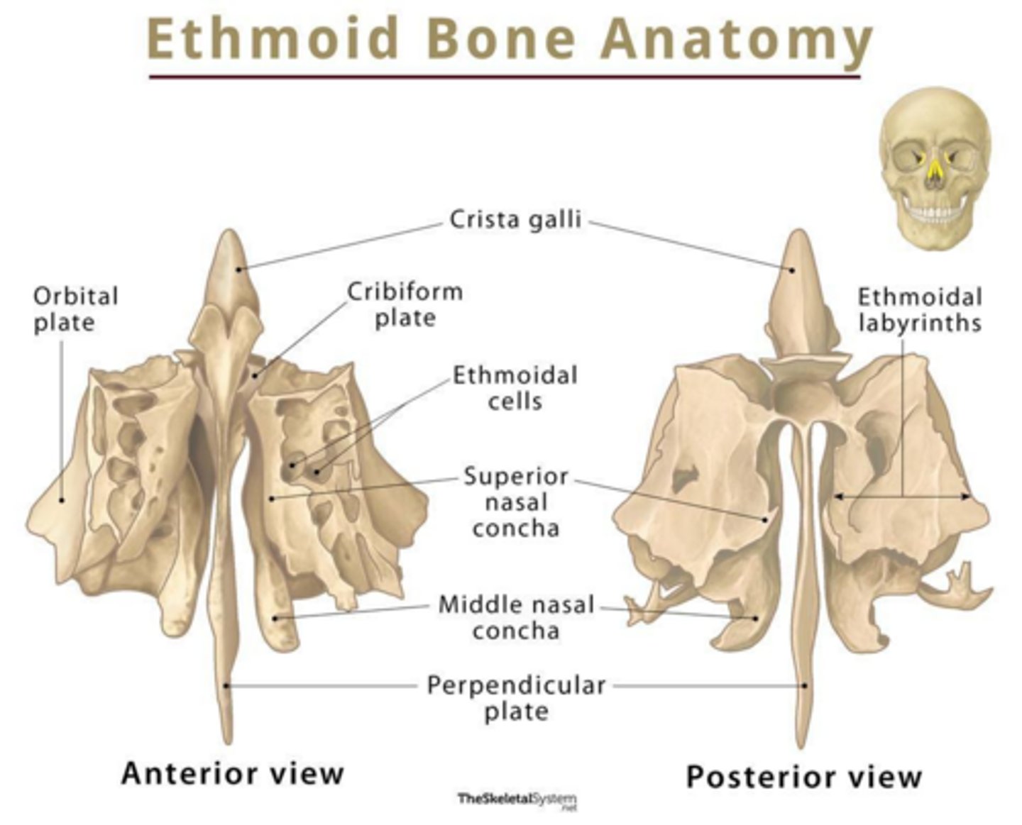
Perpendicular plate of ethmoid bone
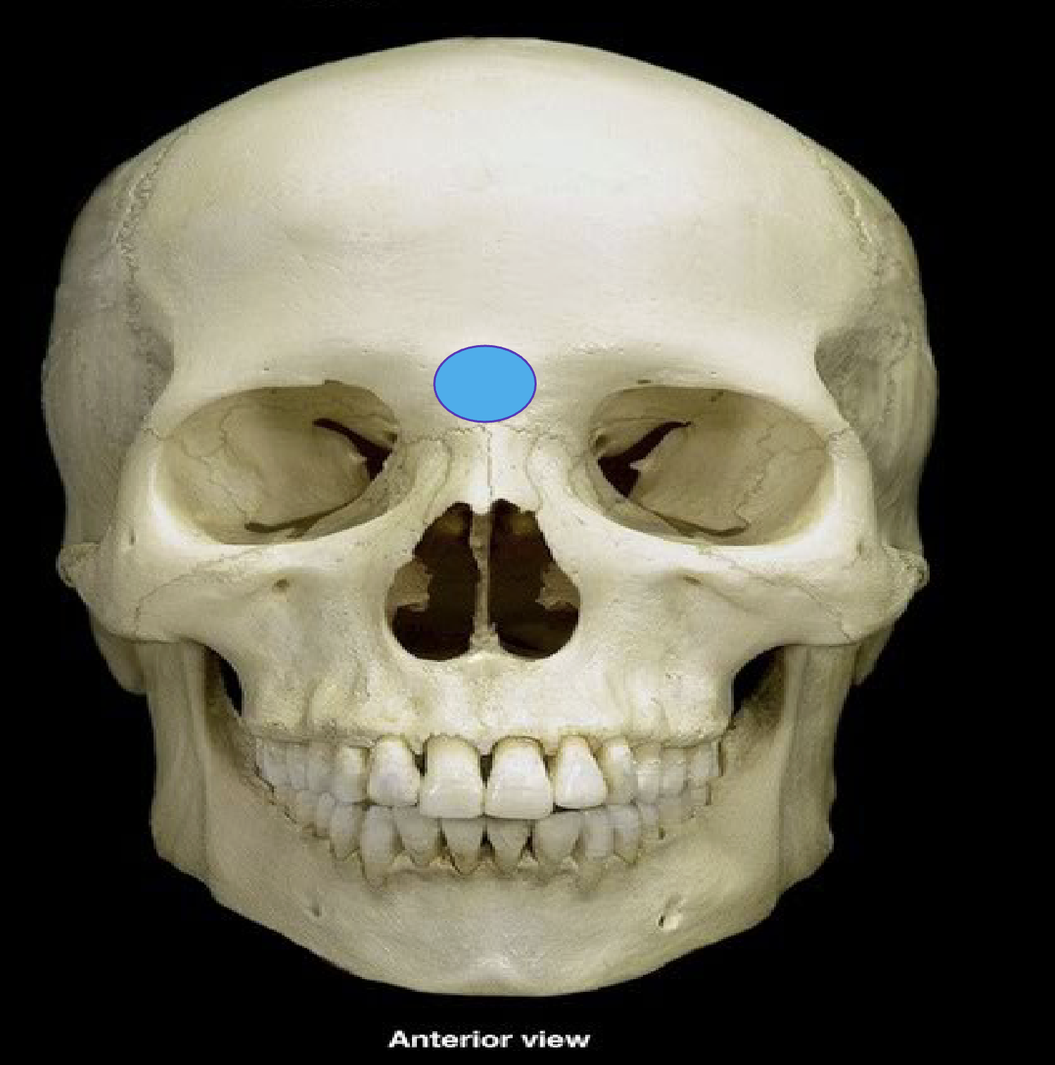
Glabella on frontal bone
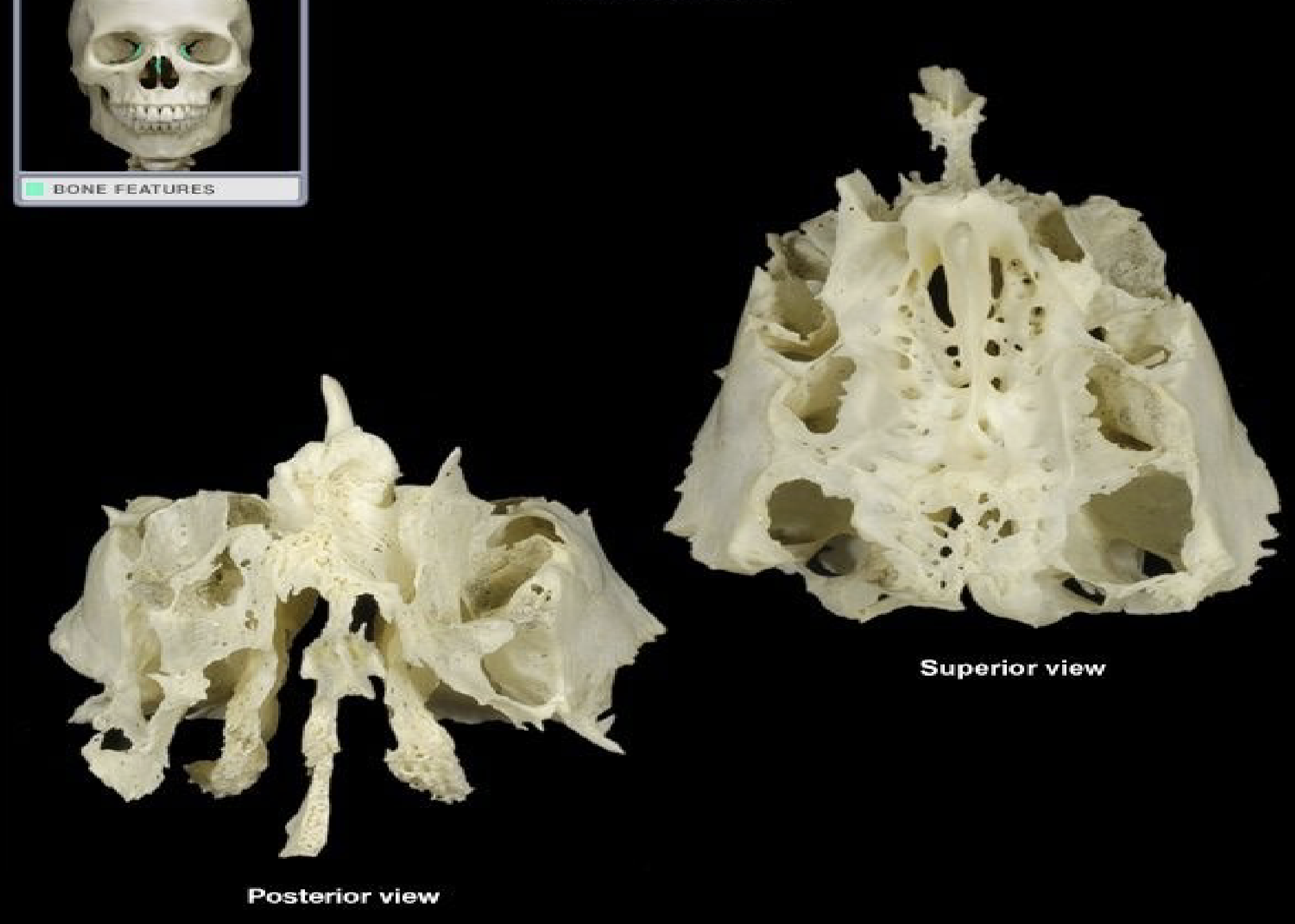
Ethmoid bone
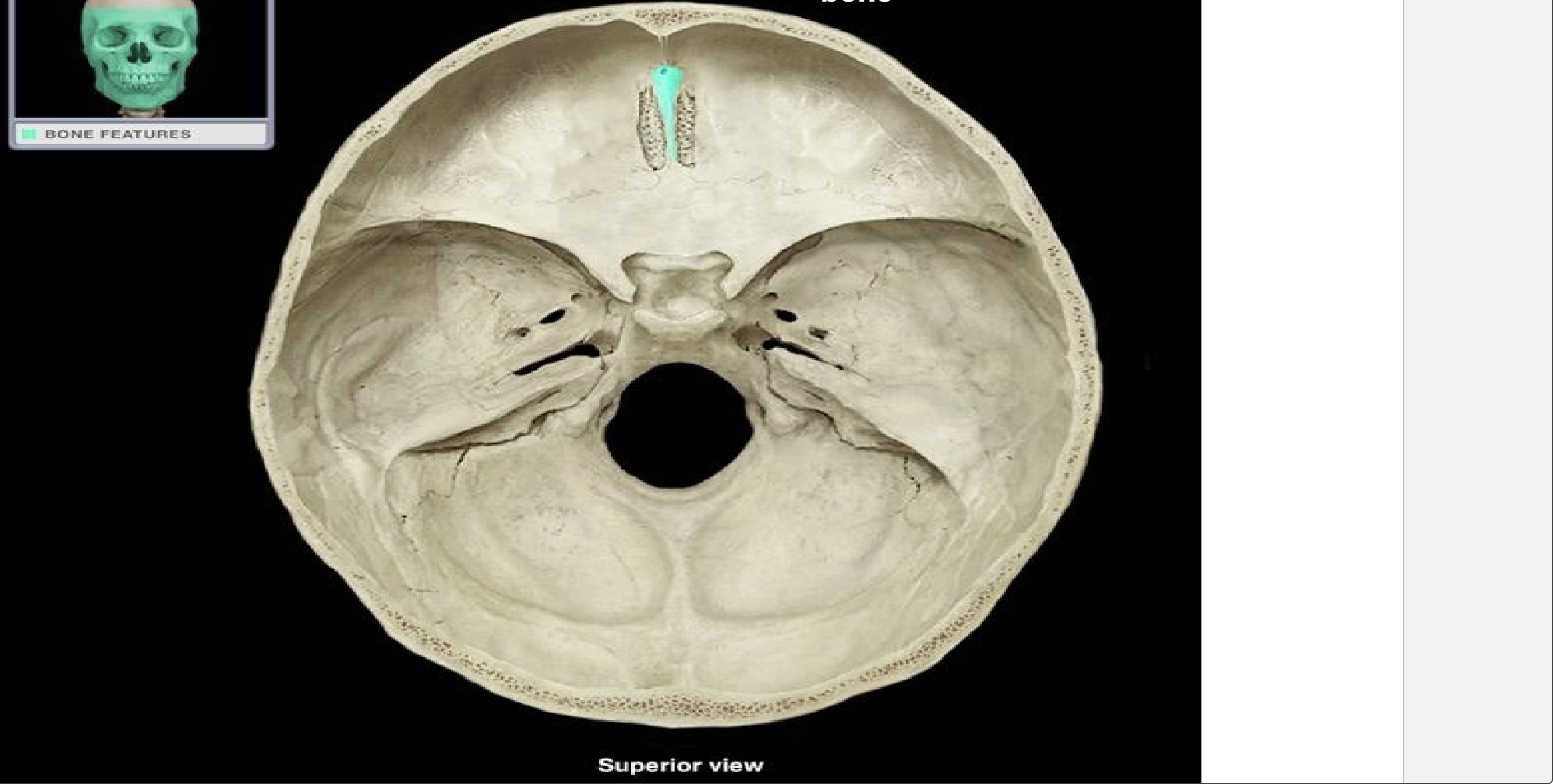
Crista galli of the ethmoid bone
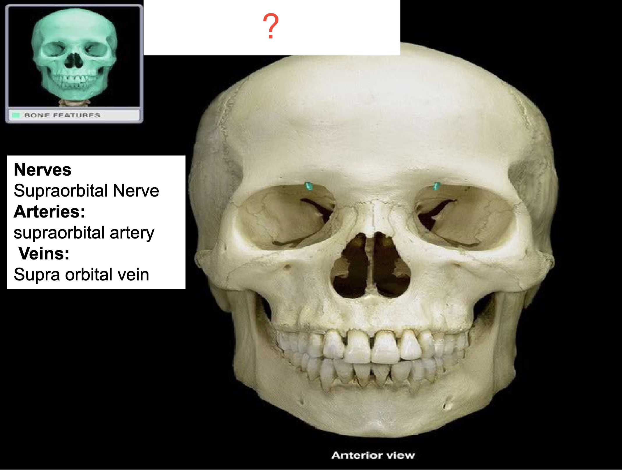
Supraorbital foramen of the frontal bone
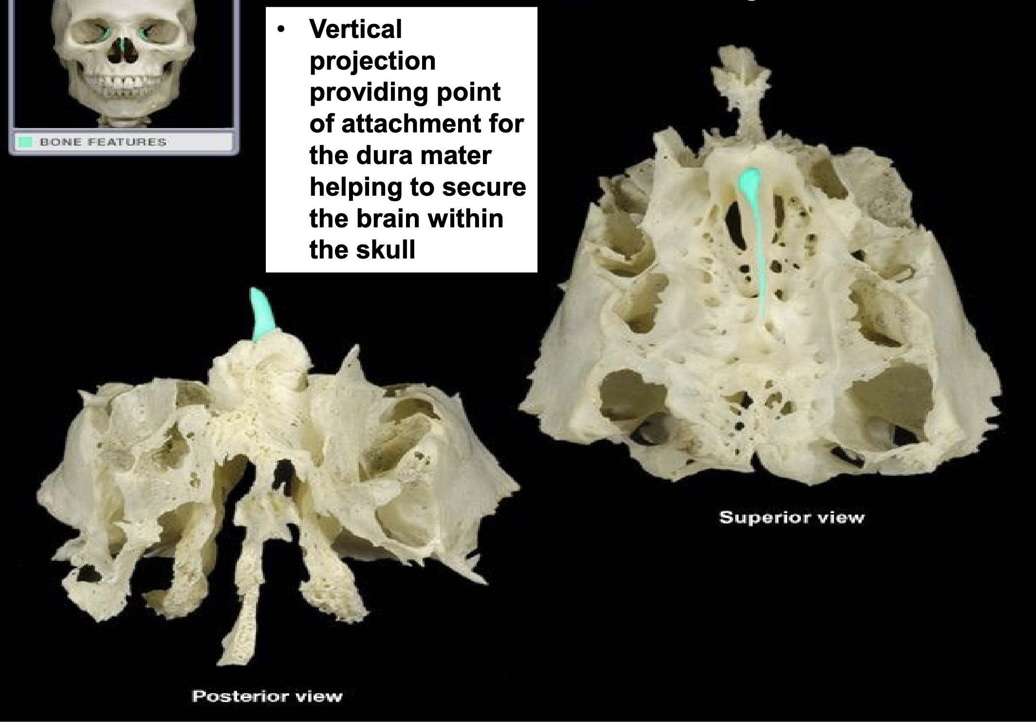
Crista galli of the ethmoid bone
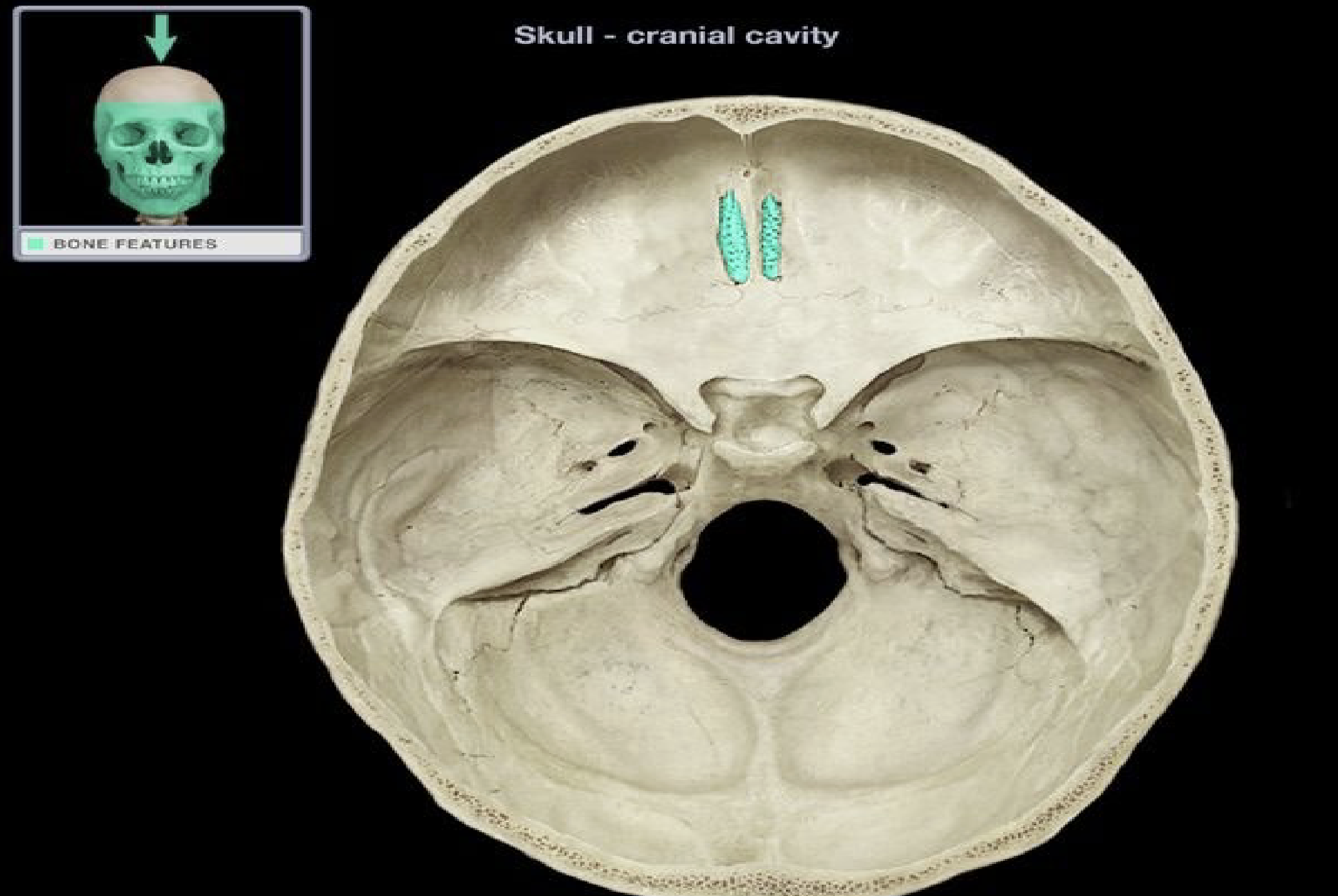
Cribriform plate of the ethmoid bone
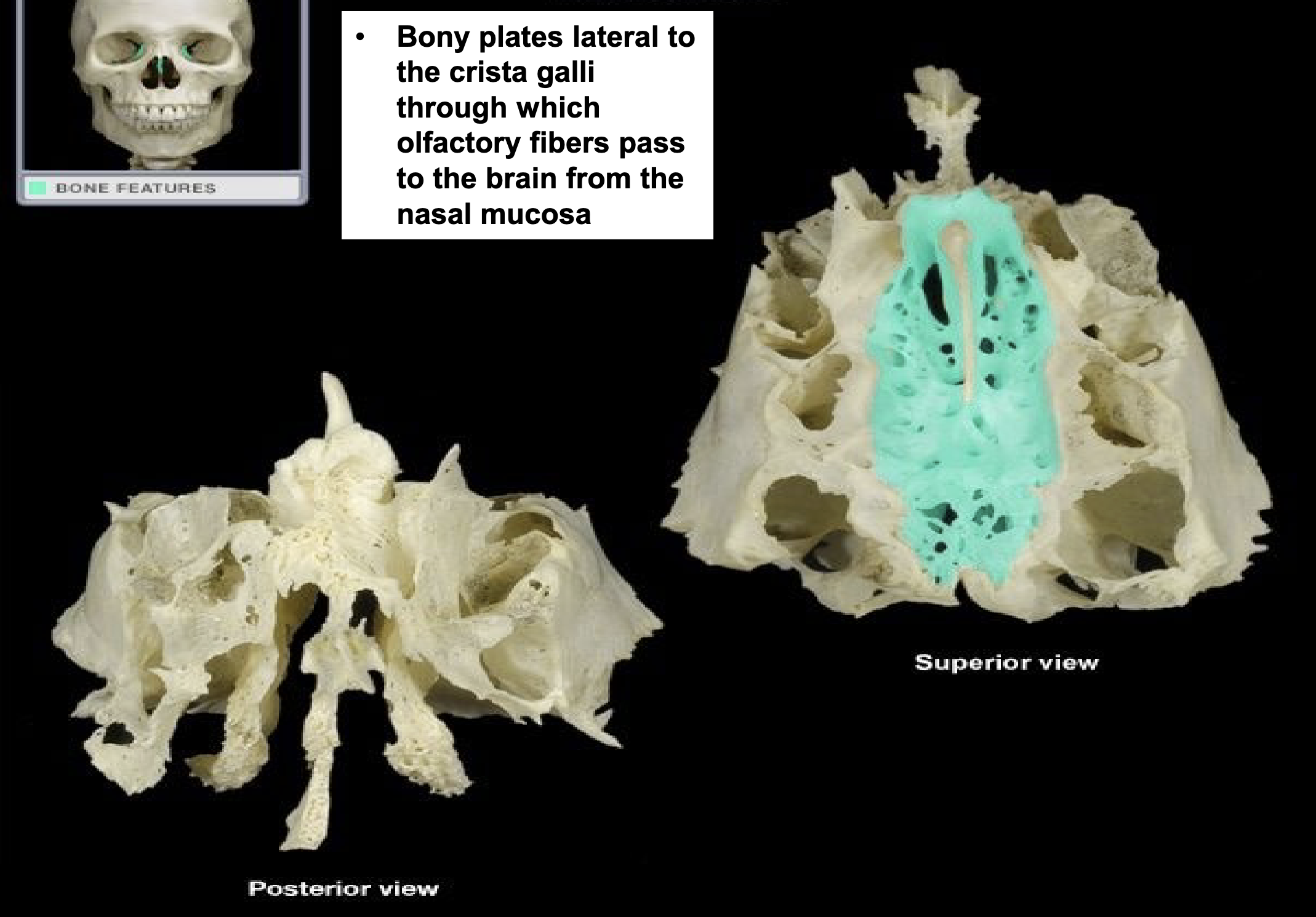
Cribriform plate of the ethmoid bone
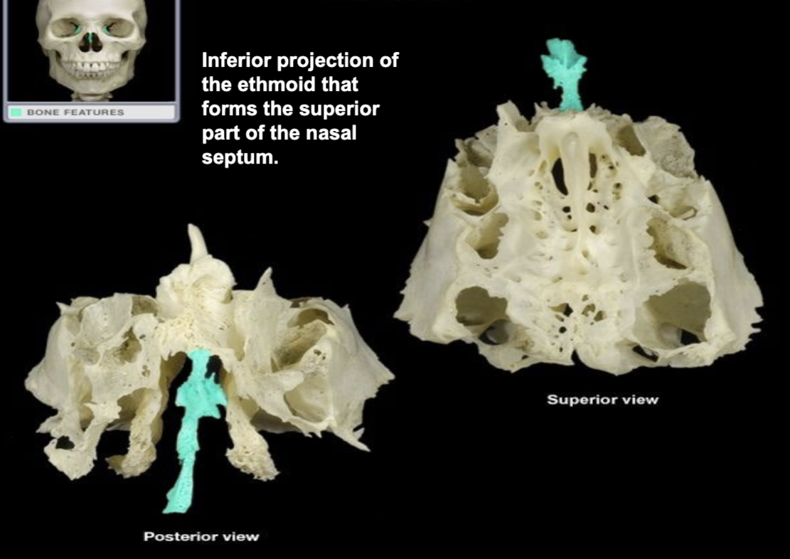
Perpendicular plate of the ethmoid bone
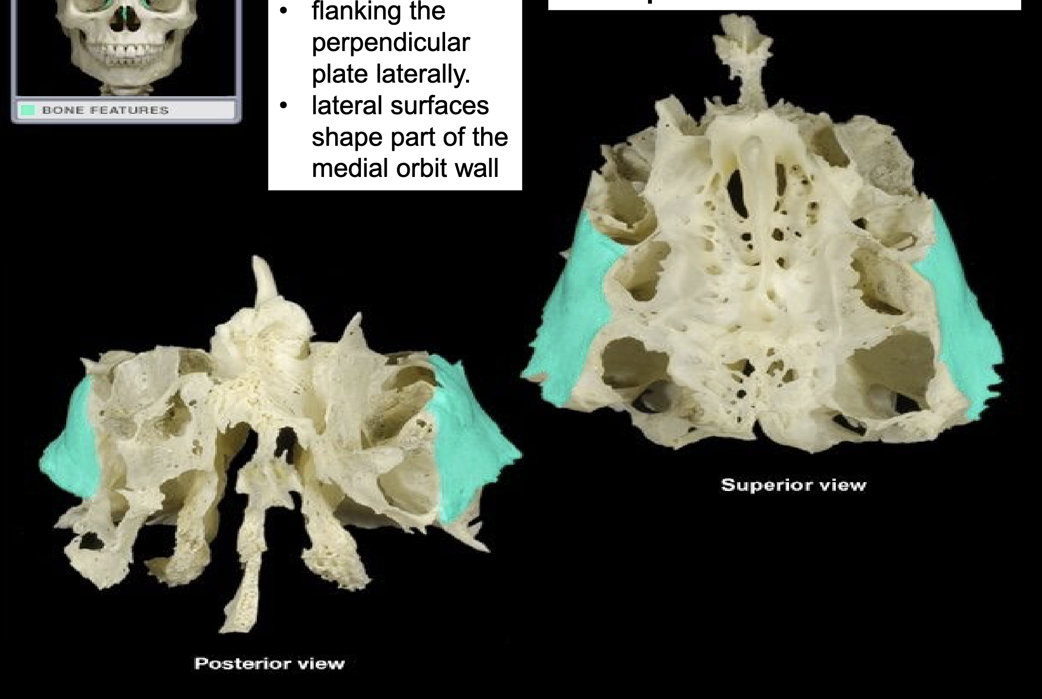
Orbital plate of the ethmoid bone
Hollow bone chambers that form sinuses
Ethmoid air cells
Groupings of ethmoid air cells on either side
Ethmoid labyrinth
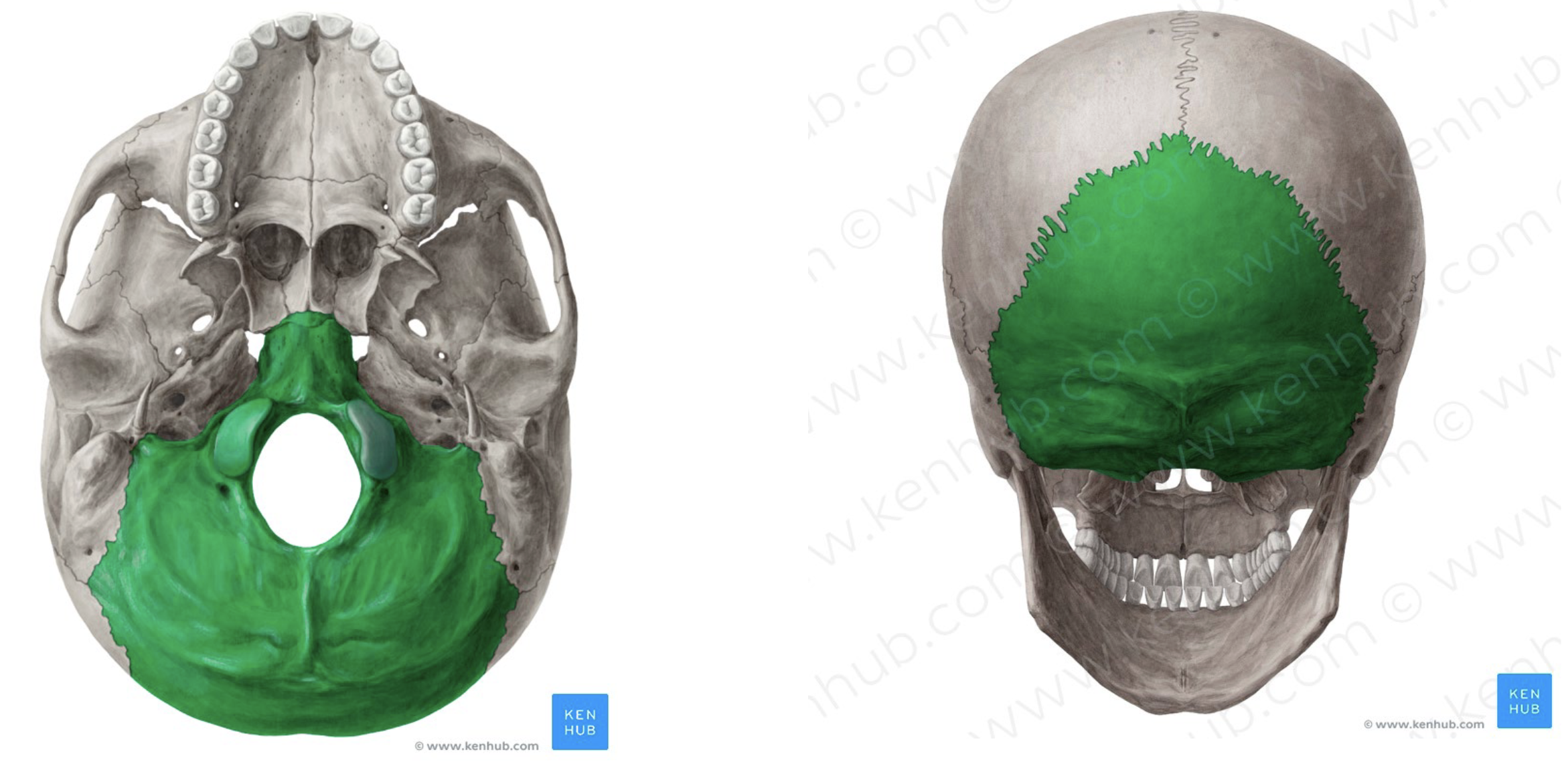
Occipital bone
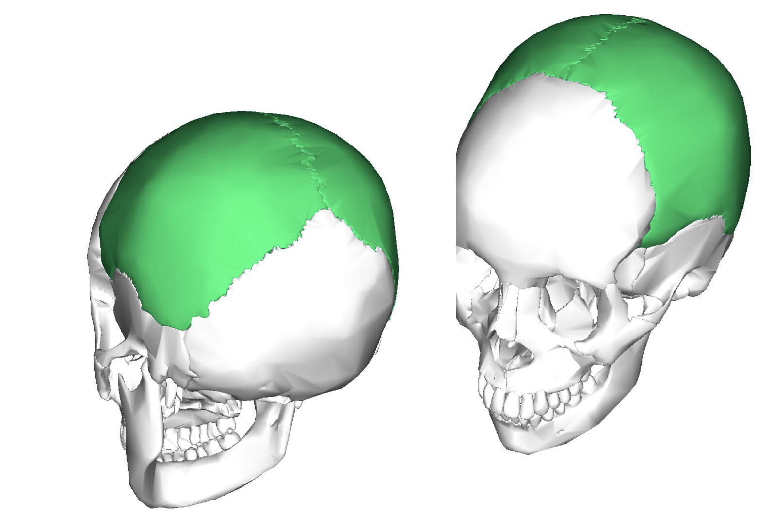
Parietal bone
Important site with several foramina that allow passage of nerves and blood vessels
Cranial fossa
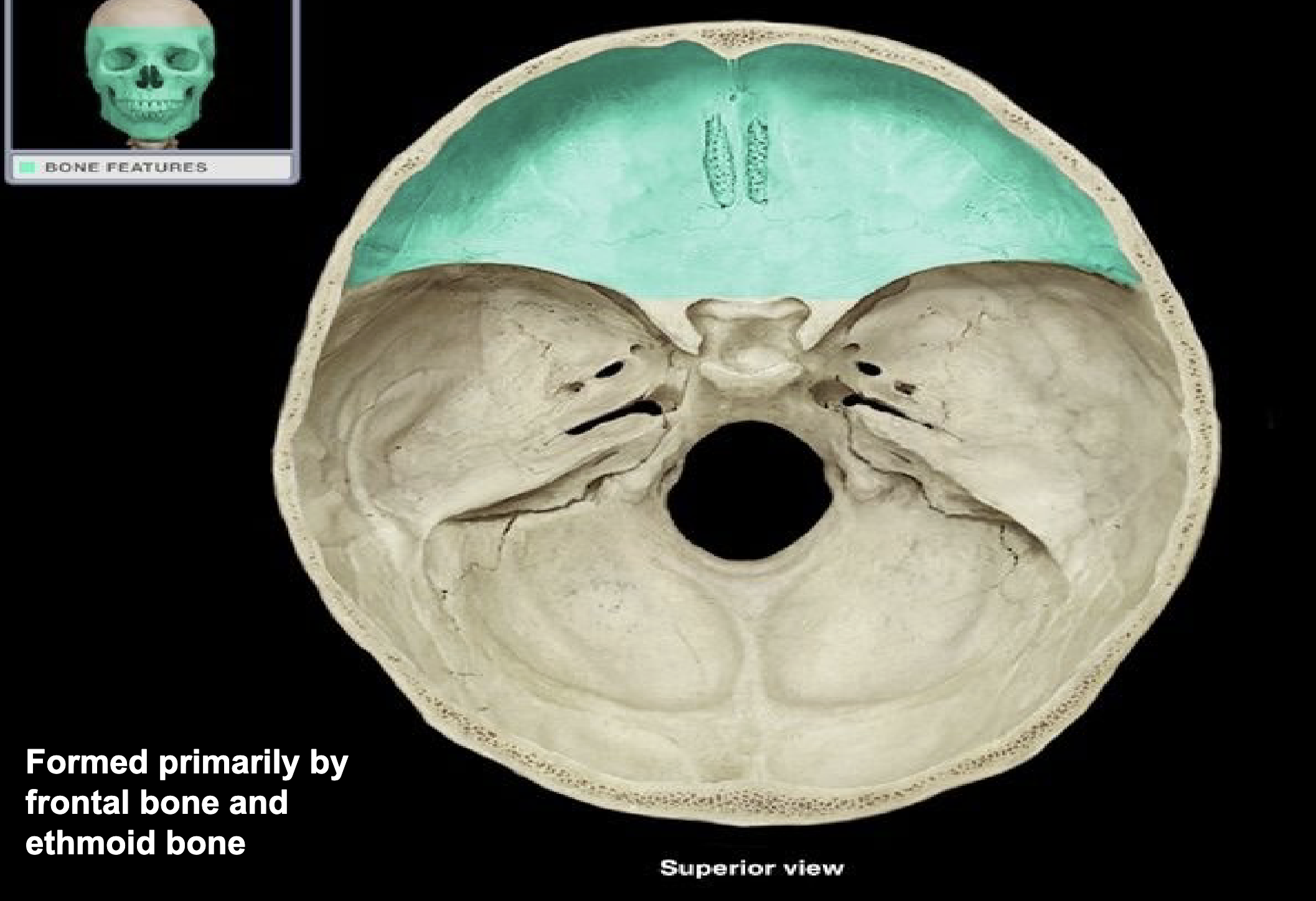
Anterior cranial fossa
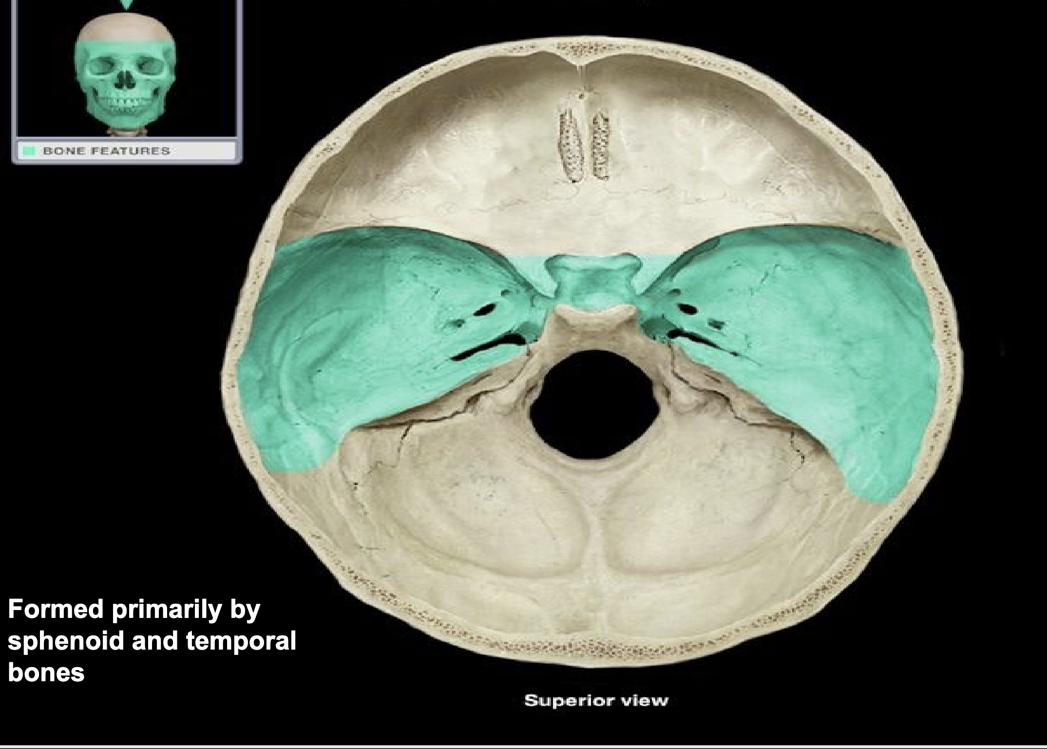
Middle cranial fossa
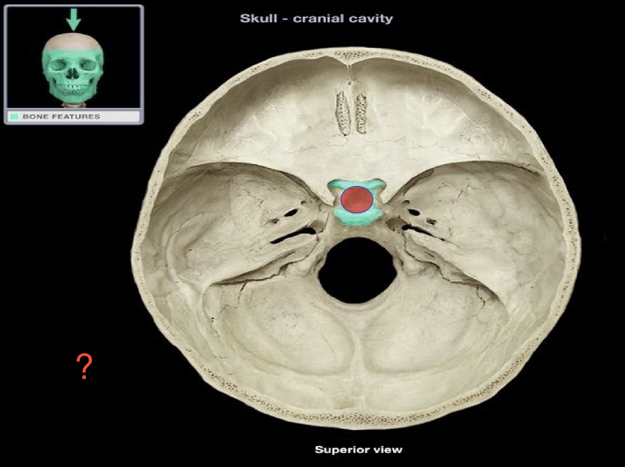
Sella turcica of sphenoid bone
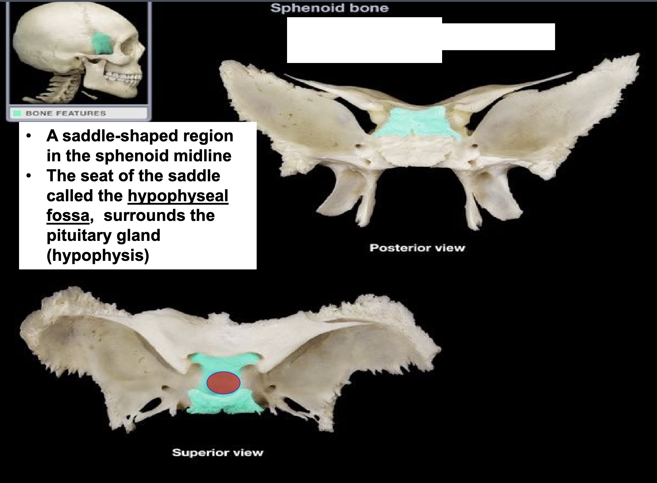
Sella turcica of sphenoid bone
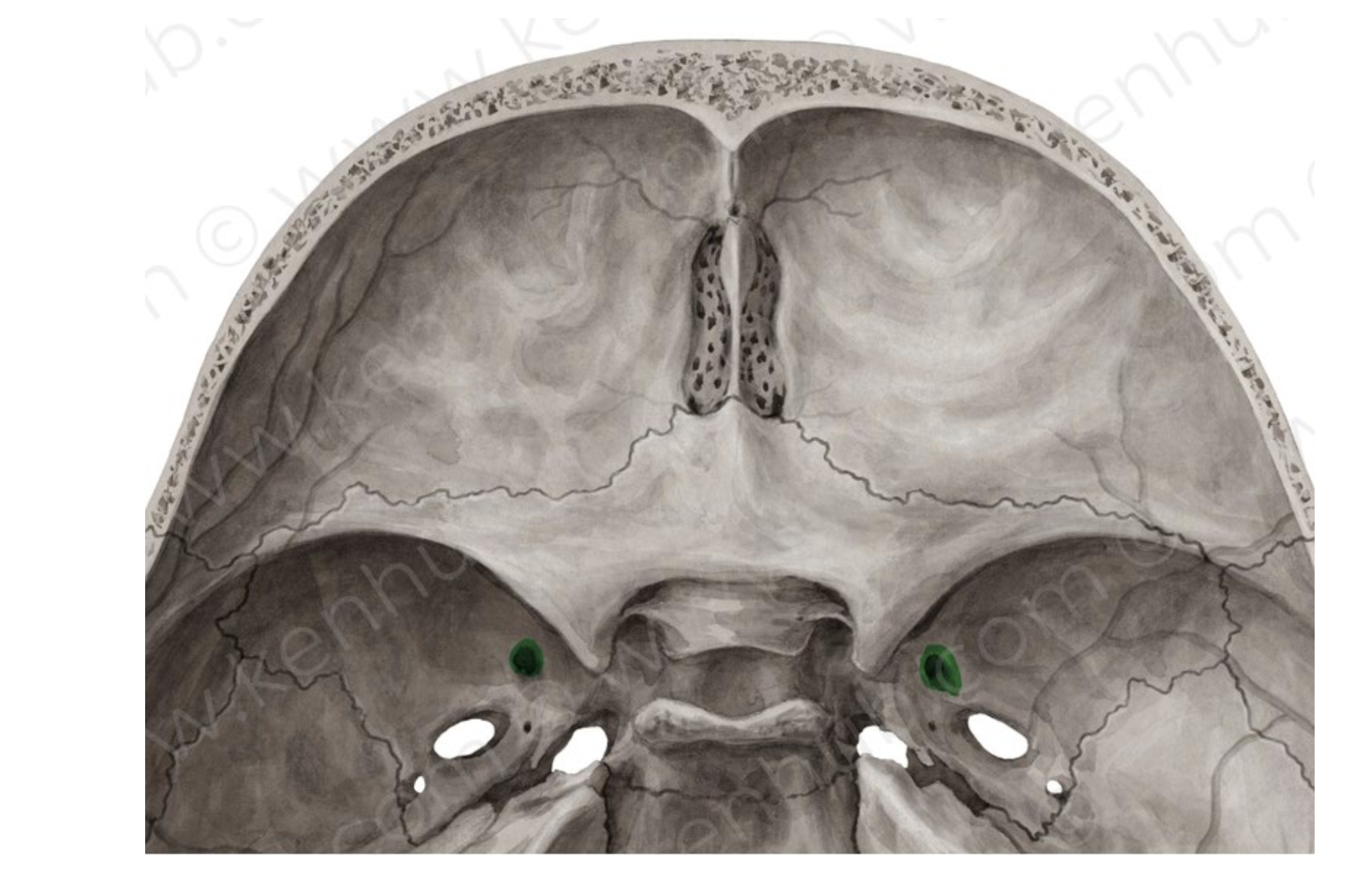
Foramen rotundum of the sphenoid bone
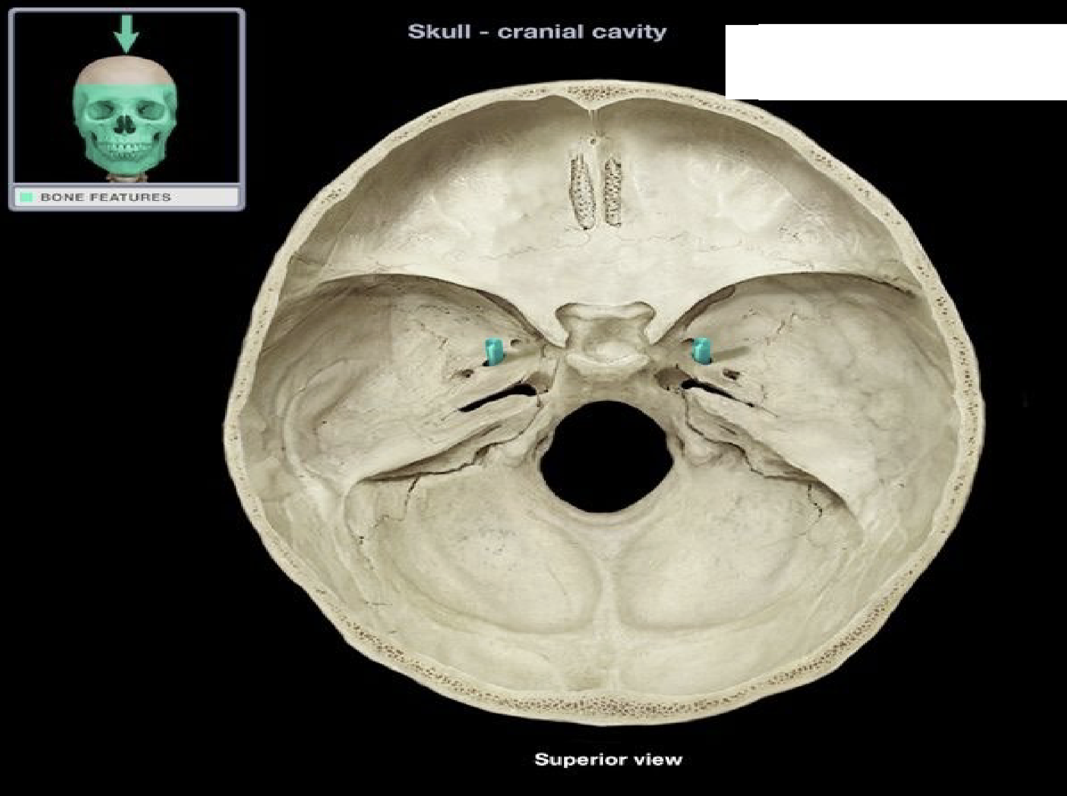
Foramen ovale of the sphenoid bone
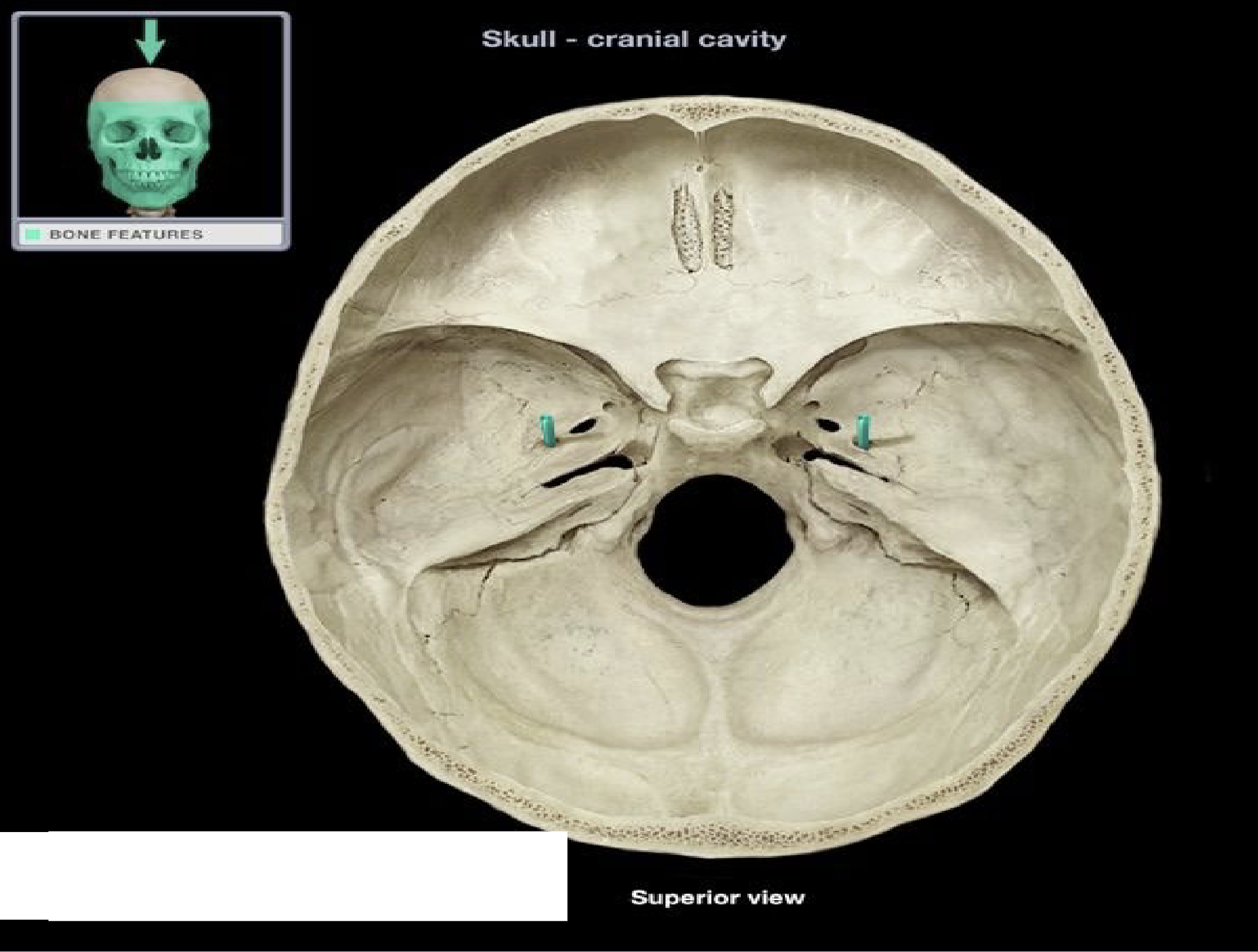
Foramen spinosum of the sphenoid bone
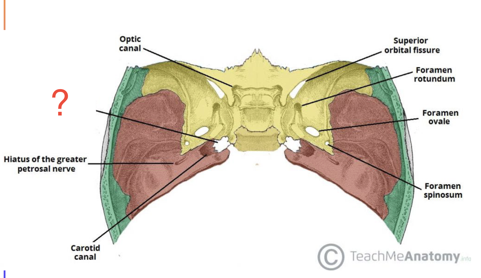
filled with cartilage in adults.
Artery of the pterygoid canal passing inferiorly through cartilage of foramen lacerum
allows internal carotid artery to enter the middle cranial fossa
Foramen lacerum
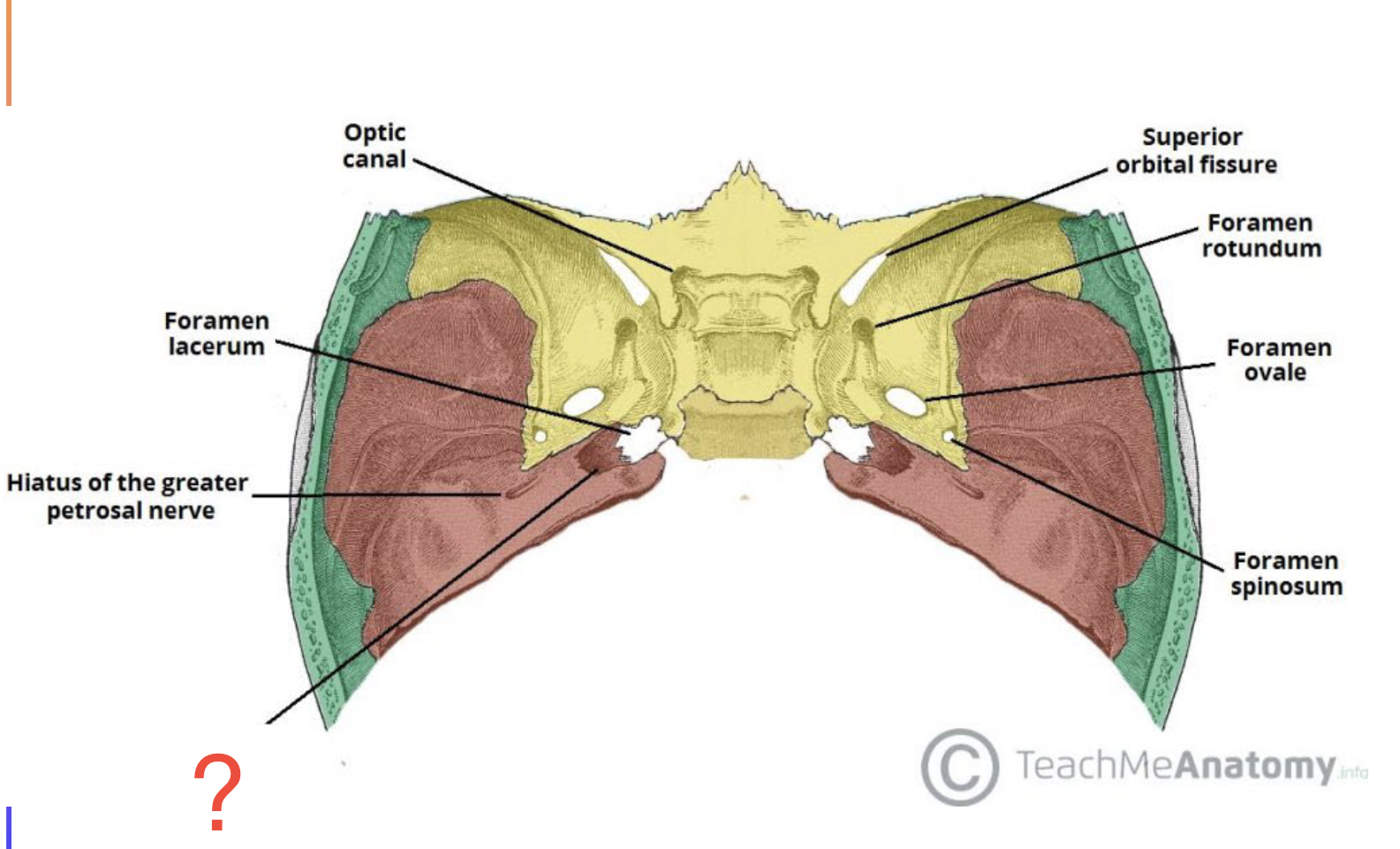
Carotid canal
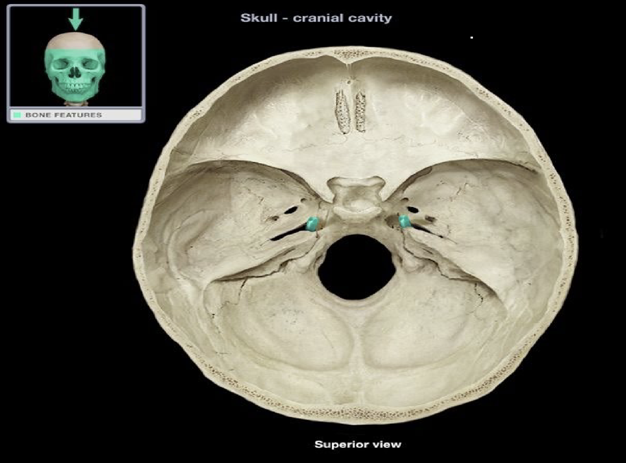
Foramen lacerum of thhe temporal bone
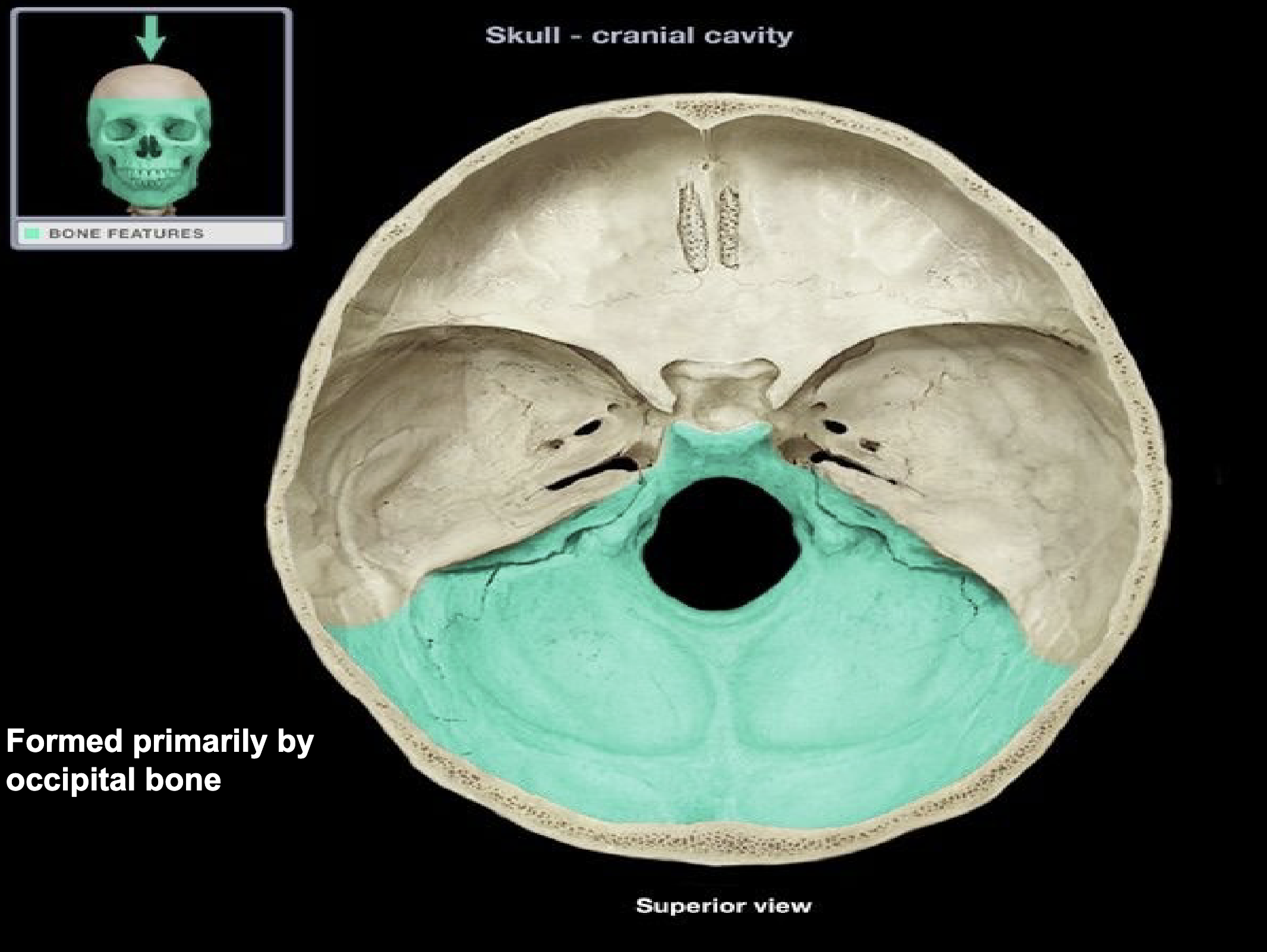
Posterior cranial fossa
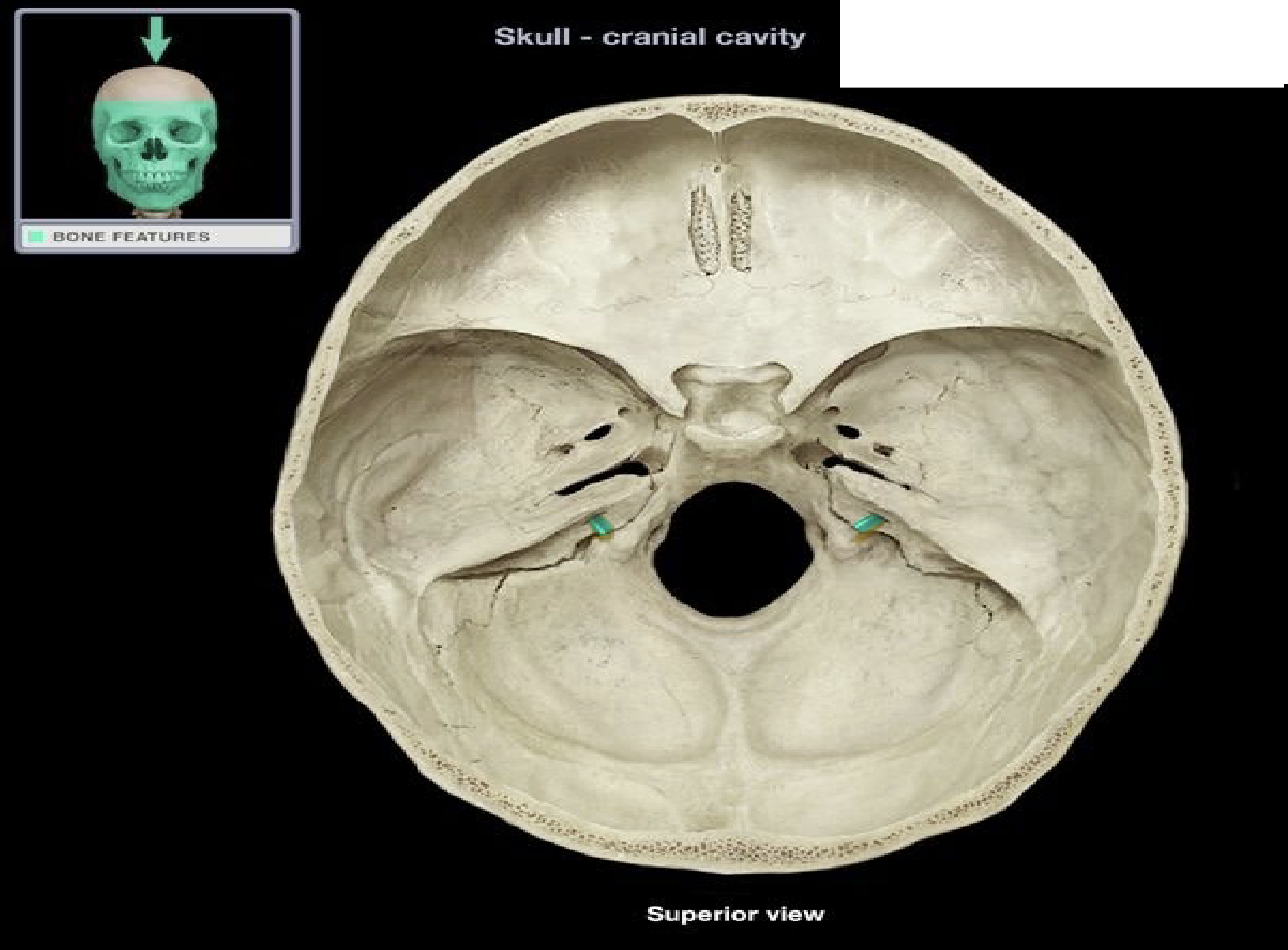
Internal acoustic meatus of the temporal bone
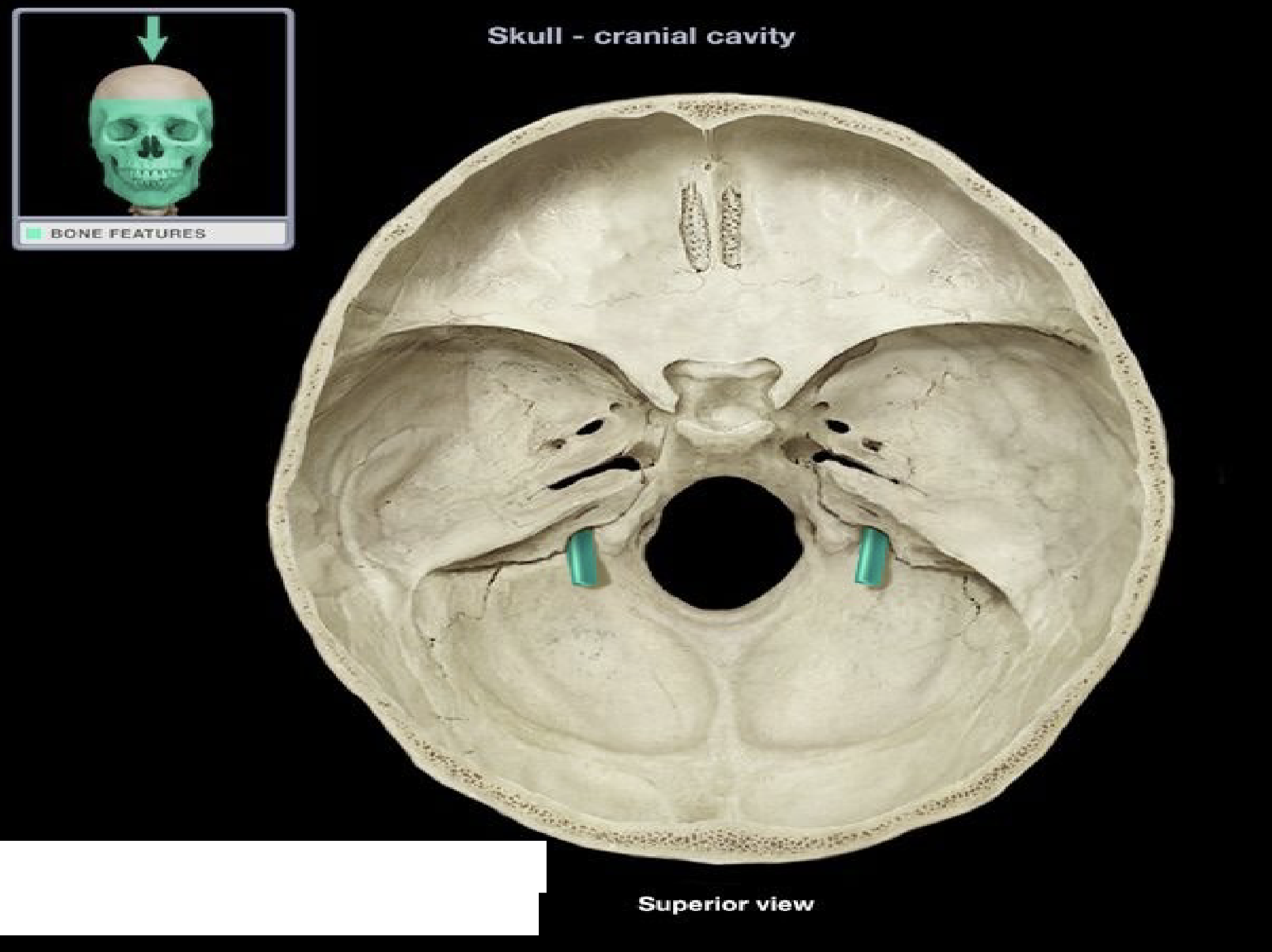
Jugular foramen of temporal bone
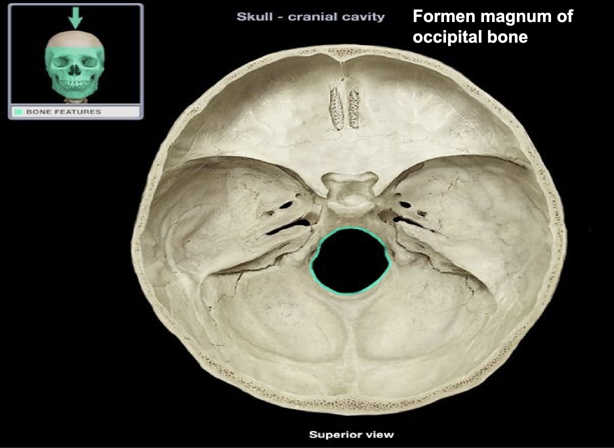
Foramen magnum of occipital bone
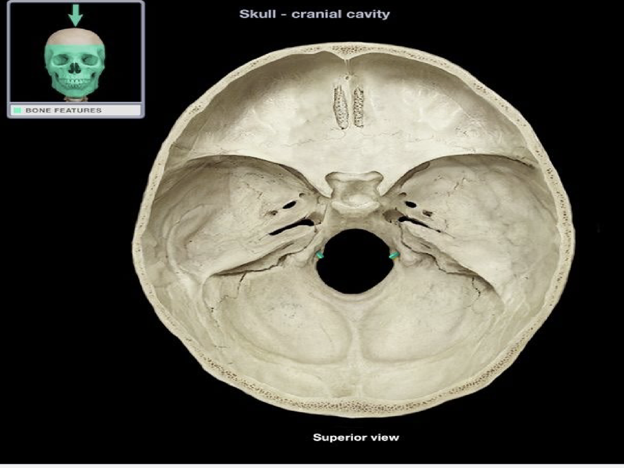
Hypoglossal canal of the occipital bone