Hematology 2 - Lecture - 13 - Plasma Cell Disorders - Complete
1/154
There's no tags or description
Looks like no tags are added yet.
Name | Mastery | Learn | Test | Matching | Spaced | Call with Kai |
|---|
No analytics yet
Send a link to your students to track their progress
155 Terms
Immature lymphoid neoplasms, Mature lymphoid neoplasms
Lymphoid neoplasm categories
Immature lymphoid neoplasms
Type of lymphoid neoplasm characterized by lymphoblastic proliferation
Mature lymphoid neoplasms
Type of lymphoid neoplasm characterized by proliferation of neoplastic lymphocytes
Immature lymphoid neoplasms
What type of lymphoid neoplasm is ALL?
Mature lymphoid neoplasms
What type of lymphoid neoplasm is CLL?
Plasma Cell Myeloma, Monoclonal Gammopathy of Undetermined Significance, Plasmacytoma, Primary amyloidosis, Heavy-Chain Diseases, Lymphoplasmacytic lymphoma, Waldenstrom macroglobulinemia
Mature B-cell neoplasms with plasma cell or plasmacytoid differentiation
Plasma cell disorders
These neoplasms are characterized by the proliferation of a single clone of immunoglobulin-producing cells or plasma cells.
M-protein
Single class or subunit of immunoglobulin that is almost always secreted in Plasma cell disorders
Paraprotein, M component
M-protein other name
M-protein
This protein, in Plasma cell disorders, is a single class or subunit of immunoglobulin or single class of immunoglobulin light chain, sometimes can even be a single type of heavy chain.
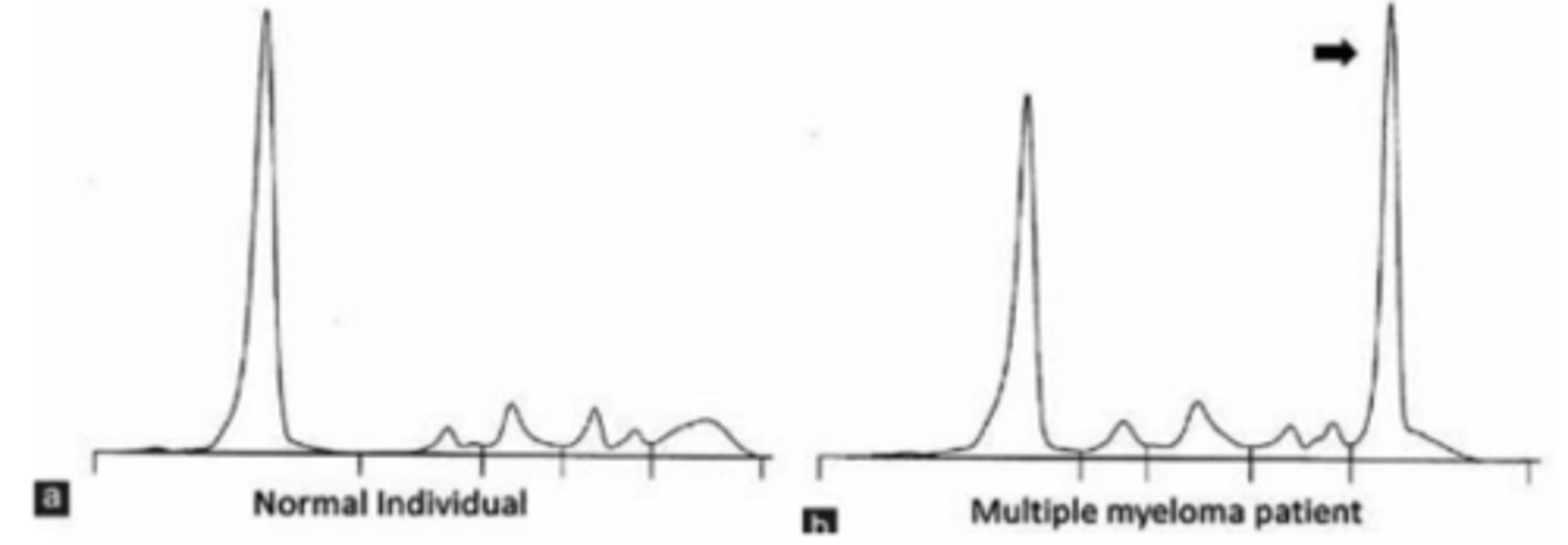
Gamma-globulin region
Plasma cell disorders can be detected as a monoclonal peak at this region on serum or urine protein electrophoresis
Albumin
Most abundant protein in the serum; always seen as the highest peak in the pattern in the leftmost
Gamma region
Region in electrophoresis where Immunoglobulins occupy
T
T/F
In normal conditions, Immunoglobulins are not as plenty and seen as just a broad-based hump, very subtle. (In electrophoresis)
M spike
In plasma cell disorders, the production of very high numbers of immunoglobulins or M proteins results in a characteristic spike at the gamma-globulin region. What do you call this spike?
No
Is an M spike specific for the presence of M-proteins? Yes/No
Monoclonal immunoglobulin
Plasma cell disorders have varying clinicopathologic features but they all have in common the production of this immunoglobulin
M-protein
Monoclonal immunoglobulins are collectively known as:
Monoclonal
Meaning of M in M-protein
2
Most monoclonal proteins consist of how many heavy chains of the same class?
2
Most monoclonal proteins consist of how many light chains of the same type?
T
T/F
In some cases, the monoclonal proteins consist only of a single light chain or fragments of heavy chains.
Renal insufficiency
Clinical manifestation in plasma cell myeloma that is related to the accumulation and deposition of M proteins
Hyperviscosity
High levels of paraprotein in the blood can also cause:
Hint: Something to do with viscosity
Unknown
Etiology of Plasma cell disorders
African-Americans
Plasma cell disorders have a genetic propensity or higher incidence in this group of people
Caucasian-Americans
Plasma cell disorders have lower incidence in African-Americans or Caucasian-Americans?
Plasma cell disorders
It is likely that these disorders result from a multi-step transformation process in which the lymphocytes or plasma cells undergo multiple genetic alterations that prevent normal cell differentiation and cell death with ultimate immortalization of the clone.
Serum or urine protein electrophoresis
Laboratory testing for M-proteins is usually accomplished by:
Gamma globulin region, Beta globulin region
M-proteins appear as a dense spike ('M-spike') at this region and occasionally in this other region too. (2 answers)
Decreased
Normal polyclonal immunoglobulins are usually (Increased/Decreased)
Bence-Jones proteins
Immunoglobulin light chains that are able to pass through the glomerular filter thus they may be detected in urine
Immunofixation
Once an M-protein is identified, it should be characterized by more refined methods as such as ___ to be able to establish what specific type of M protein is present
T
T/F
Knowing the specific identity of the protein in Plasma cell disorders has implications on the aggressiveness and the prognosis of the disease process
Plasma cell myeloma
It is an aggressive, bone marrow-based neoplasm of plasma cells associated with a monoclonal protein in serum and/or urine
Multiple myeloma
Plasma cell myeloma is also known as
Bone marrow smear examination
Laboratory diagnosis for Plasma cell myeloma
Multiple myeloma
Plasma cell myeloma is also known by this name because of the multicentricity of the lesions (multiple lesions that are spread out)
Clinical, Pathologic, Radiologic findings
The diagnosis for Plasma cell myeloma is based on a combination of:
African-Americans, Caucasians
Plasma cell myeloma is more common in ____ than in ____
Bone pain in the back or chest
Most common presenting symptom of Plasma cell myeloma, it is often associated with pathologic compression fractures of the thoracic or lumbar spine
70%
Radiographic studies show lytic lesions in what % of Plasma cell myeloma patients at the time of diagnosis
Lytic lesions
In Plasma cell myeloma, these are areas where the bone is resorbed either due to plasma cell infiltration or the interactions of plasma cells with bone cells, like osteoclasts (which facilitate the resorption of the bone).
Well-defined, punched out radiolucencies or darker areas
How are Lytic lesions in Plasma cell myeloma described as on X-rays?
Calcium
In X-rays, the whiter a bone is, the more ___ it has
Vertebral column, Ribs, Skull
Most commonly affected bones in Plasma cell myeloma
T
T/F
In Plasma cell myeloma, other areas like in the axial skeleton also have some radiolucencies seen (affected areas are spread out)
Weakness, Fatigue, Bleeding tendencies, Infections, Symptoms related to renal failure
Other physical findings that may be noted in Plasma cell myeloma
Fatigue
Physical finding in Plasma cell myeloma that is often related to anemia
Renal failure
Physical finding in Plasma cell myeloma that is most often due to the deposition of the M proteins (especially the light chains) in the renal vasculature, which causes injury and blockage
Infection
Most common cause of death in Plasma cell myeloma patients, because although the plasma cells produce large amounts of immunoglobulins, but they are non-functional.
M-protein in serum or urine, Bone marrow clonal plasmacytosis or plasmacytoma, Related organ or tissue impairment
World Health Organization diagnostic criteria for Plasma cell myeloma
>30g/L
Plasma cell myeloma
M protein concentration in serum or urine if it's an IgG (Most common)
>25g/L
Plasma cell myeloma
M protein concentration in serum or urine if it's an IgA
>1g/24hr of urine light chain
Plasma cell myeloma
M protein concentration in urine
BM Clonal Plasmacytosis or Plasmacytoma
Plasma cell myeloma
Increased number of plasma cells in the bone marrow
10%
Plasma cell myeloma - BM Clonal Plasmacytosis or Plasmacytoma
Monoclonal plasma cells usually exceed ___% of nucleated cells in the marrow but no minimal level is designated because about 5% of patients with symptomatic myeloma have.
Hypercalcemia, Renal insufficiency, Anemia, Bone lesions
Related organ or tissue impairment in Plasma cell myeloma
End organ damage
The most important criteria for symptomatic Plasma cell myeloma are manifestations of:
Anemia, Hypercalcemia, Lytic bone lesions, Hyperviscosity, Amyloidosis, Recurrent infections
Manifestations of end organ damage in Plasma cell myeloma
Lytic bone lesions
Manifestation of end organ damage in Plasma cell myeloma that is from plasma cell marrow infiltration
Anemia, Leukopenia, Thrombocytopenia
Blood cell results in Plasma cell myeloma
Leukopenia, Thrombocytopenia
Blood cell results in Plasma cell myeloma that are common during advanced stages of the disease
Normocytic, Normochromic type of anemia
Peripheral blood smear finding in most Plasma cell myeloma patients
Rouleaux formation
RBC morphology that is present in more than one half of Plasma cell myeloma patients and may be striking
Monoclonal immunoglobulin, ESR
The degree of rouleaux formation in Plasma cell myeloma correlates with the magnitude of the___ and parallels this lab test
Zeta potential
Feature of RBC that is affected due to large numbers of positively charged plasma proteins (immunoglobulins) in Plasma cell myeloma, leading to rouleaux formation
Faint purple
Rarely, this background color happens during Plasma cell myeloma when M-protein is extremely high in concentration
T
T/F
In Plasma cell myeloma, circulating plasma cells may be seen occasionally, especially when the bone marrow is overrun.
Bone marrow aspirates and biopsies
Recommended procedures from the diagnosis and to evaluate the appearance of the plasma cells in Plasma cell myeloma
Peripheral blood smears
Not the typical specimen used for Plasma cell myeloma
Visualize rouleaux formation, Morphology of the anemia
Peripheral blood smears are useful for examining these findings in Plasma cell myeloma
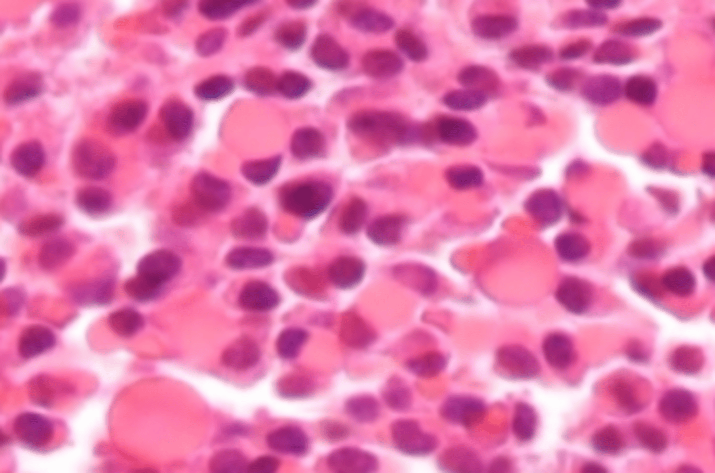
Myeloid and erythroid cells in varying stages of maturation
Normal BM finding
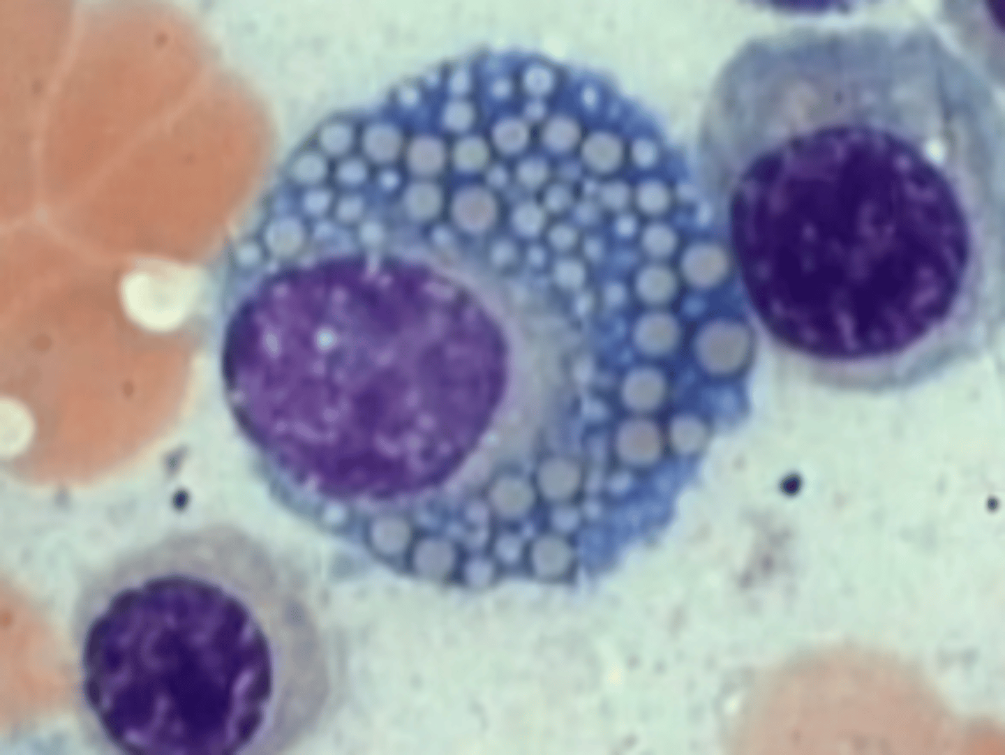
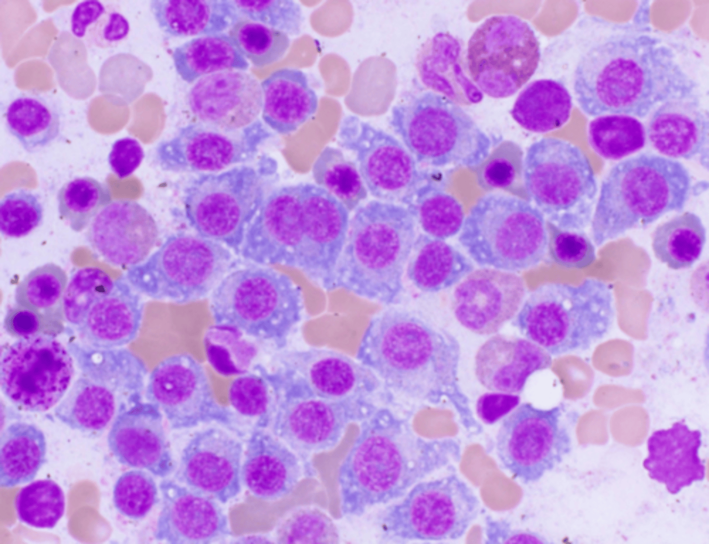
Plasma cells
These cells are strikingly increased in number in Multiple myeloma
Large with abundant basophilic cytoplasm, round eccentrically placed nuclei, lighter staining perinuclear area or area of hof
Moderate to abundant basophilic cytoplasm
Mature plasma cells in plasma cell myeloma
Cytoplasm
Larger than normal, Multinucleation or lobated, Concentric
Mature plasma cells in plasma cell myeloma
Nucleus
Dispersed, Fine reticular chromatin
Mature plasma cells in plasma cell myeloma
Nuclear chromatin
Prominent
Mature plasma cells in plasma cell myeloma
Nucleoli
High
Mature plasma cells in plasma cell myeloma
N:C Ratio
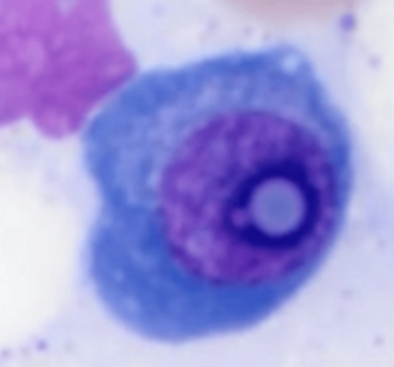
Flame cells
Plasma cells with red-tinged cytoplasm.
Hyaline inclusions, Crystalline inclusions, Vacuoles, Granules
Cytoplasmic inclusions in plasma cells in plasma cell myeloma
Plasmablastic appearance
Appearance of the neoplastic cells that has been associated with a poorer prognosis
Arterioles
Normally, plasma cells are seen clustered in ___
Marrow
In plasma cell myeloma, plasma cells may diffusely infiltrate the ___ and replace other marrow elements.
Bone marrow studies
These studies are able to give an idea on tumor burden (via percentage involvement) in Plasma cell myeloma, thus it can be a tool for prognostication and it is also used by doctors to monitor the patients' response to therapy
Bone marrow aspirates
Specimen that shows the exact morphology of the plasma cells in Plasma cell myeloma (what they look like, more mature, plasmablastic).
Plasmablasts
Plasma cell variation
Have higher N:C ratio, finer, and more reticular chromatin, and have a lot more prominent nucleoli than mature plasma cells

Flame cell
Plasma cell variation
Retaining the abundant cytoplasm and eccentric nuclear placement of a regular plasma cell, with a reddish tinge to the cytoplasm

Mott cell
Plasma cell variation
Numerous globules filling the cytoplasm, and these represent aggregates of immunoglobulins that are produced by that plasma cell
Russell bodies
Plasma cell component
Pinkish or eosinophilic cytoplasmic globules which represent produced immunoglobulins
Dutcher body
Plasma cell inclusion
Intranuclear inclusions that can easily be mistaken for nucleoli
Mott or Morula cells
Cytoplasmic changes in Plasma cells in PCM
Pale, clustered grape-like inclusions
Russell bodies
Cytoplasmic changes in Plasma cells in PCM
Round cherry-red inclusions
Flame cells
Cytoplasmic changes in Plasma cells in PCM
Vermilion red staining
Gaucher-like cells
Cytoplasmic changes in Plasma cells in PCM
Overstuffed fibrils
Flame cell
What Plasma cell variant is this?
Skip
Type "Skip"
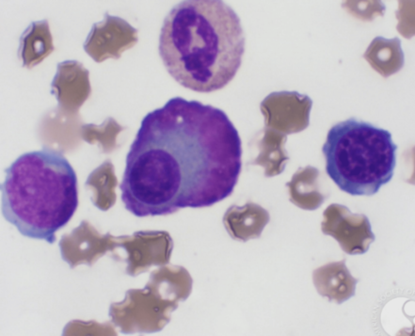
Just showing a pic of BM touch preparation showing mature and immature plasma cells, one of which is binucleate
Light chain restriction
The neoplastic plasma cells in plasma cell myeloma exhibit a monoclonal pattern of reactivity with either κ or λ light chains. This is known as:
2:1
Normal kappa(κ) to gamma(λ) light chain ratio
5:1
κ light chain monotypism K:G ratio