Module 7- Vaginal Cytology
1/50
There's no tags or description
Looks like no tags are added yet.
Name | Mastery | Learn | Test | Matching | Spaced | Call with Kai |
|---|
No analytics yet
Send a link to your students to track their progress
51 Terms
T or F, Females are born with all the ova that will be used during her lifetime
True
When do the ovaries begin functioning?
Between 8-12 months of age - at puberty
After ovulation, where are the ova transported from the ovaries?
Into the oviducts (fallopian tubes), to the respective horn of the uterus
Where does fertilization take place?
The oviducts (fallopian tubes)
Where are the fertilized ova transported to?
The uterus
What is the site of implantation and growth of fetuses?
Uterus
What are the two times that the cervix is not tightly closed to prevent a uterine infection?
During estrus and parturition
What is the cervix sealed with?
Mucus (mucous plug)
How often do dogs ovulate?
Biannual monestrus
What is the bleeding from a female dog during proestrus a result of?
Increased blood supply to the uterine wall in preparation for possible implantation
What occurs during the transition from proestrus to estrus?
Significant drop in estrogen, which reflects maturation of the follicles
Which hormone is responsible for initiating ovulation, usually occuring during early estrus?
Luteinizing hormone (LH)
Name for uses for vaginal cytology
Optimum time for breeding
Detect silent heat cycles
Predict a more accurate whelping date- mainly used in dogs and wildlife
Detect pathological diseases in the reproductive tract- VERY important
How does vaginal cytology work?
Cells grow and slough off the vaginal wall
Depending on which cells you are identifying, you can determine where an animal is in a cycle....vaginal cytology means tracking these changes
What species do we use VC for?
Mainly dogs and wildlife
Why doesn’t VC work for large animals?
Shorter heat cycle, come in and out within 15-20 hours
What do we use to synchronize heat cycles with large animals (ruminants) ?
Prostaglandin injections
What do we use to measure follicle size in horses?
Ultrasound
What methods are used for heat detection in large animals? (2)
Good record keeping
Herd management
Estrotect ( paint pouches)
Bull point marker or chin ball marker
Flehman response
What are some signs of estrus in large animals?
Walking more/ restless
animal standing to be mounted
flagging (tail off to the side)
Vulva winking
Calling/ vocalization
If offspring nursing, may get diarrhea from hormonal changes in dam
Decreased feed consumption
List 3 reasons why RVTS need to learn where animals are in their cycles as well as what normal vaginal cells look lile
More than ever for breeding wildlife
To help with client cost = $40/test
To ensure the animal is healthy before breeding
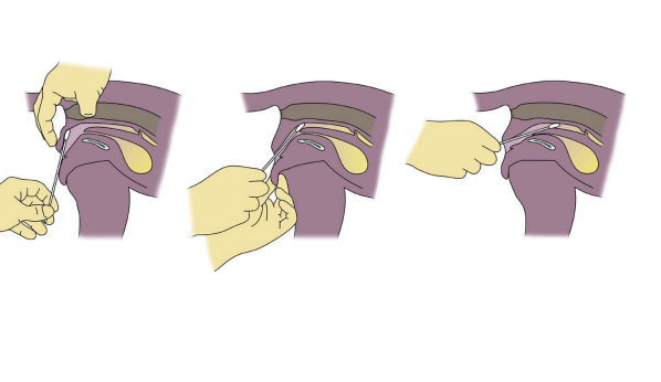
What are the 3 collection types for VC?
Swab
Labial
Pipette
List the steps for taking a VC swab
Clean the vulvar area
Vulvar labia are parted and everted if possible
Insert a premoistened cotton tipped applicator into the vagina (go dorsocranial until the advancement stops)- may use a speculum
Rotate the swab, remove and transfer to slide
Goal is to obtain sample of epithelial cells from the vagina, not the vulva
How to prepare the smear
Roll the cotton tip along the length of a glass microscope slide (2-3 tracks per slide)
List the steps of labial collection (4)
Clean the vulvar area
Massage the abdomen- trying to manipulate the cells forward
Vulvar labria are everted
Microscope slide is gently pressed against the labial mucosal area (impression smear)
When is the best/worst time to do labial collection
Best- WHen there is copious amounts of discharge
Worst- Late estrus, diestrus and estrus
Why?
Cells may be out by a day or two if you only collect from labial location
What species is commonly used for pipette VC samples?
Lab animals (rats)
How to perform Pipette VC?
Clean vulvar
Using a blunt pipette with sterile saline, introduce it the same way as the swab
Flush and recapture the saline a few times
Vaginal cells will be collected and may be placed on slide
Smear is done the same way as a blood film or a fresh drop with a coverslip
Define Proestrus
Before estrus
What cells will be mostly observed during proestrus?
Red blood cells
What clinical signs will be observed in proestrus?
Vulvar swelling and blood discharge
Define estrus
Peroid of time when a female is rexually receptive
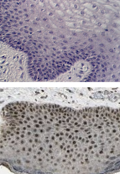
What cells will be observed on VC during estrus?
Superficial cells that are largely anucleate, have angular, folded cell margins.
Define Met-estrus
Period that starts at the end of the estrus and lasts until the beginning of diestrus
(often classified together with diestrus)
Diestrus definition?
Vulva returns to normal size, female will no longer accept the male for mating.
All signs of discharge and swelling are absent, and heat is complete.
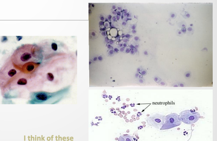
What cells characterise diestrus
Decrease in superficial cells
Increase in smaller intermediate cells
Presence of neutrophils
Anestrus definition
Period of sexual rest where the reproductive system is resting
Secretions are minimal or absent
Interestrus
Period during which the female refuses to mate - period of rest
Which are the youngest cells of the vaginal epithelium/ serve as precursors of the other epithelial cell types, located along the basement membrane and rarely are seen in exfoliative cytology
Basal Cells (epithelial)
What are the smallest cells seen on slides with a round shaped cell and a nucleus, dark blue cytoplasm, 1:1 ration of nucleus to cytoplasm
Parabasal (epithelial)
Larger than parabasal, due to the cytoplasm being larger
Round in shape (may appear multisided)
Nucleus about the same size as parabasal nucleus
Cytoplasm stains less intensely then parabasal
Intermediate Cells (Epithelial)
Largest of the vaginal epithelial cells
Nucleus(if present) is small or distorted
Large amount of cytoplasm- angular borders
Cornified- karyopyknotic- means the conversion to keratin
Superficial cells (Epithelial)
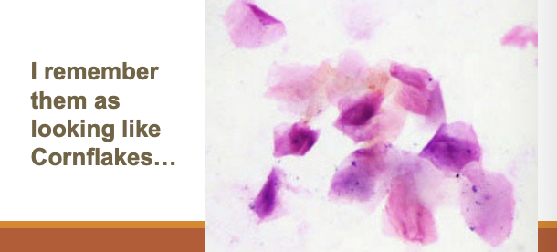
Superficial cells without nuclei often seen in large sheets or strings
Metestrus cells
Contains a neutrophil in the cytoplasm
These are present just before diestrus
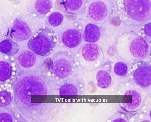
Foam Cells
Have vacuoles in the cytoplasm
May be found in prepubertal samples are should not be confused with neoplastic cells
common in parabasal cells or TVT cells (Transmissible venereal tumor is transmitted sexually, by mucous membrane contact)
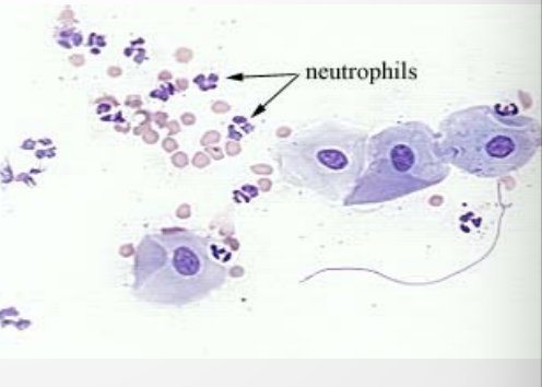
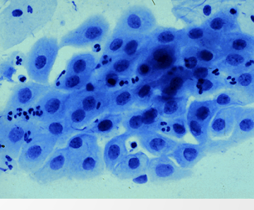
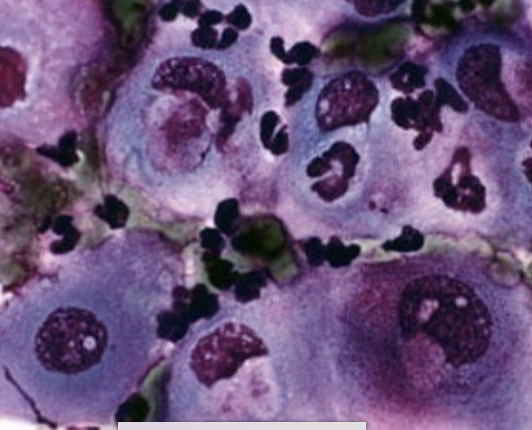
Proestrus smear, shows group of intermediate cells associated with neutrophils and red blood cells
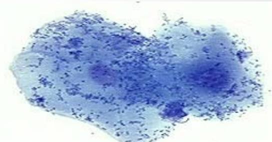
Bacteria
Often seen on vaginal smears in huge numbers, covering cells and spilling onto the background.
Dark specks covering the superficial cells in the image are bacteria
Usually have no meanin g, part of the vaginal flora, if there are no other symptoms
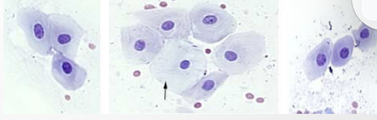
The arrow is on a non-cornified superficial cell but the other cells are...?
Intermediate cells with red blood cells
Parabasal cells
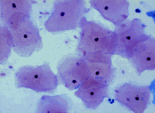
Superficial epithelial cells
Non cornified
How much saline to use for rodents with the pipette method for vaginal cytology
0.25- 0.50 cc using 3 cc syringe to infuse saline then gently removed