MBB 201 Final Exam
1/170
There's no tags or description
Looks like no tags are added yet.
Name | Mastery | Learn | Test | Matching | Spaced | Call with Kai |
|---|
No analytics yet
Send a link to your students to track their progress
171 Terms
List the main function(s) of:
Cytosol
Nucleus
Endoplasmic Reticulum
Golgi Apparatus
Lysosomes
Endosomes
Mitochondria
Chloroplasts
Peroxisomes
Cytosol→Contains many metabolic pathways; protein synthesis; the cytoskeleton
Nucleus→Contains main genome; DNA and RNA synthesis
ER→Synthesis of most lipids; synthesis of proteins for distribution to many organelles and to the plasma membrane
Golgi Apparatus→Modification, sorting, and packaging of proteins and lipids for either secretion or delivery to another organelle
Lysosomes→Intracellular degradation
Endosomes→Sorting of endocytosed material
Mitochondria→ATP synthesis by oxidative phosphorylation
Chloroplasts→ATP synthesis and carbon fixation by photosynthesis
Peroxisomes→Oxidation of toxic molecules
Why is it important that membranes form compartments within a cell?
Allows for distinct environments with different metabolic functions
Describe the evolution of the nuclear membranes and membranes of the endomembrane system (ER, Golgi, peroxisomes, endosomes, lysosomes).
∙May have arisen through invaginations of the plasma membrane
∙The interiors of the endomembrane system communicate with each other extensively
Describe the evolution of the mitochondria and chloroplasts.
∙Thought to have originated when aerobic prokaryotes were engulfed by pre-eukaryotic cells
→Mitochondria contain their own DNA and are capable of replicating this DNA and expressing RNA and protein from it
Almost all proteins begin their synthesis in the cytosol. What are the exceptions?
Some mitochondria and chloroplast proteins.
How are proteins in the cytosol that are destined for other organelles directed?
Proteins in the cytosol destined for other organelles must be directed there by a signal sequence which is dictated by the amino acid sequence.
What is a signal sequence?
∙Typically 15-60 amino acids long
→Function of the signals is dependent on the properties of the amino acids
Signal sequences are necessary and sufficient to direct a protein to a particular destination
**Don't need a signal sequence for a cytosolic protein because it is already where it needs to be
What are three ways to transport proteins into organelle compartments?
1. Transport through nuclear pores
2. Transport across membranes using protein translators
3. Transport by vesicles
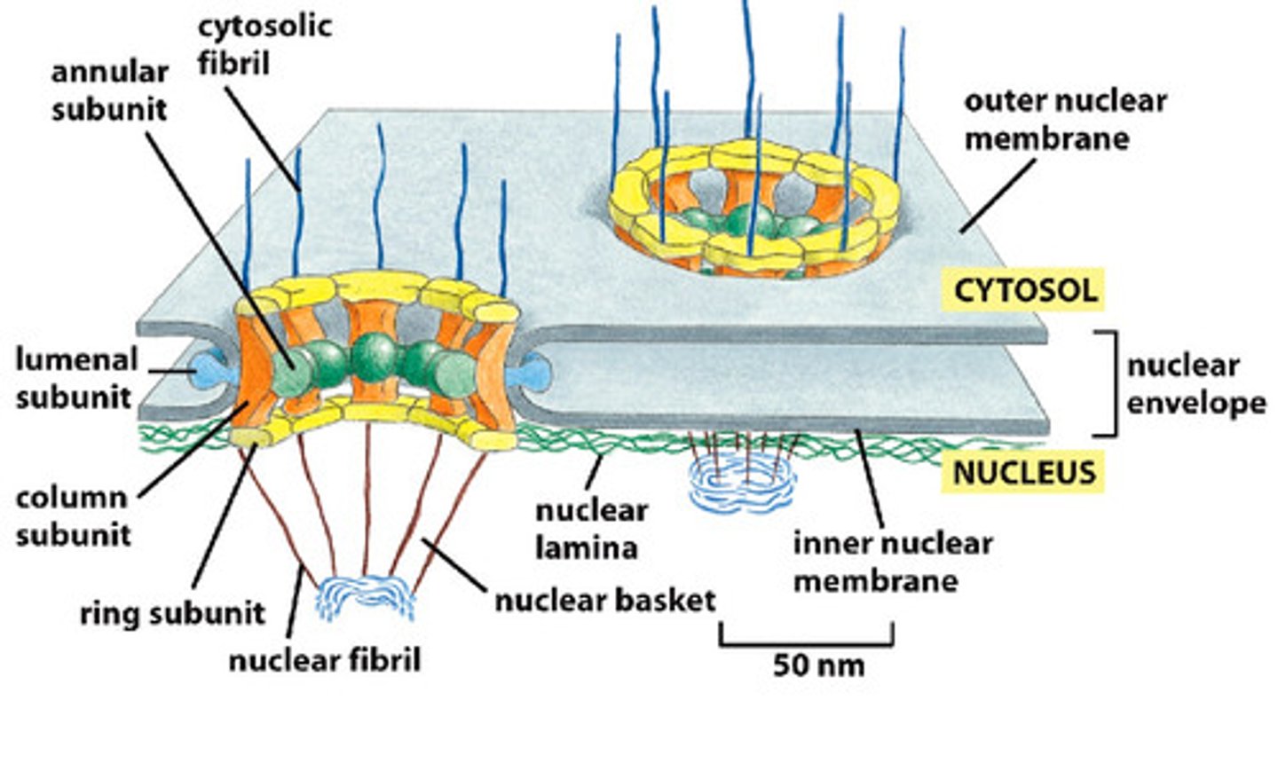
What is the nuclear envelope?
The nucleus contains a Nuclear envelope with 2 membranes
-Inner nuclear membrane
-Outer nuclear membrane (continuous with the ER)
∙Has nuclear pores to allow passage of molecules in and out of the nucleus
Describe the structure of a nuclear pore.
∙Spans across the inner and outer membrane
∙Composed of ~30 proteins, many of which are largely disordered and unstructured--creates a fibril mesh that fills the centre of the channel and prevents the passage of molecules through it
∙Small, water-soluble molecules can pass non selectively
How are cytosolic proteins transported through a nuclear pore?
∙Cytosolic proteins that are bound for the nucleus must contain a nuclear localization signal (NLS)
∙The NLS is recognized by proteins known as nuclear import receptors
→Help direct the protein through the pore by disrupting the interactions between the nuclear fibrils
Where does the energy for nuclear transport come from?
Nuclear import receptors are returned to the cytosol but require the GTPase known as Ran
∙Binding of Ran-GTP causes the dissociation of the imported protein from the receptor
∙The receptor bound to Ran-GTP can be transported back into the cytosol where GTP is hydrolyzed to GDP
→The hydrolysis frees Ran-GDP from the receptor so that is can bind to another NLS
Where does the energy for nuclear export come from?
Both import and export depend on Ran-GTP
Where are most mitochondrial and chloroplast proteins synthesized? How are the transported?
∙Most mitochondrial and chloroplast proteins are synthesized in the cytosol
∙Transported into the organelle through translocators
Explain how proteins will move across the mitochondrial or chloroplast membrane.
∙Proteins will contain a signal sequence at their N-terminus to allow their import
∙Proteins are unfolded as it is transported by a translator
∙Signal sequence is removed after translocation
∙Chaperone proteins help proteins to fold
Where do peroxisome proteins come from and how do they enter?
∙Most peroxisome proteins arrive form the cytosol
-Require a translocator to transport proteins across the membrane
-Proteins must be unfolded
∙Some proteins arrive from the ER:
-The ER-derived vesicles contains proteins that can fuse with peroxisomes to deliver their content
Proteins destined for the __________, _________, _______________, and ___________ _____________ all first enter the ER from the cytosol.
Proteins destined for the GOLGI, ENDOSOMES, LYSOSOMES, and PLASMA MEMBRANE all first enter the ER form the cytosol. Once in the lumen of the ER, it will not re-enter the cytosol.
What do all cytosolic proteins bound for the ER contain?
ER signal sequence
What is the fate of water soluble proteins in the lumen of the ER, and transmembrane proteins of the ER?
∙Water soluble proteins in the lumen of the ER:
-End up getting secreted or found in the lumen of an organelle in the endomembrane system
∙Transmembrane proteins:
-End up in the membranes of the organelles of the endomembrane system or on the plasma membrane
How do proteins move across the ER membrane?
∙Most proteins that enter the ER are threaded across the ER membrane BEFORE the polypeptide chain is fully synthesized
-Ribosomes synthesize proteins and attach to the ER membrane so that the protein can be threaded into the ER lumen as it is being synthesized
∙Regions of the ER with ribosomes attached are called rough ER
Differentiate between membrane-bound ribosomes, free ribosomes, and polyribosomes.
∙Membrane-bound ribosomes:
-Attach to the cytosolic side of the ER
∙Free ribosomes:
-Are not attached to any membrane
*Membrane-bound and free ribosomes are identical to one another
∙Polyribosomes:
-Many ribosomes bound to one mRNA molecule
How are ribosomes directed to the ER?
∙Signal recognition particle (SRP): Present in the cytosol. Binds to the ER signal sequence and the ribosome.
∙SRP Receptor: embedded in the ER membrane. Binds to SRP. Passes ribosome to a protein translocator. SRP is released.
→Protein synthesis occurs, passing the proteins through the channel in the protein translocator
How to soluble proteins move across the ER membrane?
∙The ER signal sequence causes the opening of the channel
∙The signal sequence remains bound to the channel as the rest of the protein is threaded through
∙Once the C-terminus has passed through, the signal sequence is removed by a signal peptidase on the luminal side of the ER and the protein is released into the lumen
Which category of amino acids would be most likely to be found in the stop-transfer sequence?
Non polar and hydrophobic (because the lipid bilayer is the same)
ex. Valine
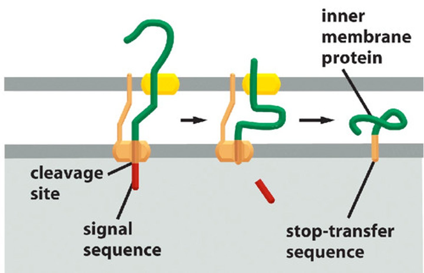
Describe how a single-pass transmembrane protein moves across the ER membrane.
∙Translocation is initiated by a start-transfer sequence. Translocation continues until a stop-transfer sequence is reached, preventing further translocation
-The translocator channel releases the growing polypeptide chain. The stop-transfer sequence forms an alpha helix and remains embedded in the membrane
∙Start-transfer sequence is cleaved
∙Orientation of the N- and C- will not change from one side of the membrane to the other
How are multi-pass transmembrane proteins created in the ER?
∙For some transmembrane proteins, the start-transfer sequence used to start protein translocation is internal
-The start-transfer sequence does not get removed like N-terminal ones do
∙Start-transfer sequences can work in conjunction with stop transfer sequences to create multi-pass transmembrane proteins
What is vesicular transport? What is its purpose?
The movement of material between organelles in the eukaryotic cell via membrane-enclosed vesicles
→Allows the transport of proteins and lipids to various parts of the cell including the endomembrane system and the plasma membrane
→Proteins in the ER are sent (at least initially) to the Golgi and may be derived to other compartments of the cell
Each intracellular compartment has a unique composition. If vesicles from one compartment fuse with another during vesicular transport, how does the cell ensure that the composition remains unique?
(ie. ER proteins stay in the ER, etc.)
Dictated by signal sequences on the protein
What are coated vesicles? What are they for?
Membrane-enclosed sacs that wear a distinctive layer of proteins on its cytosolic surface
-Helps shape the membrane into a bud and captures molecules for onward transport
∙There are several types of coated vesicles, each involved in the transport of vesicles form specific origins and destinations
Compare clathrin-coated vesicles and COP-coated vesicles.
Clathrin-coated vesicles: Found budding from the Golgi to the plasma membrane and endosomes as well as from the plasma membrane on the inward endocytic pathway
COP-coated vesicles: Found in vesicles between the ER and the Golgi, as well as from one part of the Golgi to another part
Is the adaptin used at the plasma membrane the same as the one used in the Golgi?
No, different organelles have unique compositions of proteins. This requires different adaptins to bind to different cargo receptors.
How does a vesicle form?
∙Vesicle begins as a clathrin-coated pit
∙Clathrin creates a basket like network on the cytosolic surface of the membrane. Helps to shape the membrane into a vesicle
∙Adaptins secure the clathrin to the vesicle and help select cargo molecules by binding to cargo receptors
∙Appropriate cargo proteins will have transport signals that can be recognized by the cargo receptors
∙The protein dynamin assembles a ring around the neck of the pit causing it to be pinched off
∙Once budding is complete, the coat proteins are removed and the vesicle can fuse with its target membrane
How does a vesicle know that it has reached the correct destination?
∙Rab proteins are a family of GTPases
∙Vesicles have a unique combination of Rab GTPases on the cytosolic surface of the protein as well as v-SNARE (v for vesicle)
What is tethering in terms of recognition of vesicles?
The Rab proteins are recognized and bound by tethering proteins found on the target membrane bringing the two into close proximity
What is docking, in terms of recognition of vesicles?
The v-SNARE on the vesicle interacts with complementary t-SNARE's (t for target) which firmly docks the vesicle in place
What is fusion, in terms of recognition of vesicles?
The vesicle fuses with the target membrane and the cargo protein is delivered to the interior of the organelle (or secreted if at the plasma membrane)
Does docking of a vesicle to a membrane always lead to fusing? Explain.
∙Docking does not always lead to fusion, sometimes a special stimulatory signal is required
∙The fusion of membranes is energetically unfavourable
∙Fusion occurs when the v-SNARE and t-SNARE wrap tightly around each other, winching the vesicle closer to the membrane
Formation of disulphide bonds is a common modification to proteins made in the lumen of the ER. Why aren't disulphide bonds added in the cytosol?
The cytosol is a reducing environment which reduces disulphide bonds
What is glycosylation and what is its function?
Glycosylation: covalent attachment of short branched oligosaccharides
Functions:
∙Protect proteins from degradation
∙Hold protein in the ER
∙Recognition by proteins for packaging or cell-cell interactions
→Glycosylation is rare on the cytosolic side
How are oligosaccharides added to a protein for modification in the ER?
∙Olgiosaccharides are not added one at a time- instead they are attached en bloc (all together)
∙A 14 sugar oligosaccharide is originally attached to the lipid dolichol and is transferred onto the amino group of an asparagine side chain as the peptide is translocated
∙Because they are attached to an amino, they are said to be N-linked (O-linked is less common)
Describe the ways in which proteins can exit the ER.
∙Some proteins are destined to stay in the ER and will contain an appropriate signal sequence
∙If they escape to the Golgi, they will be recognized by receptors and sent back to the ER
∙Most proteins that enter the ER are sent to the Golgi (at least initially). Move along microtubule "tracks"
∙Misfolded proteins or multimeric proteins that do not assemble properly are retained in the ER by the binding of chaperone proteins
→The chaperones assist in the folding process and prevent misfolded proteins from aggregating
∙If the protein still fails to fold, it will be exported to the cytosol where it will be degraded (proteases will degrade these proteins...specifically the proteasome
What is the unfolded protein response (UPR)?
∙If too many unfolded proteins accumulate in the ER, the unfolded protein response is triggered:
→More chaperones and quality-control related proteins are produced (don't want to be making proteins if something is wrong..also maybe you are producing proteins too fast for your chaperone proteins to help fold them)
→May inhibit protein synthesis
∙The size of the ER can be expanded to cope with the load, but if this limit is exceeded, the cell can be programmed to die
Outline the key point about the protein quality control system in the ER.
∙Chaperone proteins help misfiled proteins fold properly
∙Proteins that are misfiled are degraded in the cytosol
∙Protein complexes are checked for proper assembly before they can exit the ER
∙A chaperone protein will bind to a misfiled protein to retain it in the ER
What are cisternae and what are its distinct face?
Cisternae: flattened membrane-enclosed sacs of the Golgi
∙Has distinct faces:
→The cis face is adjacent to the ER
→The trans face points towards the plasma membrane
→The medial cisterna is in the middle
∙Vesicles from the ER enter the Golgi at the cis Golgi network
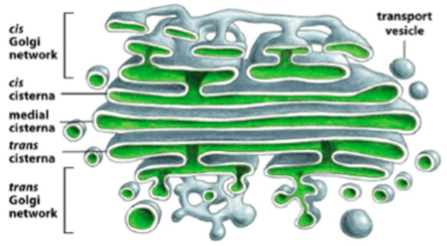
How do proteins move through the Golgi for modification?
∙Transport vesicles bud from one cisterna and fuse with the next
∙Proteins exit form the trans Golgi network (acts as a sorting centre; determines where proteins go after)
∙Proteins are further modified in the Golgi:
-Oligosaccharide chains are added, removed, and modified
∙The trans Golgi network is the main sorting station for the exocytic pathway
What is it called when vesicles from the Golgi fuse with the plasma membrane?
Exocytosis!
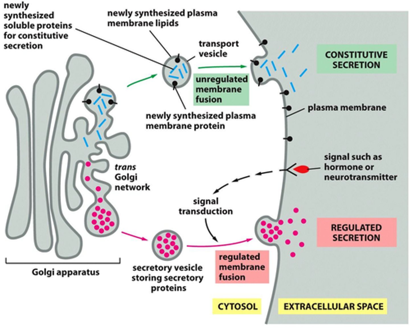
What is the constitutive exocytosis pathway?
Supplies plasma membrane with lipids and proteins- some proteins are secreted
→Entry to this pathways does not need a particular signal sequence (other than to enter the ER)
→Operates continually in all eukaryotic cells (not specialized because it is continuous)
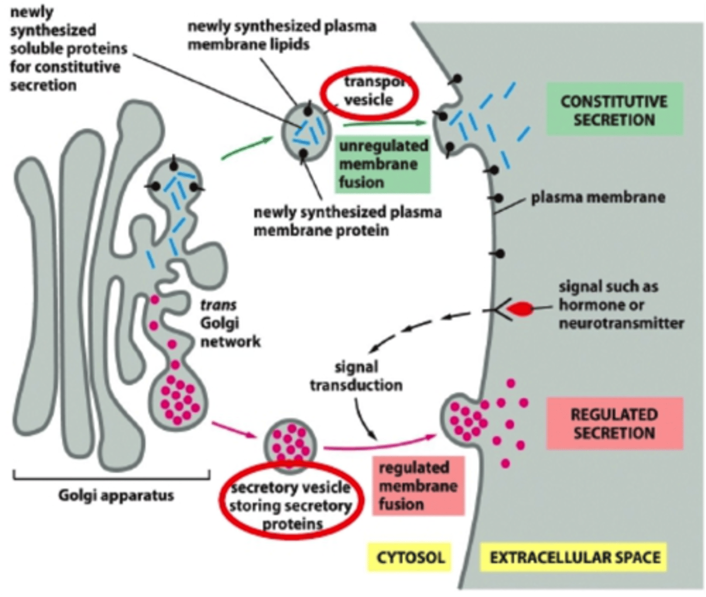
What is the regulated exocytosis pathway?
Only operates in specialized cells:
→Proteins are sorted and packed in the trans Golgi network which has conditions that cause proteins to aggregate (low pH, high Ca²⁺)
→Proteins are sorted in secretory vesicles which accumulate near the PM and wait for a signal to stimulate their fusion with the PM
→Aggregation allows secretory proteins to be at very high concentrations (allows these proteins to be in a small amount of space; will dock but not fuse until they get a signal to....so large amount of proteins will be released at the same time)
What is endocytosis? What are the two subtypes?
Endocytosis: The uptake of material through the invagination of the plasma membrane. Can be broken down into two types based on size:
1. Phagocytosis: involves the ingestion of large particles
→Mainly performed by specialized phagocytic cells (ex. white blood cells)
2. Pinocytosis: ingestion of fluid and molecules via small vesicles
→Performed by all cells
* Exocytosis delivers phospholipids to the PM, but this is balanced by removal from endocytosis
What is the basic principle of phagocytosis? What is its purpose?
∙Phagocytosis is important for the uptake of food as well as for defence against infection
∙After particles are engulfed, they are enclosed in vesicles called phagosomes
→Phagosomes fuse with lysosomes (low pH and contains lots of hydrolytic enzymes), digesting the engulfed particle
∙Example: Macrophage uptake of a bacterium
What is it about pinocytosis that allows it to continuously occur?
→Plasma membrane forms pinocytic vesicles
→Mainly carried out by clathrin-coated vesicles that pinch off and fuse with endosomes
→Indiscriminate! (takes up whatever is in the area when the vesicle is forming?
If pinocytosis is indiscriminate, how can it select what macromolecules to uptake?
Receptor-mediated endocytosis: pinocytosis that allows the selective uptake of macromolecules using specific receptors
Give an example of receptor mediated endocytosis in the body.
Ex. Receptor-mediated uptake of LDL
∙LDL receptors bind to LDL and are internalized as clathrin-coated vesicles
∙Fuse with endosomes, and are delivered to lysosomes for breakdown
∙LDL-receptor recycled to PM
Where are endosomes located at the various stages of their lifecycle?
∙Pinocytic vesicles deliver material to endosomes where they are sorted
∙Early endosomes are located near the PM mature into late endosomes by fusing with other later endosomes and are found near the nucleus
∙Endosomes maintain an acidic environment with a proton pump
∙Late endosomes mature into lysosomes
What are endosomes? What are the 3 possible paths of an endosome?
Endosomes: sorting station for the endocytic pathway
∙Possible paths:
→Recycling: returned to the PM
→Degradation: sent to lysosomes
→Transcytosis: move to a different domain of the PM
What are lysosomes?
∙Lysosomes are acidic and contain many hydrolytic enzymes involved in the degradation of macromolecules
∙Lysosomal membrane proteins are highly glycosylated on the luminal side-protects from degradation
→Contains a proton pump as well as transporters for macromolecule subunits to enter the cytosol
∙Lysosome destined proteins receive a mannose 6-phosphate tag in the ER and Golgi
What is autophagy?
Autophagy: Process by which a cell digests molecules and organelles that are damaged or obsolete
∙Process: organelle is enclosed by a double membrane, creating an autophagosomes which then fuses with a lysosome for destruction
In broad terms, what is signal transduction? How does this related to biology?
Signal Transduction: Conversion of a signal or impulse from one form to another
∙Typically in biology, signaling cells create extracellular signal molecules which are received by a target cell
-The extracellular signal is converted into an intracellular one
Cell communication can vary in terms of how __________ a message is made. Explain.
Cell communication can vary in terms of how PUBLIC a message is made.
∙The signalling molecule can take on a large variety of forms including: proteins, peptides, amino acids, nucleotides, steroids, fatty acid derivatives and gases
What is endocrine signalling? Give an example.
∙The most "public" signalling system
→Endocrine cells produce signal molecules known as hormones which are delivered through the bloodstream
→Signal can be broadcast to the entire body
→Ex. Insulin and glucagon are hormones secreted to regulate blood sugar levels
What is paracrine signalling? Give an example.
→Signalling cels produce signal molecules known as local mediators which diffuse locally through the extracellular fluid
→Signal is limited and can only be delivered to nearby cells
→If the signalling cell responds to their own signal, this is a form of paracrine signalling known as autocrine signalling
→Ex. Cancer cells secrete local mediators that promote their own survival
*2nd most public form of cell communication
What is neuronal signalling?
→Signals can be delivered very quickly over long distances (>1m!) however instead of broadcasting a signal widely, the signal is sent to specific target cells
→Signal is transmitted along a neutron in the form of an action potential. The electrical signal is converted into a chemical signal in the form of a neurotransmitter at the nerve terminals
→The neurotransmitter binds to the receptors on the target cell which can be converted back into an electrical signal
*3rd most public form of cell communication
What is contact dependent signalling?
→Most intimate and short range of all
→No signalling molecule is secreted. Instead, physical contact is made between molecules embedded in the plasma membrane of the signalling cell and receptors on the target cell
Give an example of contact dependent signalling.
Lateral inhibition in Drosophila
-Nervous system begins as a sheet of epithelial cells
-Some cells begin differentiating into neurons
-Each developing neuron produces an inhibitory signal protein which when bound by receptors on neighbouring cells prevents them from developing into neurons
Which type of cell signalling travels the largest distance?
Endocrine!
How do cells respond to a signal?
∙Cells respond selectively to a picture of signals
-The ability of a cell to respond is dependent on whether or not it has an appropriate receptor
∙Even if two cells have the same receptor, they may respond in different ways
∙The extracellular signal alone is not the message: the information conveyed is also dependent on how the target cell receives and interprets the signal
Can a cell receive multiple extracellular signals? Explain.
Yes, cells will contain a limited set of receptor proteins that will respond to different extracellular signals
∙A combination of signals can evoke a response that is different from the sum of the effects of each individual signal
∙Cells are programmed to kill themselves in the absence of signals
Are cell responses to extracellular signals fast or slow?
Responses to extracellular signals can be (relatively) fast or slow depending on what needs to happen
-Responses range in the millisecond range to several hours
What are the two general classes of extracellular signal molecules?
1. Molecules that do not cross the plasma membrane and bind to surface receptors
→Usually large and/or hydrophilic
2. Molecules that cross the plasma membrane and enter the cytosol and bind to intracellular receptors
→Usually smaller and/or hydrophobic
What kind of receptors do steroid hormones bind to? Give an example.
∙Hydrophobic molecules that can cross the plasma membrane
∙Bind to nuclear receptors: receptors that when bound to ligand can enter the nucleus and initiate transcription
→Receptor can initially be found in the cytosol or nucleus
Ex. Cortisol
What is nitric oxide and how does it work?
∙Nitric oxide (NO) is a gas that can diffuse across the plasma membrane and bind to proteins like guanylyl cyclase forming GMP (cGMP)
∙Only works locally because it is quickly converted into nitrates and nitrites
∙NO produced in endothelial cells causes smooth muscle cells to relax, causing blood vessels to dilate
Examples:
→Nitroglycerine is used to treat angina because it is converted into NO
→VIAGRA works by blocking the enzyme that degrades cyclic GMP prolonging the NO signal (ie. muscle relaxation)
What does the binding of an extracellular signal to a receptor generate?
∙Binding of the extracellular signal to the receptor generates an intracellular signalling response using intracellular signalling molecules
∙Intracellular molecules activate effector proteins which cause a cellular response
∙Most extracellular signal molecules bind to cell surface receptors
What are the 4 functions of intracellular signalling pathways?
1. Relay signals onwards
2. Amplify signals
3. Detect signals from more than one intracellular signalling pathway and integrate them
4. They can distribute the signal to effector proteins causing a response
*Responses are often modulated by feedback inhibition
What are molecular switches? What are the two classes?
Molecular switches: signalling protein that toggles between active and inactive states in response to a signal
→It is important to be able to control both the activation and inactivation
Two classes:
1. Proteins activated or inactivated by phosphorylation
2. GTP-binding proteins
What is the largest class of molecular switches? Explain.
∙Protein phosphorylation is the largest class of molecular switches
∙Involves protein kinases which phosphorylates proteins, and protein phosphatases which dephosphorylate proteins
∙Phosphorylation can either activate or inactivate a protein
∙Two main types:
1. Serine/threonine kinases
2. Tyrosine kinases
**All have hydroxyl groups on their side chain (polar uncharged)
What is the significance of a phosphorylation cascade?
∙Many molecular switches controlled by phosphorylation are also protein kinases!
∙The phosphorylation of one molecular switch causes it to phosphorylate another molecular switch allowing the transmission, amplification, distribution and regulation of signals
∙Ex. MAP kinase (MAPK)
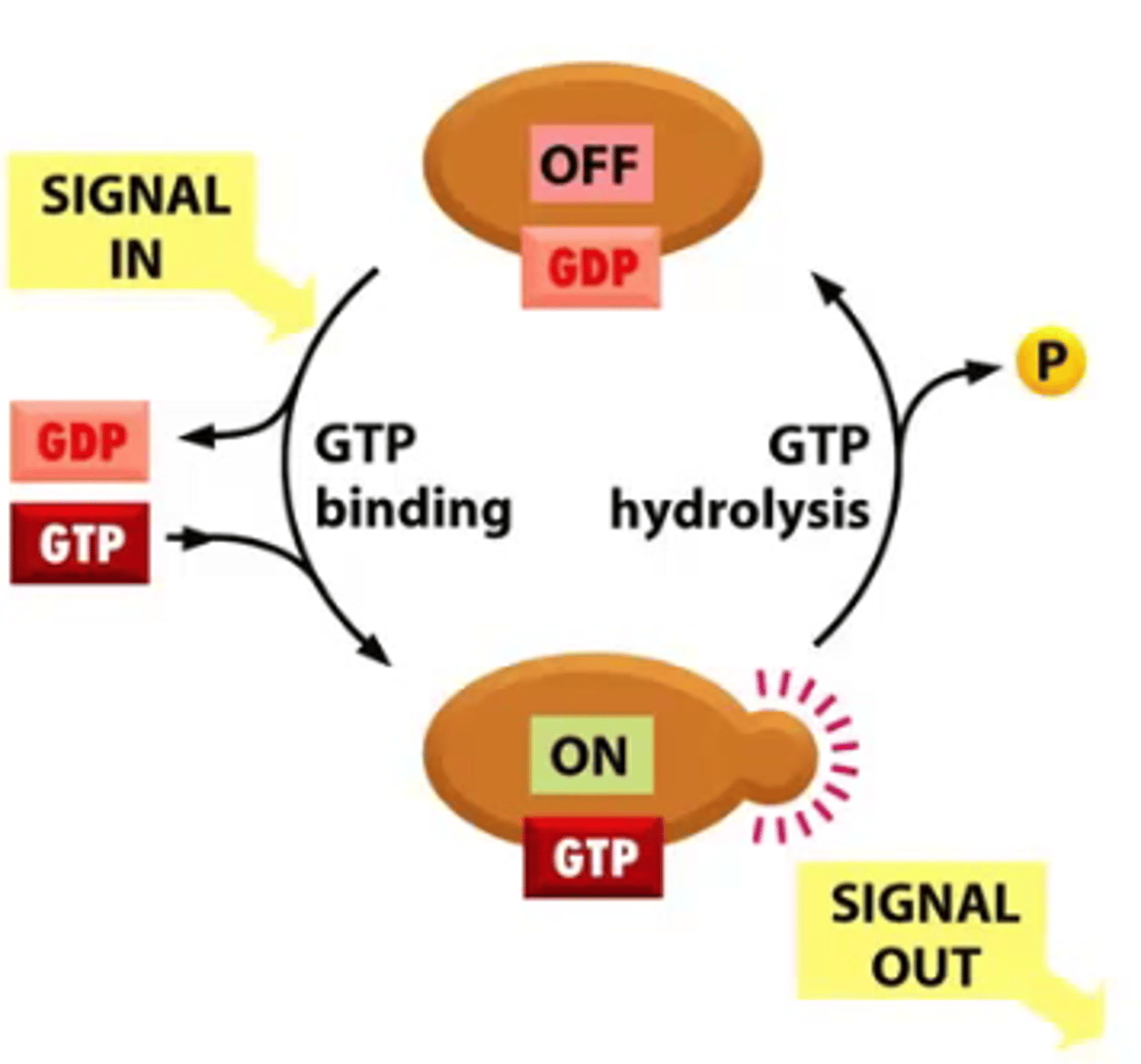
What are GTP-binding proteins?
∙Toggles between active and inactive depending on whether they have GTP or GDP bound
∙GTP-binding proteins possess GTP-hydrolyzing (GTPase) activity
→GTP binding proteins are active when they are bound to GTP
∙Two main types of GTP-binding proteins:
1. Large, trimeric GTP-binding proteins (AKA: G-proteins)
2. Monomeric GTPases
What two sets of regulatory proteins aid monomeric GTPases?
1. Guanine nucleotide exchange factors (GEFs):
→Activate proteins by exchanging GDP for GTP
2. GTPase-activating proteins (GAPs):
→Inactivate proteins by promoting GTP hydrolysis (causing inactivation of GTP binding proteins)
What are the 3 major classes of cell-surface receptors? Why are there more receptors than there are extracellular signals?
1. Ion-channel-coupled receptors
2. G-protein-coupled receptors
3. Enzyme-coupled receptors
There may be more than one receptor for each signal, and they may belong to different classes
Differentiate between ion-channel-coupled receptors, G-protein-coupled receptors, and enzyme-coupled receptors.
∙Ion-channel-coupled receptors can receive a chemical signal (ex. a neurotransmitter) and transduce it into an electrical signal by opening the ion channel and causing a change in membrane potential
∙G-protein-coupled receptors activate membrane bound, trimeric GTP-binding proteins causing the activation (or inactivation) of an enzyme or an ion channel in the plasma membrane
∙Enzyme-coupled receptors can act as an enzyme or associate with enzymes in the cell
What is the largest family of cell surface receptors in humans and what functions do they have?
∙GPCRs are the largest family (>700 in humans)
∙Variety of functions:
-Binds to hormones, local mediators and neurotransmitters
∙Composed of a single polypeptide chain that spans the lipid bilayer 7 times
∙Ancient, can be found in prokaryotes
What does the binding of an extracellular signal molecule to a GPCR do?
∙Binding of an extracellular signal molecule to a GPCR causes it to change conformation
∙This in turn activates a trimeric G-protein which results in the transmission of a signal
∙There are several G-proteins-each is specific for a set of receptors and target enzymes or ion channels
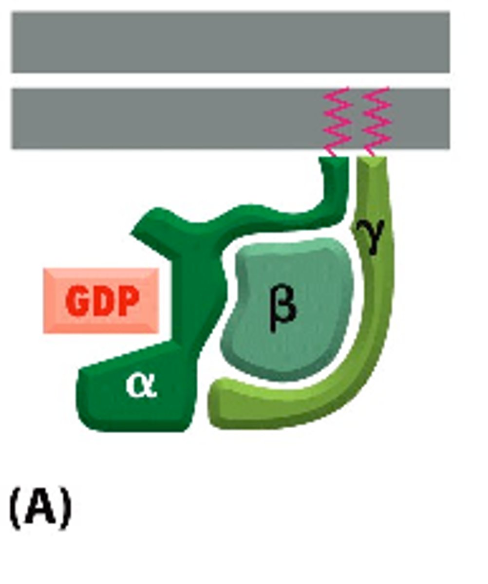
What are the subunits of a trimeric G-protein?
∙Each trimeric G-protein is made up of an α, β, and γ subunit.
-α and γ are tethered to the plasma membrane by a short lipid tail
When unstimulated, α is bound to GDP
What happens upon stimulation of a GPCR by a signal molecule?
∙Upon stimulation by a signal molecule, the GPCR changes conformation causing the associated G-proteien to decrease its affinity for GDP
∙GDP dissociated and is exchanged for GTP
∙Sometimes, the G-protein subunits dissociate (this doesn't always happen) and are switched "on"
∙G-proteins interact with target enzymes or ion channels
What dictates the length of response of a GPCR?
∙The amount of time that the G-protein subunits are "switched on" dictates the length of the response
∙The subunits will remain on when GTP is bound to the α subunit
How is a GPCR switched off?
∙The α subunit contains a GTPase activity which can hydrolyze the GTP to form GDP
∙The α subunit reassembles with the βγ complex
∙The protein is returned to its original, inactive state
How do G-proteins and ion channels contribute to the regulation of heart rate?
∙Acetylcholine binding to GPCRs of heart pacemaker cells activates the G-protein Gi
∙The βγ subunit binds to a K⁺ ion channel, causing it to open
∙Channel closes when GTP is cleaved and the subunits re-associate with one another
Acetylcholine binds to a GPCR on the heart muscle, making the heart beat more slowly. The activated receptor stimulates a G protein, which opens a K+ channel in the plasma membrane. What would enhance this effect of acetylcholine?
Addition of a high concentration of a nonhydrolyzable analog of GTP (will keep alpha subunit active won't be hydrolyzed)
What are the two most common enzyme targets of G-proteins?
1. Adenylyl Cyclase:
∙Produces cyclic AMP (cAMP)
2. Phospholipase C
∙Produces inositol triphosphate (IP₃) and diacylglycerol (DAG)
∙Adenylyl cyclase and phospholipase C are activated by different G-proteins
∙cAMP, IP₃, and DAG are examples of second messengers
Outline the roles of adenylyl cyclase and cAMP phosphodiesterase in the cAMP signalling pathway.
Adenylyl Cyclase:
∙Generates cAMP from ATP, releasing Pii
∙The alpha subunit of the G-protein Gs is responsible for the activation of adenylyl cyclase
cAMP phosphodiesterase:
∙Converts cAMP to AMP using water
Give an example of a cAMP signalling pathway.
Caffeine
∙Works by inhibiting cAMP phosphodiesterase, keeping cAMP levels high
∙Note: intracellular cAMP levels are normally very low
∙Effects of cAMP vary from cell to cell, but in general, it prepares the body for action
How does cAMP lead to glycogen breakdown?
cAMP exerts most of its effects by activating cAMP-dependent protein kinase (PKA):
∙PKA is normally inactivated by binding to a regulatory protein
∙Binding of cAMP to PKA releases the regulatory protein
∙PKA can then phosphorylate other proteins (acting glycogen phosphorylase in skeletal muscle)
→Glycogen breakdown is an example of a relatively fast response
What are the slow responses of cAMP?
∙cAMP can also cause the activation of gene expression-a relatively slow response
∙PKA phosphorylates transcriptional regulators which a can initiate transcription
Give an example of a bacterial toxin that affects G-proteins.
Ex. Cholera toxin
∙Modifies the alpha subunit of Gs so that it can no longer hydrolyze GTP
∙The G-protein is constantly active!
∙Result: in the intestine, this can result in prolonged outflow of Cl⁻ and water=dehydration
What is phospholipase C? What does it do?
Phospholipase C: enzyme that cleaves an inositol phospholipid that is a part of the membrane. This generates:
-Inositol 1, 4, 5-triphosphate (IP₃)
∙Released into the cytosol
-Diacylglycerol (DAG)
∙Remains embedded in the membrane
-Both products are important in signalling
Outline the inositol phospholipid pathway.
∙IP₃ binds to and opens Ca²⁺ channels embedded in the ER membrane
-Free Ca²⁺ is released into the cytosol which can act on other proteins
∙DAG recruits a cytosolic protein to the plasma membrane known as protein kinase C (PKC)
-Activation of PKC required the binding of Ca²⁺
-PKC phosphorylates several intracellular proteins
What is the significance of calcium signalling?
∙Ca²⁺ is important in a lot of signalling events
→ex. muscle contraction, secretion
∙Cytosolic Ca²⁺ levels are maintained at very low levels compared to the extracellular fluid as well as the interior of organelles like the ER
∙When Ca²⁺ channels open, Ca²⁺ rushes down its electrochemical gradient
What is the relationship between calcium and calmodulin?
∙Ca²⁺ binds to specific proteins in order to exert their effects. The most common of which is calmodulin
∙Calmodulin binds to four Ca²⁺ ions, inducing a conformational change allowing it to interact with other proteins like Ca²⁺/calmodulin-dependent protein kinases (CaM-Kinases)
Briefly describe the GPCR signalling in photoreceptors.
∙Light activates the GPCR known as rhodopsin
∙Rhodopsin activates the G-protein known as transducin
∙The alpha subunit of transducin activates cyclic GMP phosphodiesterase (reduces cGMP levels by converting into GMP)
∙Affects the state of cation channels, leading to a signal being relayed to the brain
Why is it important for photoreceptors to be able to amplify signals and adapt?
∙Photoreceptors can amplify a signal so that visions possible even in dim conditions
-Allows you to see in bright sunlight
-Strong light reduces Ca²⁺ concentrations which is required by signal amplification enzymes