Path bone section
1/86
There's no tags or description
Looks like no tags are added yet.
Name | Mastery | Learn | Test | Matching | Spaced | Call with Kai |
|---|
No analytics yet
Send a link to your students to track their progress
87 Terms
most common oral scc site
lateral tongue
2nd most common oral scc site
floor of the mouth
list 3 fibro-osseous lesions
fibrous dysplasia
PA C-O dysplasia
ossifying fibroma
what is a periapical cyst / radicular cyst
the most common odontogenic tumor
it is an asymptomatic cyst arising from the periapical granuloma
it is located on the apex of any teeth and is ass. w/ non-vital teeth
what is a periapical granuloma
it is a painful asymptomatic cyst located on the apex of any teeth and is as w/ non-vital teeth
it has a diffuse lucency = widened PDL space
what is acute/chronic osteomyelitis
it is a painful bone infection characterized by necrotic bone and ill-defined radiolucency
it is as w/ Paget’s disease, BIS, and radiation therapy
common location = mandible
what is amelogenesis imperfecta and the 3 types
it is characterized by genetic abnormal enamel
hypocalcified a.i → decreased enamel density
hypomaturation a.i → decreased enamel density
hypoplastic a.i → thin enamel
what is dentinogenesis imperfecta
a genetic condition characterized by blue translucent teeth, short roots, and no pulp
multiple radiolucencies = PA abscesses
what is dentin dysplasia and its 2 types
type 1 → most common; normal crown + short roots + no pulp; PA abscesses
type 2 → rare; normal crown + normal roots + thistle pulp; primary teeth have an amber crown appearance
list the odontogenic epithelial tumors (3)
1. ameloblastoma
2. peripheral ameloblastoma
3. adenomatoid odontogenic tumor
ameloblastoma (+ common location, associations/radiographic appearance, population)
common locations: posterior mandible
clinical features: asymptomatic; unerupted teeth
radiographic: uniocular, multiocular (soap/bubble honeycomb),
appearance: bone expansion, resorbs + displaces teeth
population: young>>middle aged, wide range
what is a peripheral ameloblastoma (NOT ON STUDY GUIDE)
it is a rare presentation of an ameloblastoma on the gingiva. most odontogenic cysts/tumors will have a peripheral apperance
it is not as invasive and has little to no bony involvement
what is an adenomatoid odontogenic tumor (AOT) / odontogenic adenomatoid tumor (OAT)
it an encapsulated benign tumor
AOT/OAT (+ common location, associations/radiographic appearance, population)
common locations: maxilla, anterior (80%), ass w impacted tooth (75%)
radiographic: uniocular radiolucency w opaque snowflakes
population: 70% young ppl <20; females
list the epithelial/mesenchymal mixed tumors (5)
1. odontoma
2. compound odontoma
3. complex odontoma
4. ameloblastic fibroma
5. ameloblastic fibro-odontoma
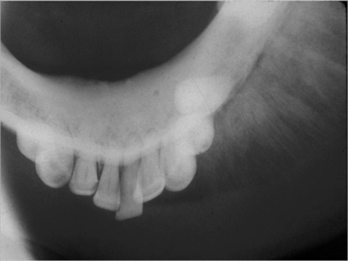
what is an odontoma
it is the most common odontogenic tumor; it is composed of enamel, dentin, cementum, and pulp tissue
two types are compound odontoma + complex odontoma
what is a compound odontoma
it appears as a cluster of small teeth, toothlets, denticles
compound odontoma (+ common location, associations/radiographic appearance, population)
common locations: anterior jaws
clinical features: asymptomatic; ass w impacted/unerupted teeth
Radio: cluster of mini teeth
population: young 10-20yrs
what is a complex odontoma
it contains odontogenic tissue w no small teeth
complex odontoma (+ common location, associations/radiographic appearance, population)
common locations: posterior jaws
clinical features: asymptomatic; ass w/ impacted/unerupted teeth
radiographic: unilocular radiopaque mass w radiolucent rim
population: young adults (10-20)
what is an ameloblastic fibroma
it is a benign unencapsulated tumor
what is an ameloblastic fibroma (+common location, clinical features, radiographic, population)
common locations: posterior mandible
clinical features: ass w unerupted teeth (75%)
radiographic: uniocular/multiocular radiolucency
population: young 10-20yrs; males
what is an ameloblastic fibro-odontoma
a tumor w features of an ameloblastic fibroma and odontoma
ameloblastic fibro-odontoma (+ common location, associations/radiographic appearance, population)
common locations: posterior mandible
clinical features: asymptomic; ass w impacted tooth
radiographic: well-defined radiolucency w radiopaque component
population: children
AOT/OAT impacts this gender population. ameloblastic fibroma impacts this gender population
AOT/OAT: females
AF: males
all epithelial tumors (except peripheral ameloblastoma) are ass w impacted, unerupted teeth or both. which are impacted by what
impacted teeth
-ameloblastoma
-AOT/OAT
-ameloblastic fibro-odontoma
both impacted/unerupted
-odontomas
unerupted teeth
-ameloblastic fibroma
--------------------
impacted
impacted
(2) impacted/unerupted
unerupted
impacted
define cyst **
a sac or cavity that is lined w epithelium and enclosed w CT
describe the basic info section of cysts **
there are two types of cysts - developmental cysts which include intraosseous and extraosseous cysts. the other are inflammatory cysts such as periapical cysts and residual cysts
developmental cysts are broken down further into non-odontogenic and odontogenic. if it is an odontogenic cysts this means they develop from the dental lamina, rests of malassez, and REE
what is the most common cyst **
a periapical cyst
what is the second most common cyst **
dentigerous cyst
list the intraosseous odontogenic developmental cysts (5)
1. dentigerous cyst
2. primordial cyst
3. odontogenic keratocyst (OKC)
4. lateral periodontal cyst
5. calcifying odontogenic cyst
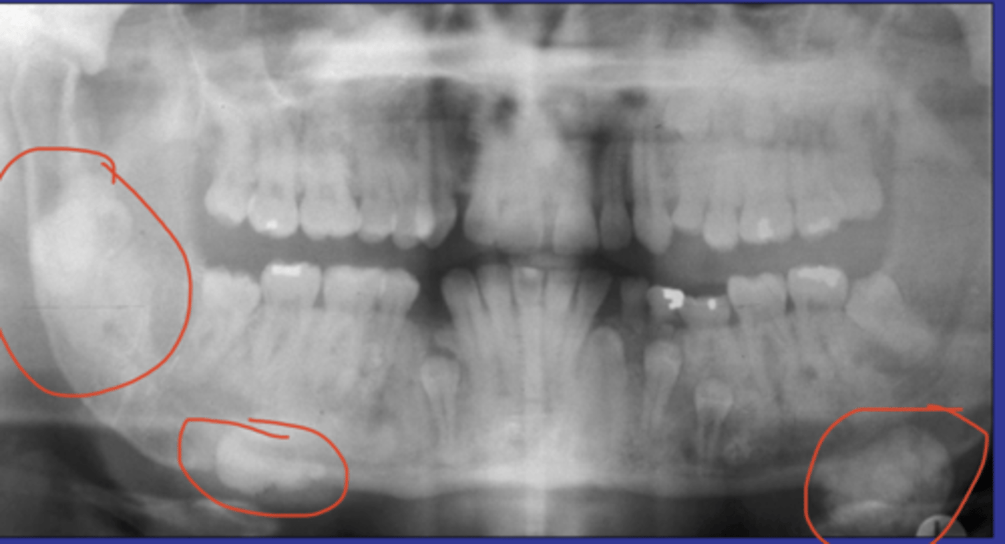
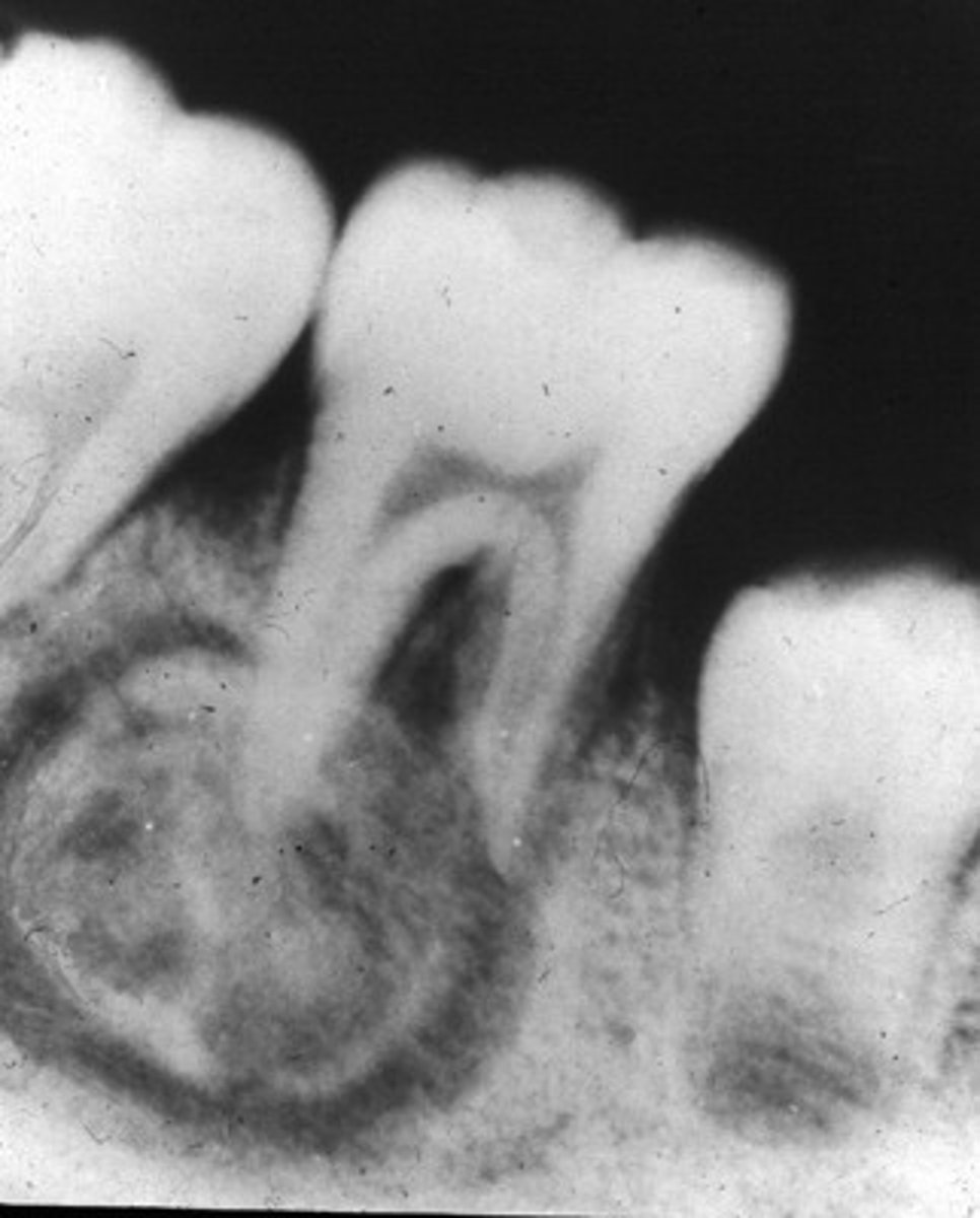
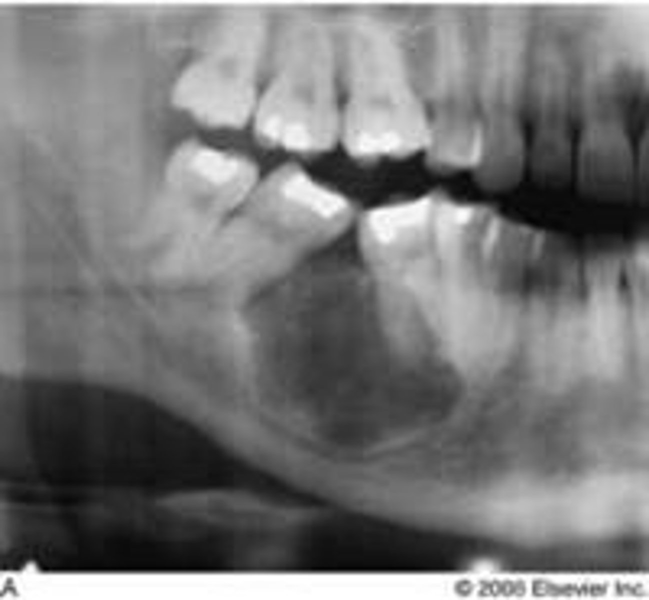
what is a dentigerous cyst
it is a cyst which forms around the crown of an impacted tooth
dentigerous cyst / follicular cyst (+ common location, associations/radiographic appearance, population)
common locations: mand 3rd crown; the crown of any unerupted tooth
clinical features: if large it may displace teeth
radiographic: well-defined uniocular radiolucency
population: young adults; males
what is a primordial cyst
it is a cyst which arises instead of a tooth
primordial cyst (+ common location, associations/radiographic appearance, population)
common locations: mand 3rd molars; posterior to developing 3rd molar
radiographic: uniocular/multiocular well-defined radiolucency
prognosis: most are histologically OKC
population: young adults
what is an odontogenic keratocyst / keratocystic odontogenic tumor **
it is a benign cyst but it can grow to be super aggressive; ass/ w Gorlin SX/NevoidBCCSx
OKC (+ common location, associations/radiographic appearance, population)
common locations: posterior mandible
clinical features: asymptomatic; displacement and resorbs teeth structure
radiographic: well-defined uniocular (when small)/multiiocular radiolucency
population: young adults 20-30s; males
what developmental cyst is considered the "great mimicker" **
OKC
what is a lateral periodontal cyst
it is a cyst which develops on the lateral aspect of the root surface of other vital teeth
lateral periodontal cyst (+common locations, clinical features, XR, population
common locations: mand canine/premolar area
clinical features: asymptomatic; adjacent teeth are vital
radiographic: uniocular/multiocular grape like radiolucency on the lateral aspect of a root
population: middle-aged adults (50-60s); males
which developmental cyst has a uniocular/multiocular grapelike radiolucency
lateral periodontal cyst
similarities of the 4 developmental cysts
-all these developmental cysts appear on the mandible
-require surgical removal
-require surgical excision
-arise from the dental lamina
-uniocular/multiocular
-impacts males
-minimal-high recurrence
-young adults
-resorbs/displace teeth
mandible locations (mand 3rd+crown, mand 3rd+posterior of 3rd, posterior mand, mand can/prem area)
surgical removal = dentigerous cyst, primordial cyst, and gingival cysts
surgical excision = everything else; OKC also requires curettage
dental lamina = OKC and lateral periodontal cyst
uniocular radiolucency = dentigerous cyst and COC; everything else is uniocular/multiocular
impacts males = primordial cysts, OKC, and lateral periodontal cyst
minimal recurrence = dentigerous cysts;
high recurrence = primordial cysts and OKC
young adults = everyone except lateral periodontal cysts + gingival cysts (50-60s); OKC and COC are YA (20-30s)
resorbs/displace teeth = dentigerous cysts, OKC, COC
what is a calcifying odontogenic cyst (COC) / Gorlin cyst
it is a nonaggressive developmental cyst
COC / Gorlin cyst (+ common location, associations/radiographic appearance, population)
common locations: anterior portion of jaws (65%)
clinical features: asymptomatic; slow growing; ass w impacted tooth; histologically = ghost cells
radiographic: uniocular radiolucency +/- opacities; resorption + displacement of roots
population: young adults (20-30s)
list the extraosseous cysts
eruption cysts
gingival cysts
list the non-odontogenic intraosseous developmental cysts (3)
1. nasopalatine cyst
2. median palatal cyst
3. globumaxillary cyst
what is a nasoplaotine cyst / incisive canal cyst
it is a cyst within the nasopalatine canal
nasopalatine cyst / incisive canal cyst (+ common location, associations/radiographic appearance, population)
common locations: nasopalatine canal/papilla bt central incisors and hard palate
clinical features; vital teeth
radiographic: well-defined "heart shaped" radiolucency
population: middled-aged (40-60s); males
what is the median palatal cyst
it is a cyst which arises at the midline of the hard palate, posterior the nasopalatine canal cyst
medial palatal cyst (+ common location, associations/radiographic appearance, population)
common locations: midline of the heart palate
clinical features: swelling at the midline of the hard palate
population: any age
what cyst is not an acceptable term/dx and was once believed to be a developmental fissural cyst
globulomaxillary cyst
list the nonodontogenic intraosseous cysts (3)
1. nasopalatine duct cyst
2. median palatal cyst
3. globulomaxillary cyst
what is the histology and radiography of a globulomaxillary cyst
histology: OKC, periapical cyst, lateral periodontal cyst
radio: well-defined radiolucency bt the maxillary lateral incisor/canine
list the nonodontogenic extraosseous cysts (6)
1. nasolabial cyst
2. branchial cleft cyst / cervical lymphoepithelial cyst - oral lymphoepithelial cyst
3. thyroglossal tract cyst
4. epidermoid cyst
5. dermoid cyst
6. stafne bone cyst
why is a stafne bone cyst not a real cyst
it is not a real cyst. it is a radiolucent lesion that has no epithelium lining and it is asymptomatic
stafne bone cyst / stafne defect / static bone cyst (+ common location, radiographic appearance, tx, population)
common locations: posterior mandible and inferior the mandibular canal
radio: uniocular lucency / well-defined cyst like radiolucency
TX: none
population: men 80-90%
little notes abt non-odontogenic developmental cysts - intraosseous (3) + extraosseous (6)
-nasopalatine duct cyst + stafne duct cyst affect males. nasolabial cyst affect females
-their populations are almost paired:
--middle aged+men
--any age
--middle age
--YA
--YA
--any age
--birth/young
--men
list the bone diseases (4)
1. osteogenesis imperfecta
2. paget disease
3. fibrous dysplasia
4. cherubism
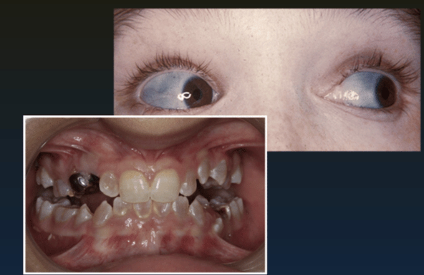
what is osteogenesis imperfecta
it is a group of inherited disorders where an abnormal or deficient amount of collagen produces insufficient bone
osteogenesis imperfecta (+clinical features, population
genetic
• blue sclera of eyes + multiple bone fractures + joint hyperextension
• teeth = dentinogenesis imperfecta
• population: children
what is another name for pagets disease and what is it
osteitis deformans. it is chronic benign disease that is usually polyostotic
pagets disease / osteitis deformans
-cause
-clinical features
-oral features
-radiographic
-prognosis
-population
cause: unknown
clinical features: painful gradual enlargement of bones; a positive alkalaine phosphatase in blood is dx
oral features: jaws are mostly affected esp the MAXILLA; dentures appear too tight
radiographic: extensive hypercementosis; mixed lesion w cotton wool opacities and round lucencies
prognosis: guarded bc of risk of osteosarcoma
population: adults >40; males
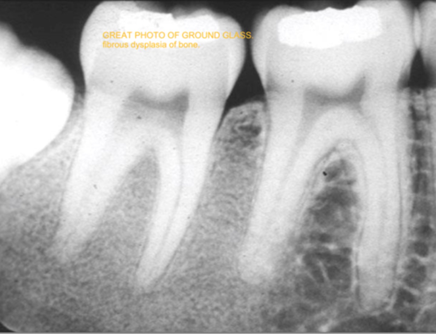
what is fibrous dysplasia
it is an uncommon chronic benign bone disease where bone is replaced w CT; it is usually monostotic but it may be polyostotic
fibrous dysplasia
-cause
-clinical features
-oral features
-radiographic
-prognosis
-population
cause: unknown
clinical features: painless gradual enlargment of bone replaced by fibrous CT (paget's is gradual enlargement presenting w pain)
oral features: maxilla is affected (like paget's); bone expansion/displacement
radiographic: ground glass/orange peel appearance
prognosis: guarded bc of 25-50% recurrence
population: teens/YA <20s
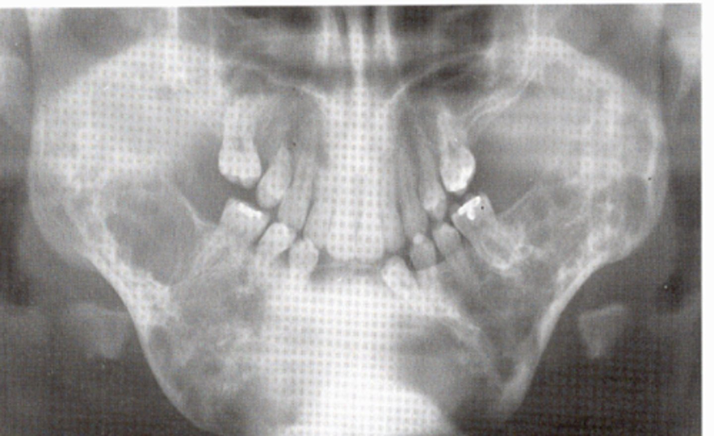
what is cherubism
it is a rare genetic disorder named for the appearance of plump cheeks
cherubism
-cause
-clinical features
-oral features
-radiographic
-population
cause: inherited autosomal dominant
clinical features: plump cheeks; upturned eyes; histologically GIANT CELL LESIONS
oral features: posterior mndible; usually symmetric
radiographic: bilateral multiocular lucencies
population: children
bone disease are the only section that ask for specifically abt clinical features and [ ]
[ ]
oral features
list bone lesions (4)
1. central giant cell granuloma
2. traumatic bone cyst
3. periapical c-o dysplasia
4. idiopathic sclerosis / dense bone island
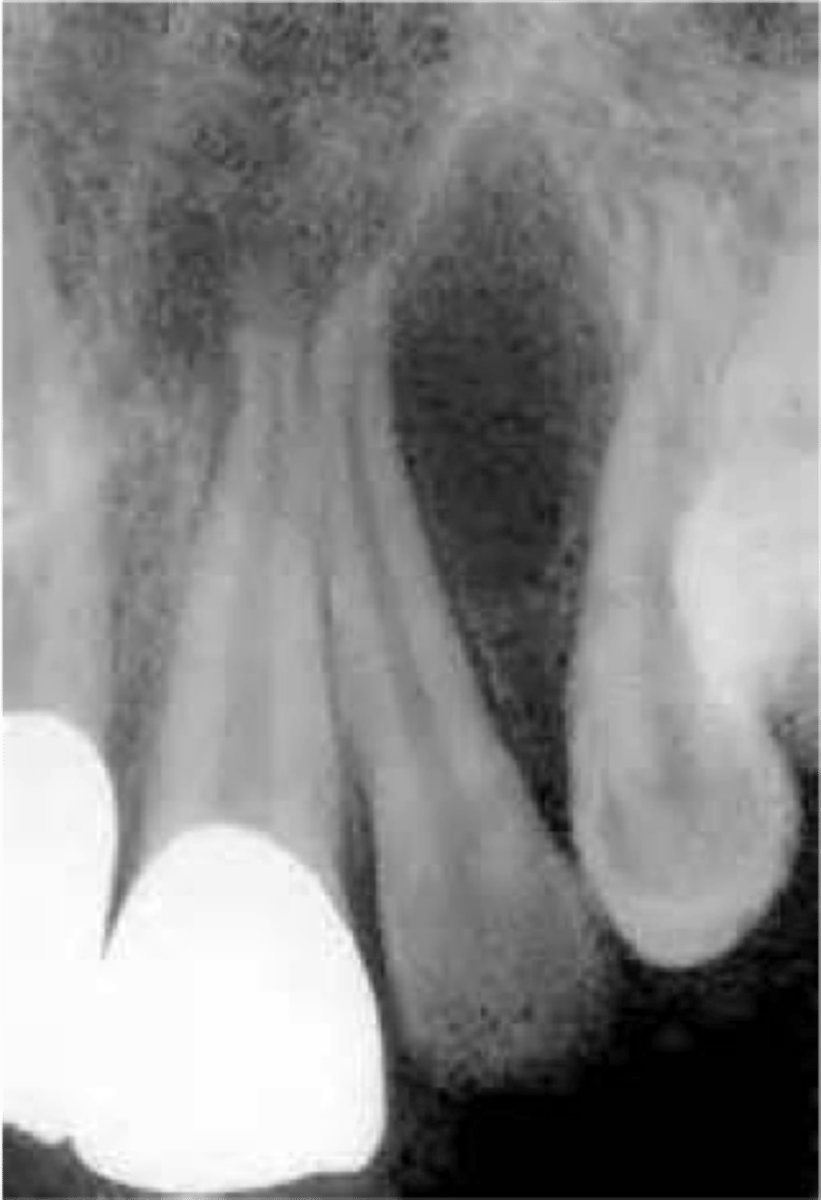
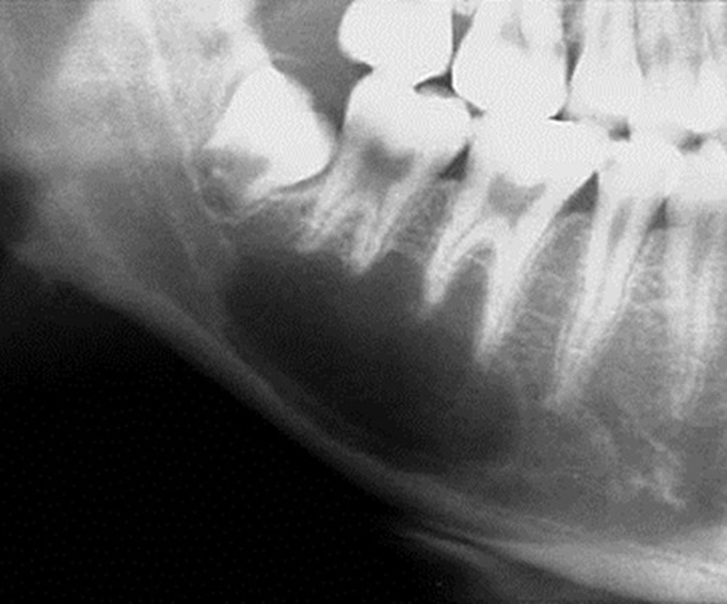
central giant cell granuloma
-common locations
-clinical features
-radiographic
-population
common locations: anterior jaws and mostly the mandible; cross the midline from canine to canine
clinical features: it is a slow growing expansion; histologically we see giant cells similar to hyperparathyroidism/browns tumors
radio: it starts as a uniocular lesion, then develops into a multilocular lucency; it may cause expansion+divergence
population: YA <30s
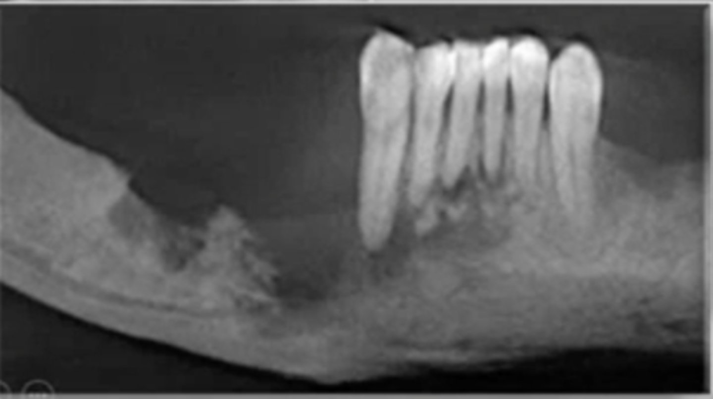
why is a traumatic bone cyst not a true cyst
there is no epithelial lining
traumatic bone cyst / solitary bone cyst
-common locations
-radiographic
-population
common locations: mandible only
radiographic: well-defined uniocular radiolucency which scallops bt roots of adjacent vital teeth; surgery reveals an empty cavity
population: children
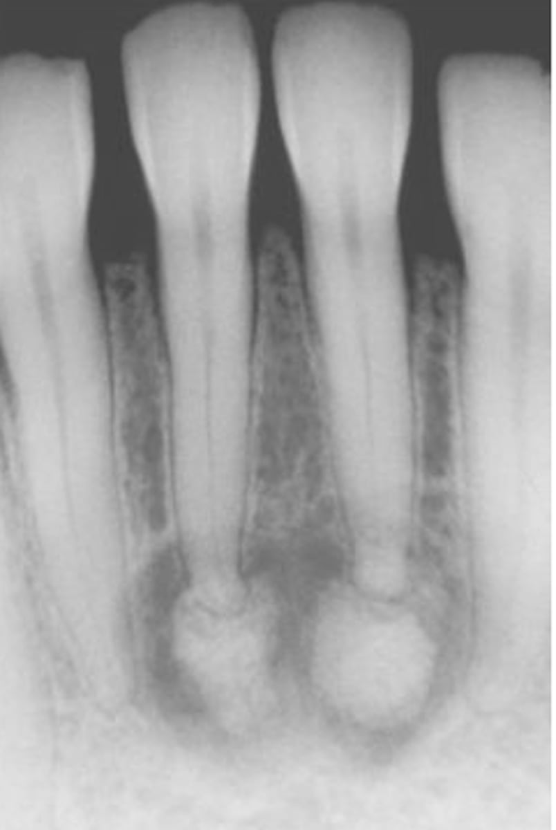
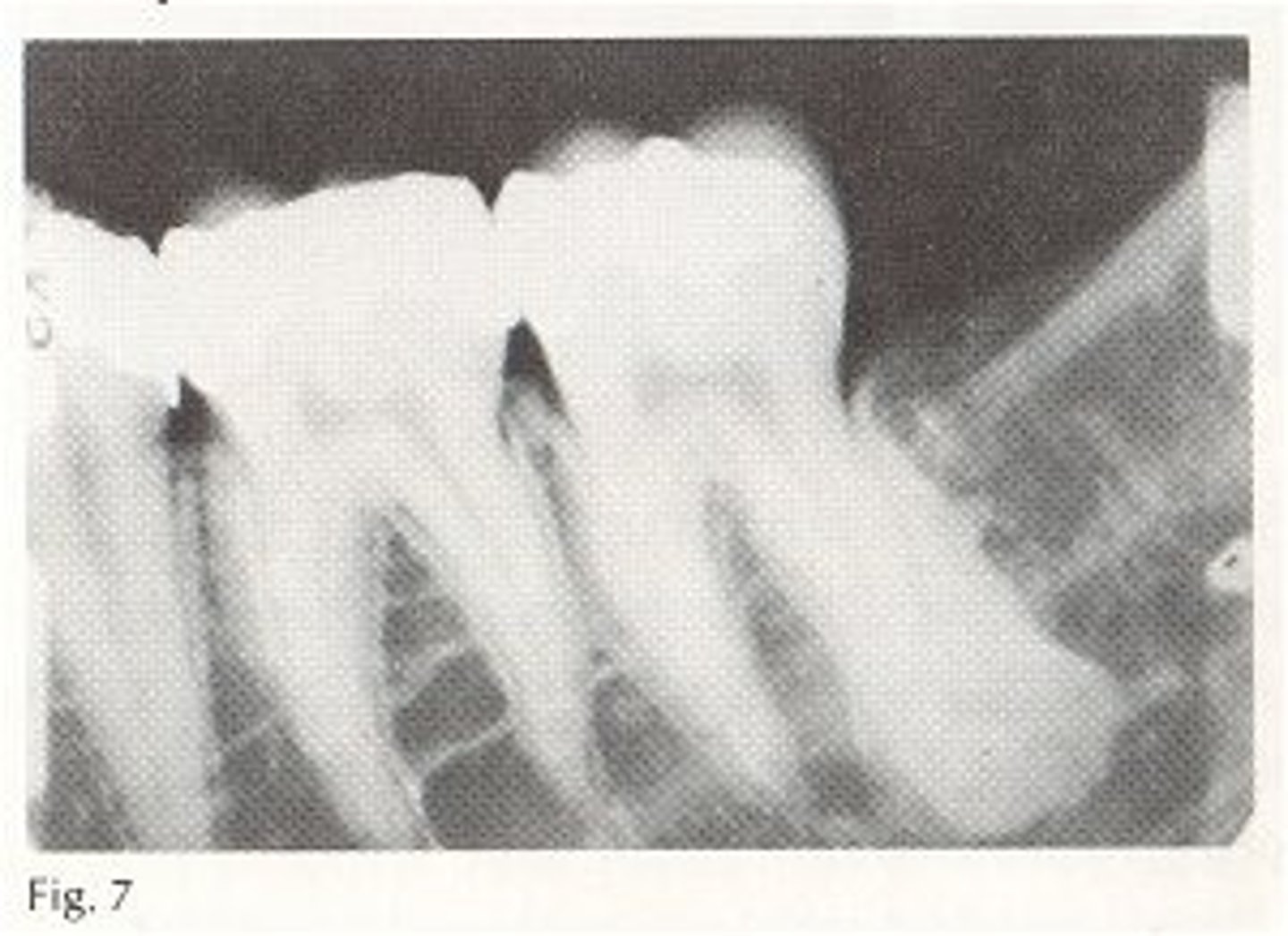
periapical cemento-osseous dysplasia (periapical C-O dysplasia)
-common locations
-clinical features
-radiographic
-population
common locations: anterior mandible of periapical region
clinical features: asymptomatic; vital teeth
radiographic: early lesions are radiolucent, then they become mixed, then they become radiopaque; the PDL is still intact
population: middle aged adults; females 14:1; black females
what is idiopathic osteosclerosis / dense bone island
it is an isolated area of lamellar bone within a medullary space
idiopathic osteosclerosis / dense bone island
-common locations
-radiographic
-population
common locations: posterior mandible 90%
radiographic: it is a focal area of radiopacity which often affects PA/interradicular bone; involves vital teeth
population: arises in children 10-20yrs
list the bone tumors (6)
1. osteoma
2. cementoblastoma
3. ossifying fibroma
4. osteosarcoma
5. chondrosarcoma
6. metastatic disease
which of the bone tumors are from benign neoplasms (3)
1. osteoma
2. cementoblastoma
3. ossifying fibroma
which of the bone tumors are from malignant neoplasms (3)
1. osteosarcoma
2. chondrosarcoma
3. metastatic disease
osteoma
-clinical features
-radiographic
-population
it is a neoplasia; ass w/ gardners sx (multiple osteomas, intestinal polyps, supernumerary teeth)
common locations: mandible
radiographic: a well-defined radiopaque mass
population: YA
cementoblastoma
-common locations
-clinical features
-radiographic
-population
common locations: mand 1st molar
clinical features: it presents w pain
radiographic: it is a dense radiopaque mass that binds w the apex; it is a sclerotic mass w a thin radiolucent rim; vital teeth are involved
population: children + YA <30s
ossifying fibroma
-common locations
-clinical features
-radiographic
-population
common locations: posterior mandible
clinical features: expansion + divergence
radiographic: well-defined unilocular radiolucency w opaque flecks
population: YA 20-30s
what is an osteosarcoma
it is a malignancy of bone producing cells
osteosarcoma (+location, features, radio, prognosis, population)
common locations: head and jaws 8%
clinical features: there is usually pain at first
radiographic: mixed opaque lesion w ill-defined borders; symmetrical widening of the PDL; sunburst pattern of bone growth above the crestal bone
TX: it metastasizes to the lung/brain
prognosis: death
population: YA; males
what is chondrosarcoma
it is magignancy of cartilage
chondrosarcoma (features, population)
a slow growing painless cancer characterized by loose teeth, ill-defined lucency + opacities
• population: older adults (60-70); males
what is metastatic disease
it is a tumor which arises from the distant site of a metastasis of a carcinoma of another organ
what is the most common malignancy of bone
metastatic disease
metastatic disease (features, population)
moth-eaten, bone expansion cancer found on the gingiva and posterior mandible
• population: older adults (60-70s)