Lab V: Hand II, Vertebral Column II
1/44
There's no tags or description
Looks like no tags are added yet.
Name | Mastery | Learn | Test | Matching | Spaced | Call with Kai |
|---|
No analytics yet
Send a link to your students to track their progress
45 Terms
define ulnar deviation
when the median nerve is damaged and ADduction occurs
define radial deviation
when the ulnar nerve is damaged and only ABduction occurs
what action can the 3rd digit NOT do?
ADduction of the hand
What finger is the only one that can circumduct?
only the thumb
extension/flexion in fingers vs thumb
extension/flexion in the fingers occur on the sagittal plane while in the thumbs, it occurs in the coronal plane
ABduction/ADduction in the fingers vs in the thumbs
ABduction/ADduction in the fingers occurs in the coronal plane while in the thumbs, it occurs in the sagittal plane
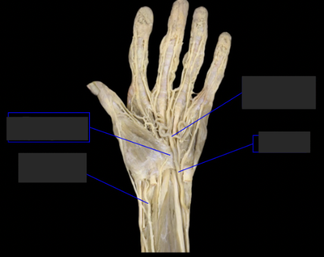
label the tagged areas (left to right, top to bottom)
transverse carpal ligament, radial artery, superficial palmar arterial arch, ulnar artery
superficial palmar arterial arch: where does it branch from? what does it anastomose with and for what? what does it give rise to?
superficial palmar arterial arch branches from the ulnar artery. It anastomoses with the deep palmar arterial arch for collateral circulation. It gives rise to the palmar digital arteries.
Superficial goes with what artery and deep goes with what artery?
superficial goes with ulnar and deep goes with radial: SU (your) DR
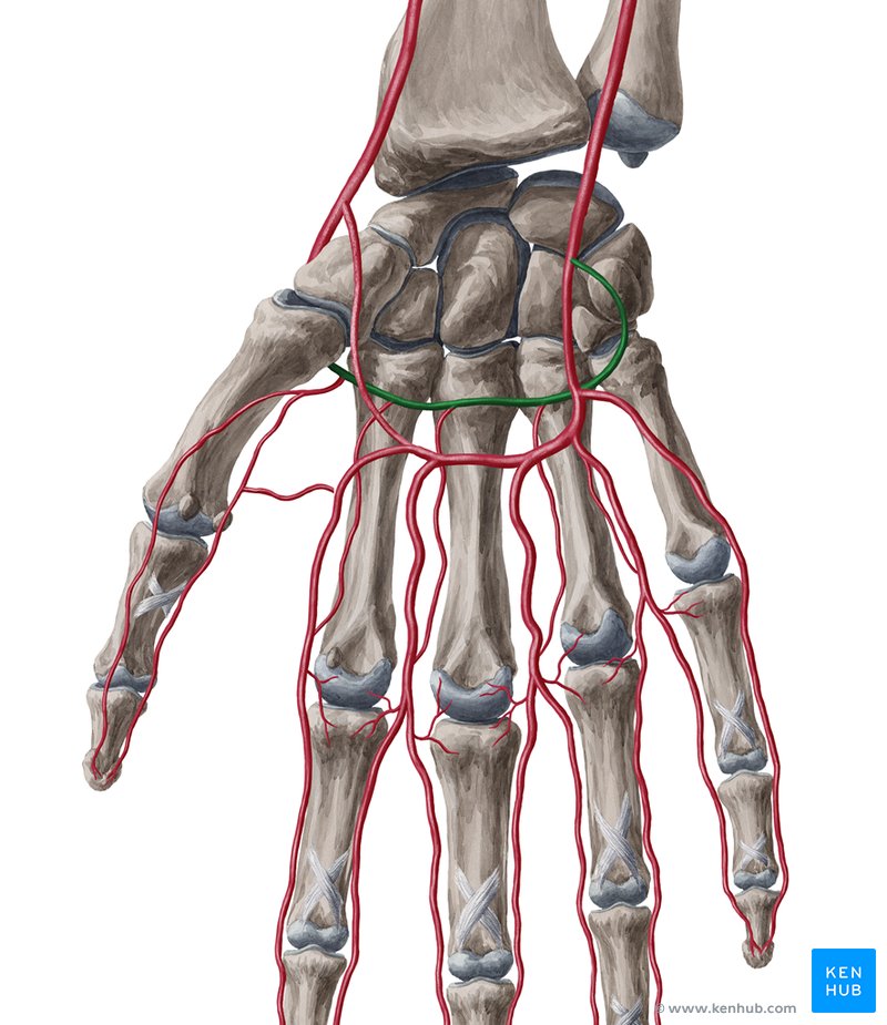
deep palmar arterial arch (green): where does it branch from? what does it anastomose with? about where can we find it?
Deep palmar arterial arch branches from the radial artery. It anastomoses with the superficial palmar arterial arch for collateral circulation. it can be found around the palmaris longus. It looks like a straight line and is pale.
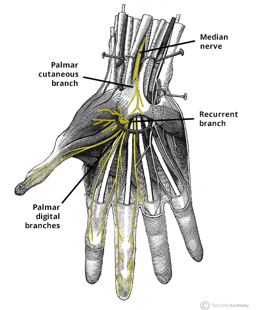
Median nerve (in palm): Where is it located? Where does cutaneous (sensory) branch to? Recurrent (motor) branch to what? Where does it branch to?
The median nerve is deep to the flexor retinaculum, under the digitorum superficialis going towards the radius and next to carpi radialis. Cutaneous branches to digits 1-3 and lateral half of 4. Recurrent branches to thenar muscles (distal digit 1). Branches to lumbricals 1 and 2 on the radial side.
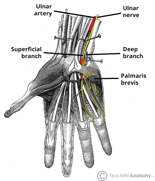
Ulnar nerve (in palm): Where is it located? What does it run with? Where does cutaneous branch to? Where does motor branch to?
The ulnar nerve is superificial to flexor retinaculum and runs with the ulnar artery. It is on top/diagonal to the artery around the palmar arterial arch. Cutaneous branches to digit 5 and the medial half of digit 4. Motor branches to all other intrinsic hand muscles.
What do the intrinsic muscles of the hand act on?
The intrinsic muscles of the hand act on the digits
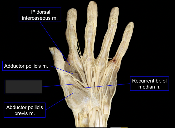
What is this tagged area? What is the muscle, origin, insertion, innervation, and main actions?
ABductor pollicis brevis
origin: flexor retinaculum, scaphoid, and trapezium
insertion: proximal phalanx of thumb
innervation: median nerve
main action: ABducts the thumb, aids in opposition
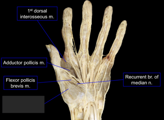
what is this tagged area? What is the muscles, origin, insertion, innervation, and main actions?
Flexor pollicis brevis
origin: flexor retinaculum and trapezium
insertion: proximal phalanx of the thumb
innervation: median nerve
main actions: flexes thumb, aids in opposition
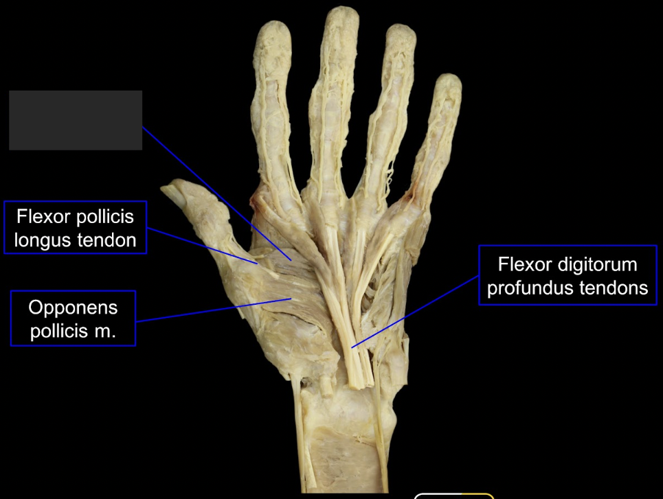
what is this tagged area? What is the muscles, origin, insertion, innervation, and main actions?
opponens pollicis (deep muscle btwn flexor and ABductor)
origin: flexor retinaculum and trapezium
insertion: lateral side of 1st metacarpal
innervation: median nerve
main actions: opposition
what intrinsic muscles are part of the ulnar/recurrent n. branch?
the thenar muscles: ABductor pollicis brevis, Flexor pollicis brevis, Opponens pollicis
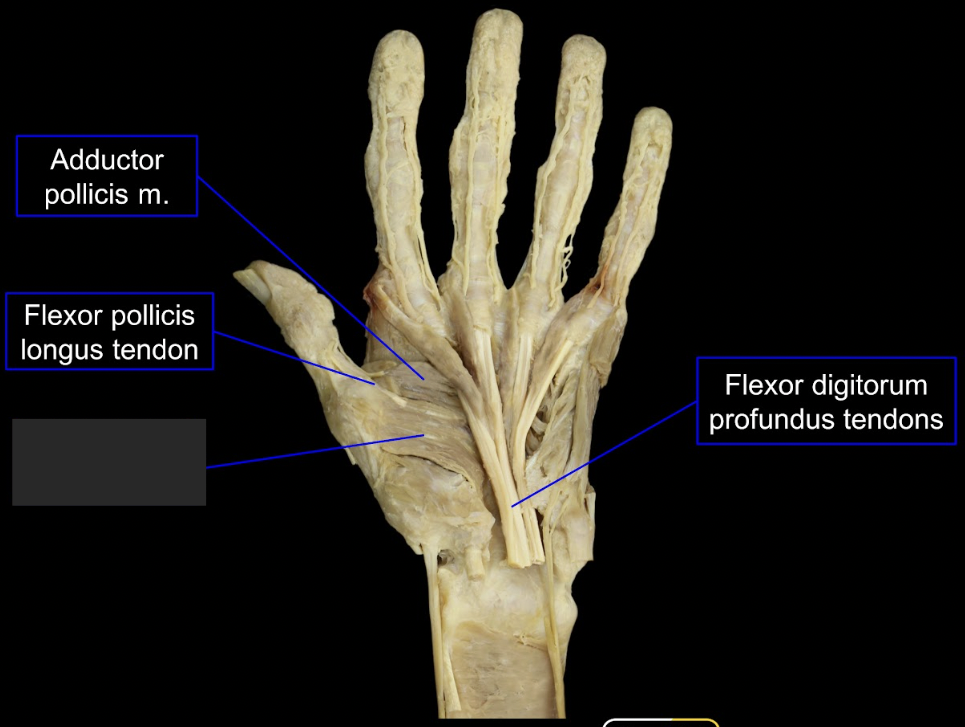
what is this tagged area? What is the muscles, origin, insertion, innervation, and main actions?
ADductor pollicis middle to thumb, superficial on thumb area)
origin: 2nd and 3rd metacarpal bones
insertion: medial side of the proximal phalanx of thumb
main action: ADducts thumb, assists opposition
innervation: ulnar nerve

what is this tagged area? What is the muscles, origin, insertion, innervation, and main actions?
ABductor digiti minimi (outside of pinky)
origin: pisiform bone
insertion: medial side of proximal phalanx of digit 5
innervation: ulnar nerve
main actions: ABducts digit 5
what is this tagged area? What is the muscles, origin, insertion, innervation, and main actions?
flexor digiti minimi (lateral to flexor minimi)
origin: hamate bone and flexor retinaculum
innervation: ulnar nerve
insertion: medial side of proximal phalanx digit 5
main actions: flexes proximal phalanx of digit 5

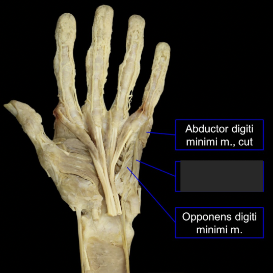
what is this tagged area? What is the muscles, origin, insertion, innervation, and main actions?
Opponens digiti minimi (in btwn ABductor and Flexor)
origin: hamate and flexor retinaculum
insertion: medial border 5th metacarpal
innervation: ulnar nerve
actions: opposes 5th digit to thumb
What muscles act on the 5th digit?
Hypothenar Muscles: ABductor digiti minimi, Flexor digiti minimi, Opponens digiti minimi
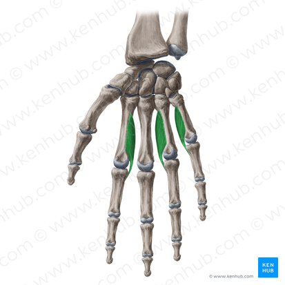
what is this tagged area? What is the muscles, origin, insertion, innervation, and main actions?
Lumbricals (bulbly, lateral to tendon, along digitorum)
origin: tendons of flexor digitorum profundus
insertion: lateral sides of extensor expansion digits 2-5
innervation: ulnar nerve/recurrent nerve branch digits 1-2
main actions: flex MCP joints, extend IP joints
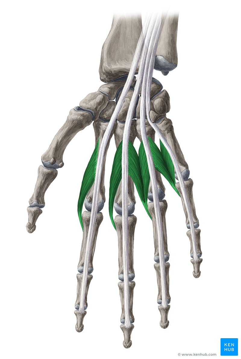
what is this tagged area? What is the muscles, origin, insertion, and main actions?
Dorsal Interossei (flush against the bone, in between the metacarpals)
origin: adjacent sides of two metacarpal bones
insertion: extensor expansion and base of digits 2-4 (2 muscle on digit 3)
main actions: Flex MCP joints, extend IP joints, ABduct digits 2,3, 4
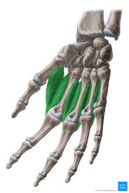
what is this tagged area? What is the muscles, origin, insertion, and main actions?
palmar interossei (goes over and under)
origin: palmar surface of 2nd, 4th, and 5th metacarpals
insertion: extensor expansions of digits 2,4,5
main actions: flex MCP joint, extend IP joints, ADduct digits 2,4,5
(think about which digits ADduct, and which don’t)
palmar aponeurosis: origin, insertion, main actions
origin: palmaris longus tendon
insertion: digits 2-5
main actions: protects deeper structures
significance (what it does and results in + attachment) of extensor expansion
Extensor expansion is attached to dorsum of proximal phalanges 2-5. It receives insertion of lumbrical and interosseous muscles. It allows the lumbricals and interossei to flex the MCP joints and extend the IP joints (at the same time)
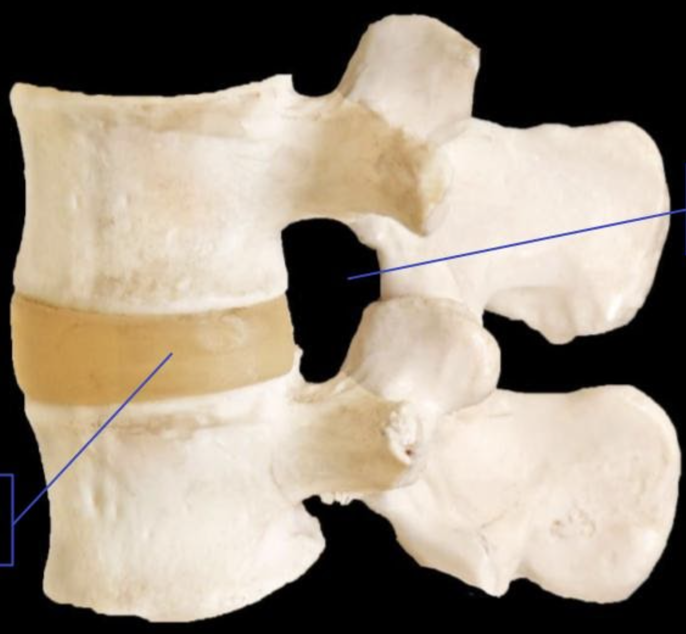
Label the tagged areas from left to right
intervertebral disc and intervertebral foramen
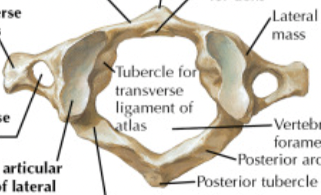
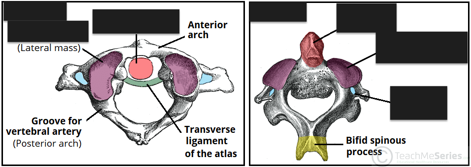
what part of the vertebral column is this bone from and in what view?
C1, superior view
what part of the vertebral column is this bone from?
thoracic (giraffe shaped)
what part of the vertebral column is this bone from?
lumbar (rhino shaped)
what part of the vertebral column is this bone from and in what view?
C2, anterior view
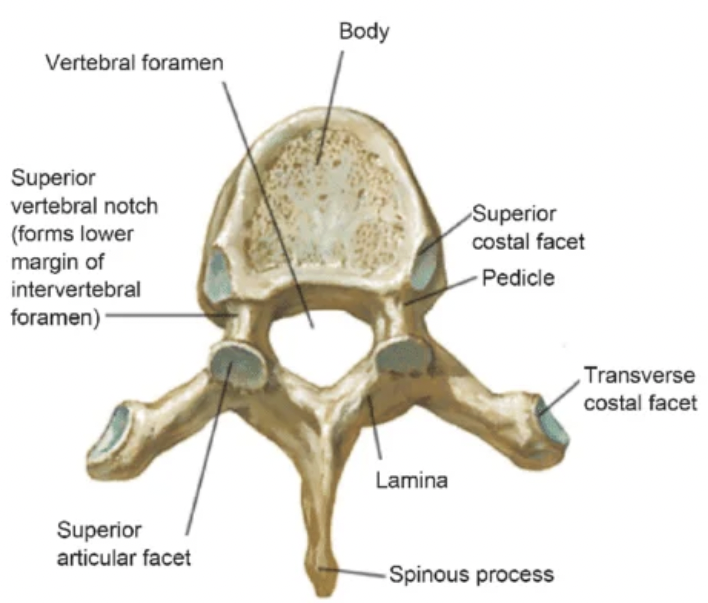
name the 4 tagged areas from top to bottom
vertebral body, vertebral foramen, transverse process, superior articular facet
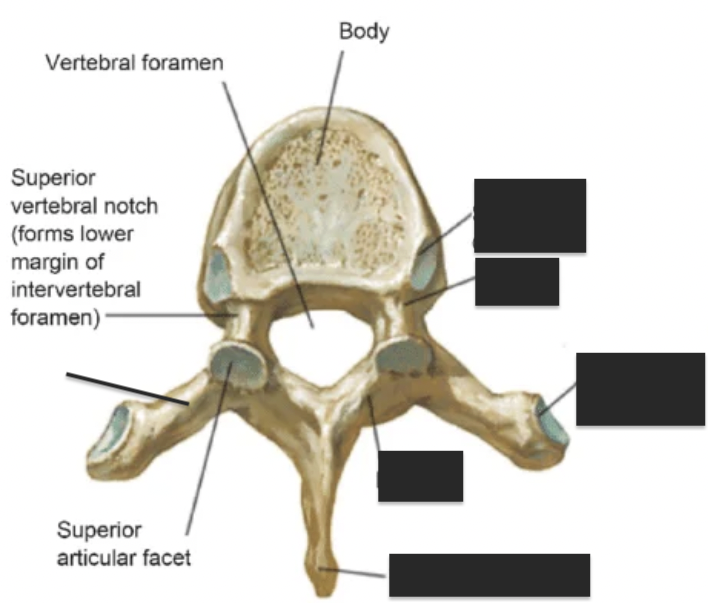
costal facet vs articular facets
costal facets or demifacets, are specialized surfaces on the thoracic vertebrae that articulate with the ribs to handle respiration. articular facets are general joints that aid in movement.
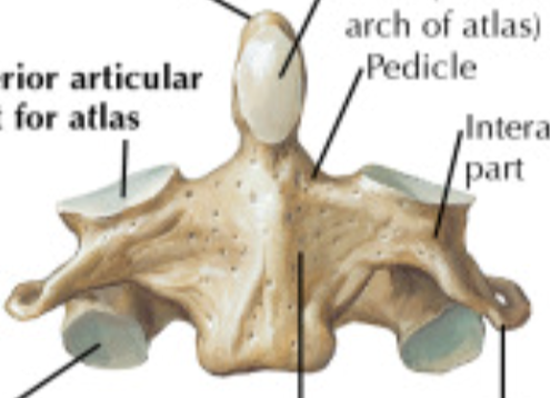
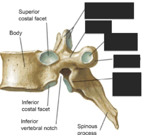
what are the 5 tagged areas?
superior costal facet, pedicle, transverse costal facet, lamina
pedicle vs lamina
pedicles are on the anterior side and connect the vertebral body to the posterior elements. laminae are on the posterior side and connect the pedicles to the spinous process.
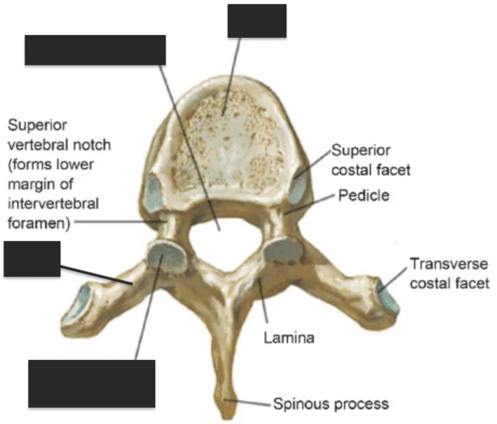
name the 4 tagged areas from top to bottom
superior costal facet
body
inferior costal facet
spinous process
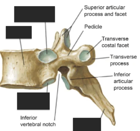
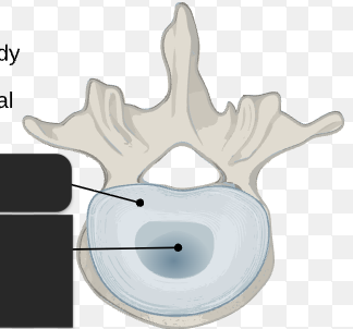
name the 6 tagged areas from top to bottom
superior articular process, superior articular facet, pedicle, transverse costal facet, transverse process, inferior articular process
what are the individual marked areas called? what are these referred to as?
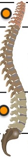
cervical and lumbar curvature; secondary curvature
what are the individual marked areas called? what are these referred to as?
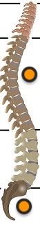
thoracic and sacral curvature; primary curvature
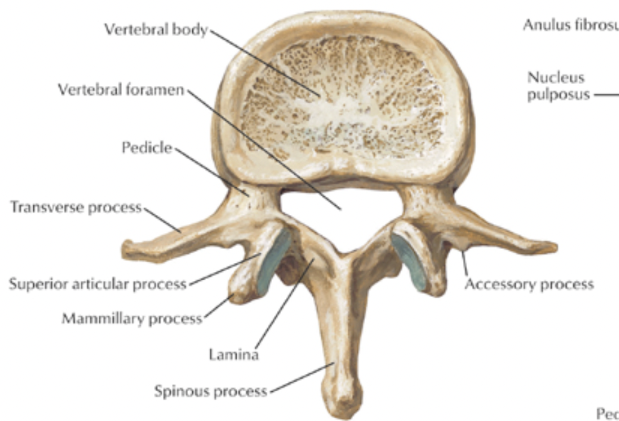
label the 2 structures. These compose the ___.
annulus fibrosus, nucleus pulposus. intervertebral disc
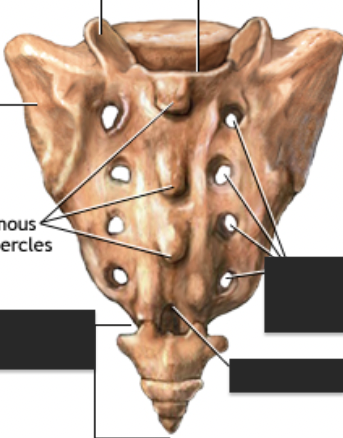
label the structures/areas.
sacrum: sacral foramina, sacral hiatus. coccyx
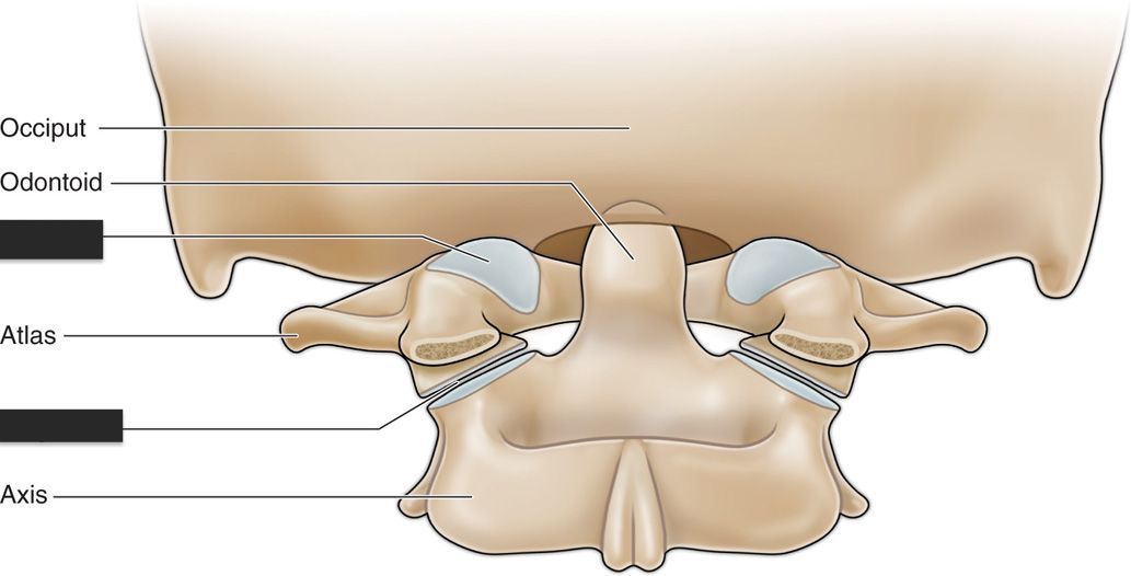
label the tagged areas.
atlanto-occipital joint and atlantoaxial joint

name the left structure and tagged areas (left to right).
Atlas or C1 vertebra; superior articular facet, odontoid process of C2
name the right structure and tagged areas (left to right).
axis or C2 vertebra; odontoid process (dens), superior articular facet, transverse foramen