IA/P 8 TEST (Facial Bones)
1/64
There's no tags or description
Looks like no tags are added yet.
Name | Mastery | Learn | Test | Matching | Spaced | Call with Kai |
|---|
No analytics yet
Send a link to your students to track their progress
65 Terms
The axiolateral projection is used to demonstrate the mandible. How is the head positioned to demonstrate the ramus of the mandible?
True Lateral
Which of the following skull types is considered average in size and shape?
Mesocephalic
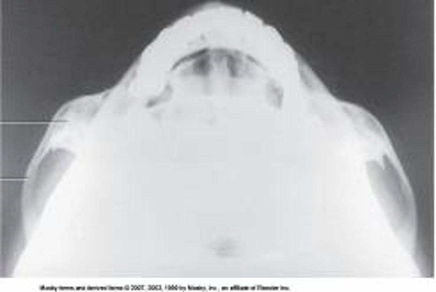
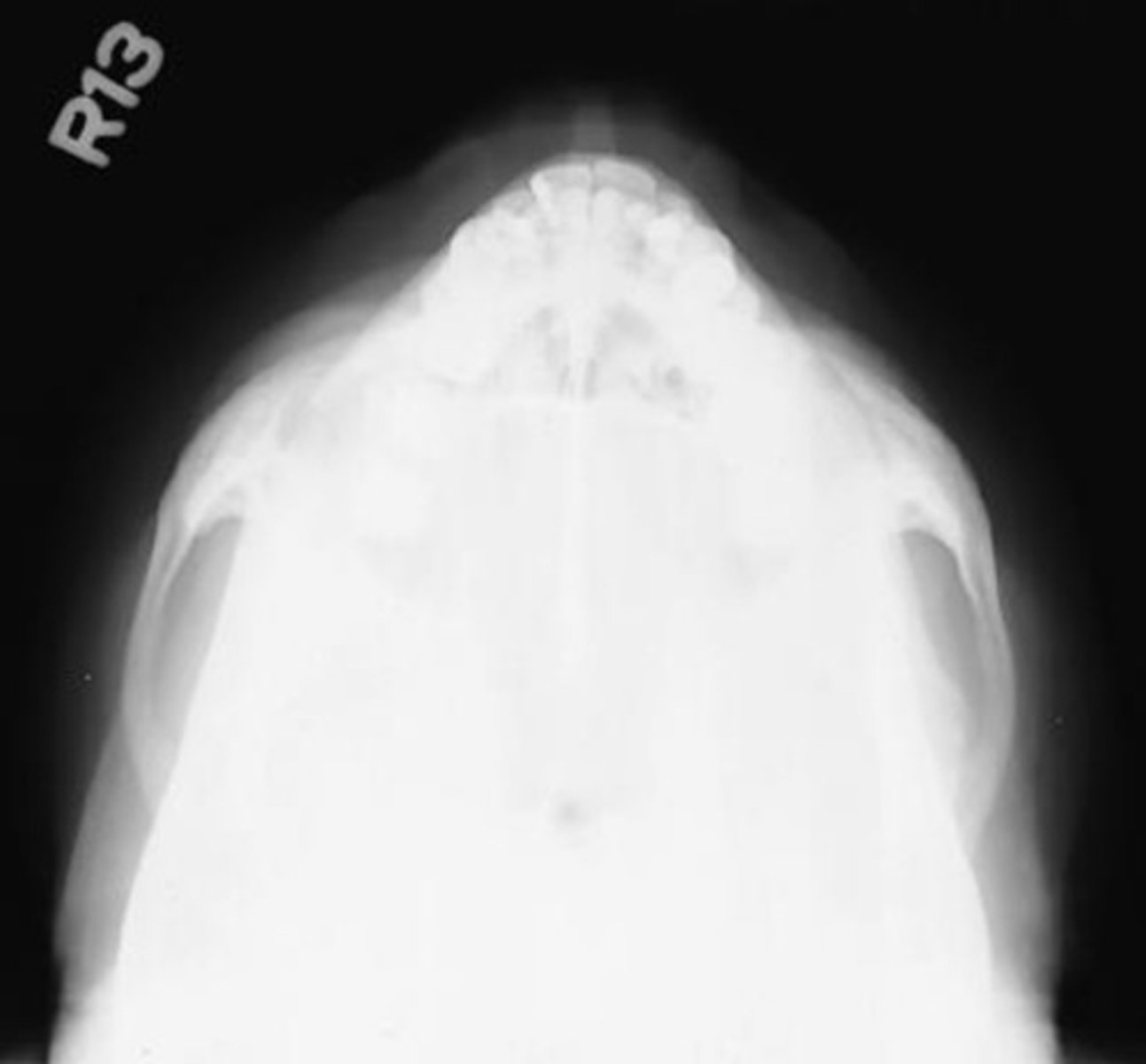
What projection and anatomy is demonstrated in the image below?
SMV of the zygomatic arches
For the tangential projection of the zygomatic arch, the central ray is directed perpendicular to the _____ line.
infraorbitomeatal
Which of the following is true regarding the lateral projection of the nasal bones?
1. MSP is parallel with the tabletop.
2. Both sides are done for comparison.
3. The interpupillary line is perpendicular to the tabletop.
1, 2, and 3
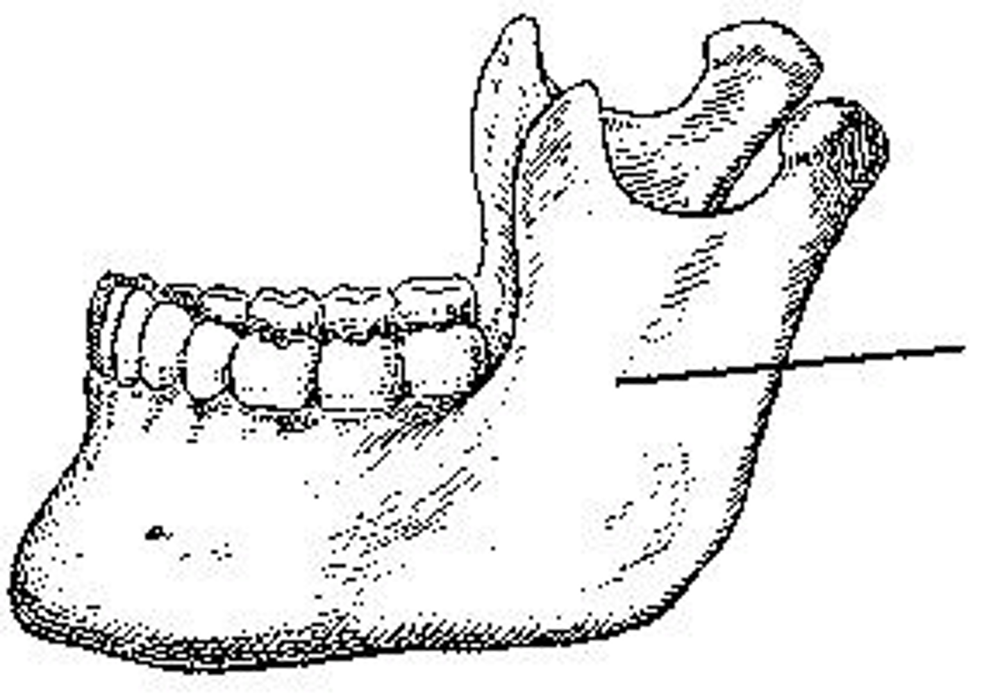
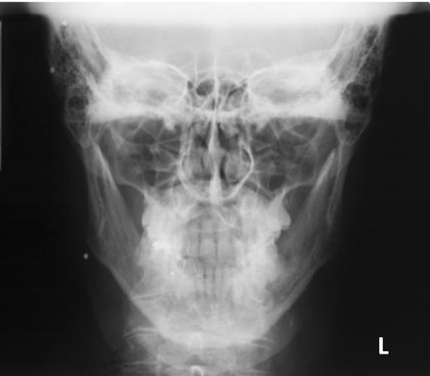
What part of the mandible is identified?
Body
At which level will the central ray be placed for the tangential projection of the zygomatic arch?
At a level 1 inch posterior to the outer canthi
What is the central-ray angulation for the PA axial projection of the mandibular rami?
20 to 25 degrees cephalad
Which of the following projections will clearly demonstrate any medial or lateral displacement of fractures of the mandibular rami?
1. PA
2. PA axial
3. Axiolateral oblique
1 and 2
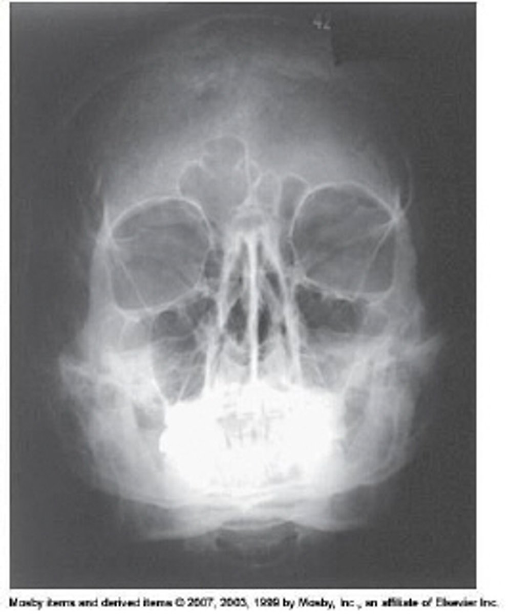
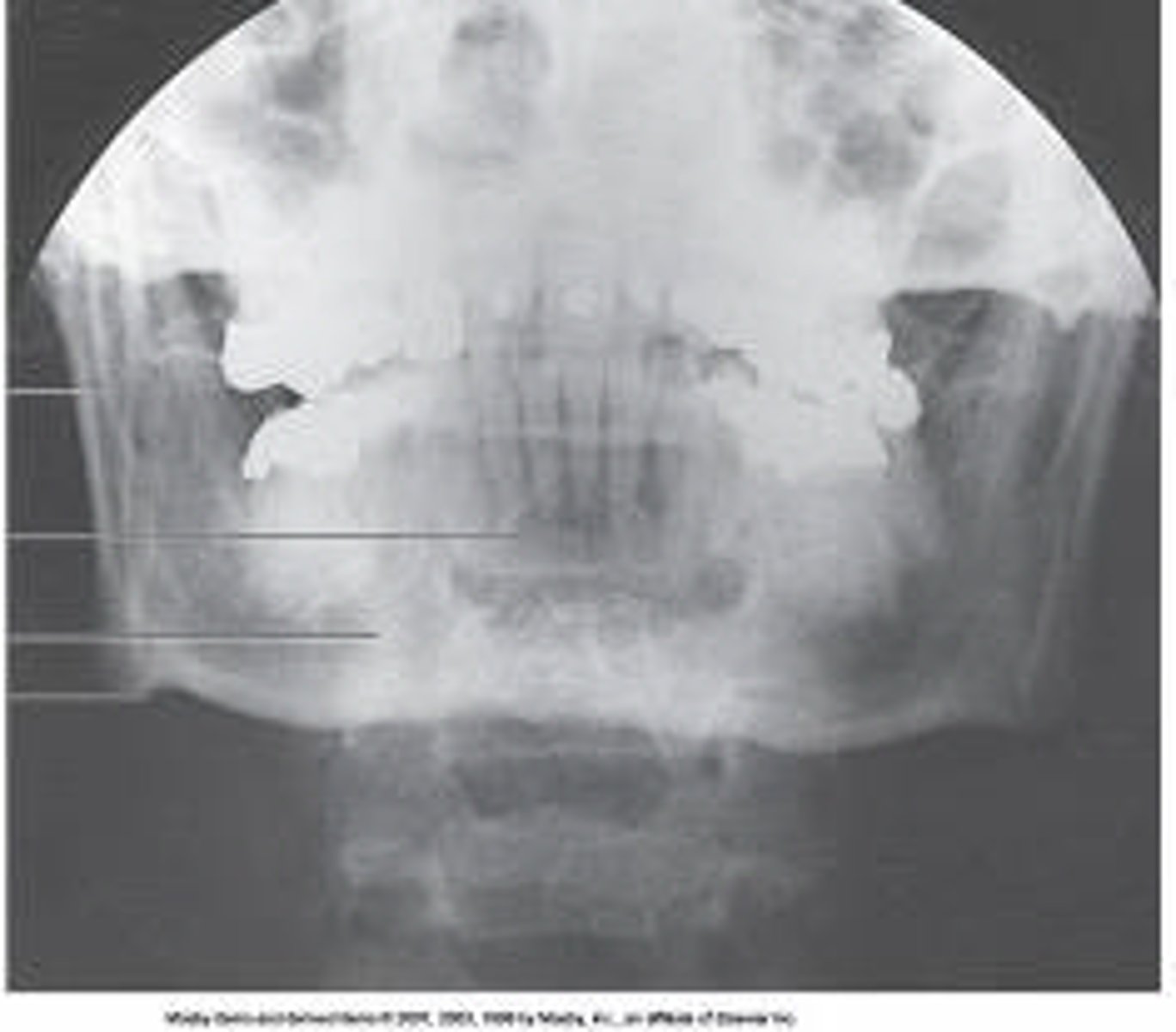
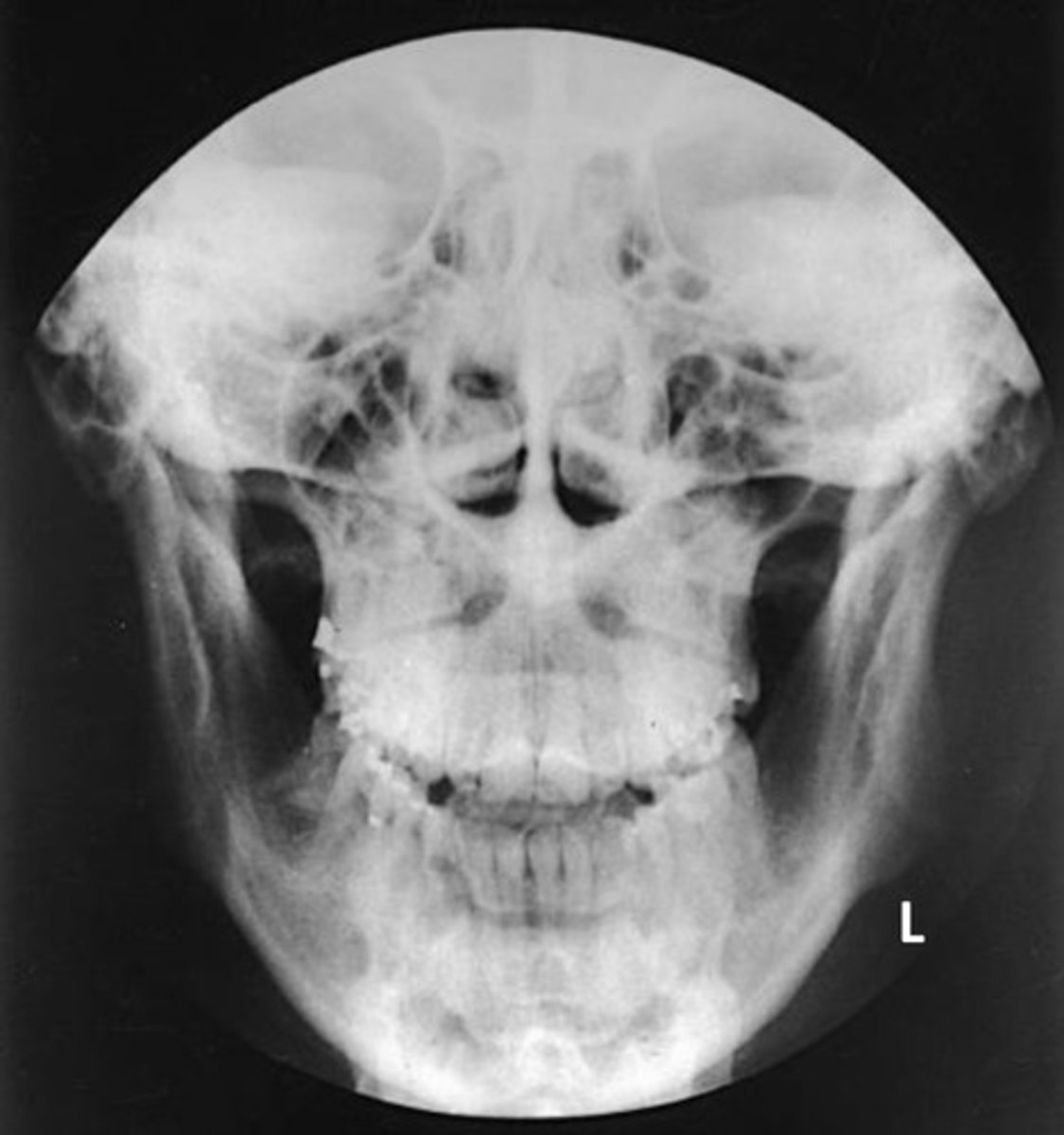
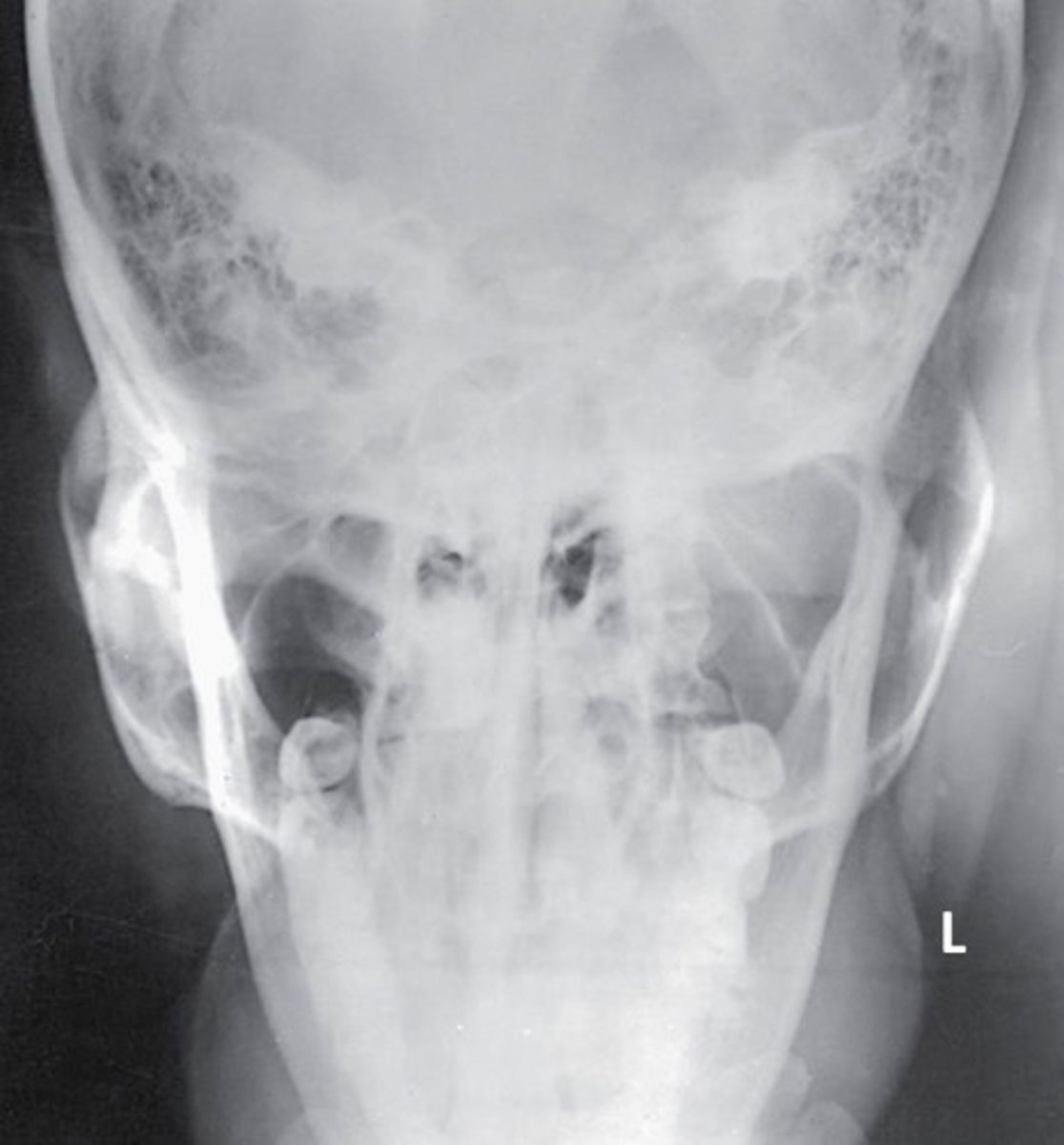
What projection and method is demonstrated?
Parietoacanthial (modified Waters)
Which of the following is(are) true regarding positioning for the Waters method for the facial bones?
1. The orbitomeatal line forms a 37-degree angle with the plane of the IR.
2. The mentomeatal line is perpendicular to the IR plane.
3. The coronal plane is perpendicular to the IR plane.
1 and 2
Which of the following is (are) true regarding positioning for the Waters method for the facial bones?
1. The orbitomeatal line forms a 37-degree angle with the plane of the IR.
2. The mentomeatal line is perpendicular to the IR plane.
3. The coronal plane is perpendicular to the IR plane.
1 and 2
The axiolateral oblique projection is used to demonstrate the mandible. How is the head positioned to demonstrate the body of the mandible?
30 degrees toward the IR
The parietoacanthial projection of the facial bones is commonly called the _____ method.
Waters
The axiolateral oblique projection is used to demonstrate the mandible. How is the head positioned to demonstrate the symphysis of the mandible?
45 degrees toward the IR
Which reference line is perpendicular to the IR for the PA and PA axial mandibular rami?
OML
The parietoacanthial projection (Waters method) of the facial bones is often modified so that there is less angulation of the facial bones. For this modification, the orbitomeatal line is adjusted to _____ degrees.
55
Several methods area available to perform the axiolateral oblique projection of the mandible to demonstrate the symphysis, body, or ramus. What is the central-ray angulation for all of these projections?
25 degrees cephalad
Which of the following is placed perpendicular to the image receptor for the acanthoparietal projection (reverse Waters method) of the facial bones?
Mentomeatal line
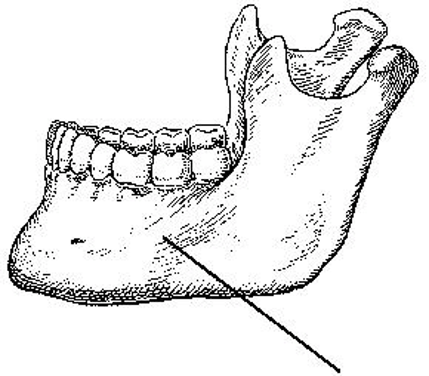
What portion of the mandible is identified?
Ramus
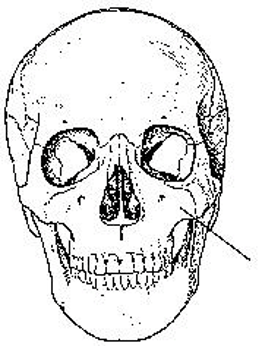
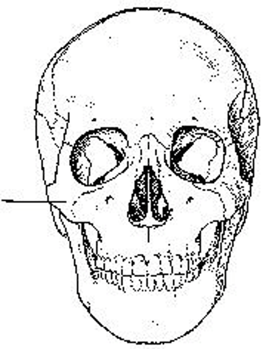
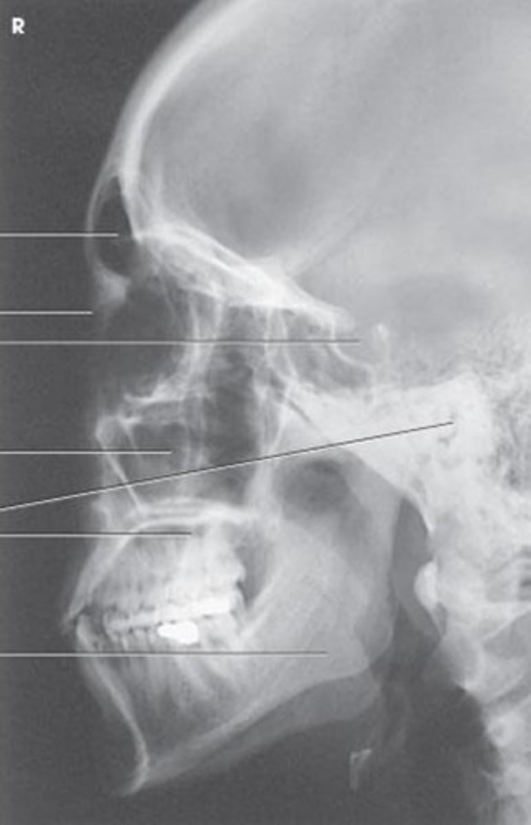
What bone is identified?
Maxilla
What projection and anatomy is demonstrated?
PA of the mandibular body
What bone is identified?
Zygoma
For a lateral projection of the facial bones, the central ray will enter:
halfway between the outer canthus and the EAM
For the Waters method for the facial bones, the orbitomeatal line is placed at what angle to the IR?
37 degrees
At which level will the central ray be placed for the SMV projection of the zygomatic arches?
At a level 1 inch posterior to the outer canthi
Which of the following lines is placed as nearly parallel to the image receptor as possible for the SMV projection of the zygomatic arches?
IOML
Which of the following is centered to the image receptor for a parietoacanthial projection of the facial bones?
Acanthion
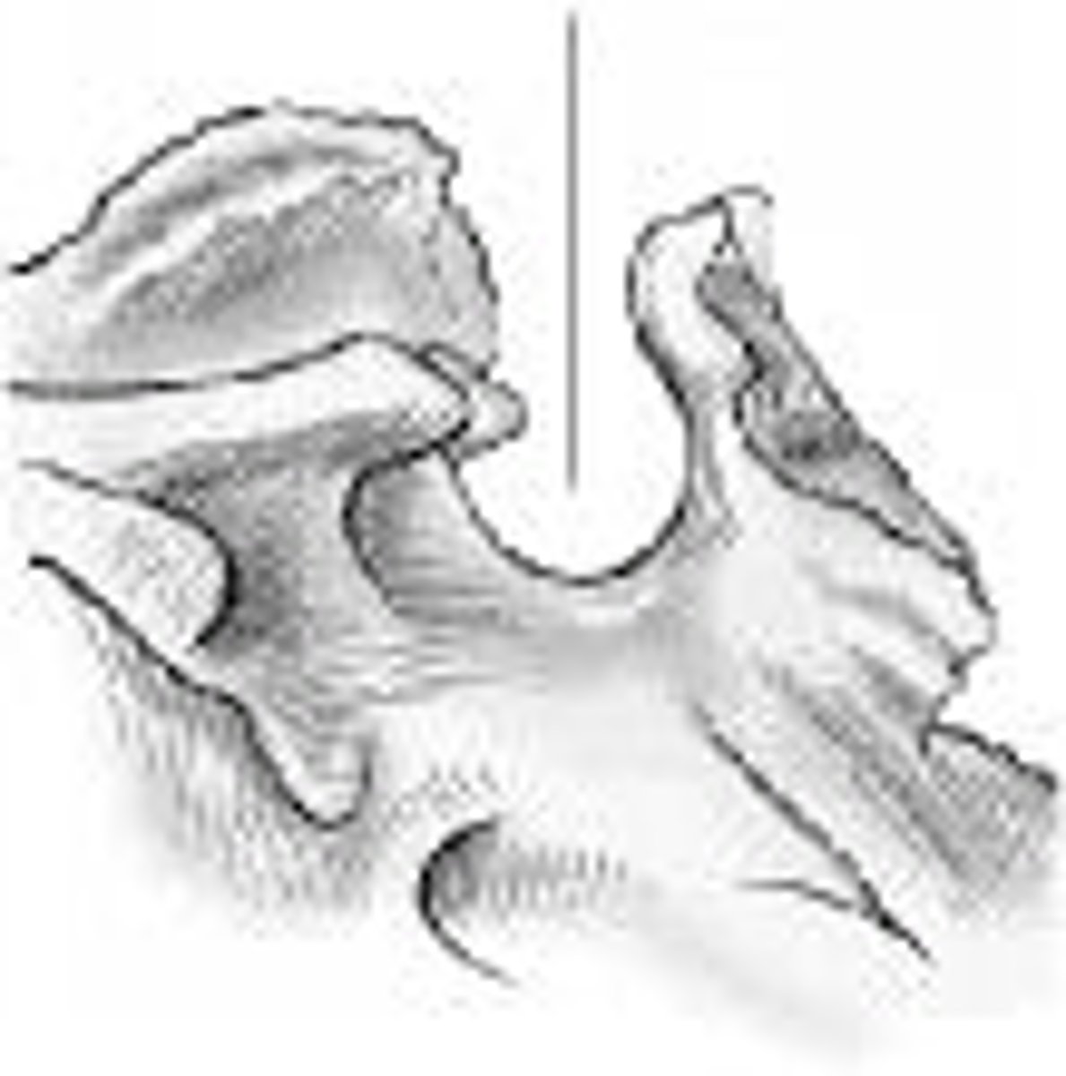
What part of the sphenoid is identified?
sella turcica
Which of the following is placed perpendicular to the front edge of the IR for a lateral projection of the facial bones?
Infraorbitomeatal line
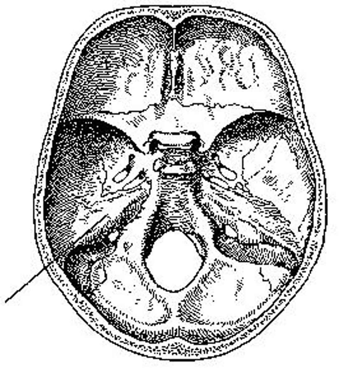
What part of the cranial base is identified?
petrous portion
Inflammation of bone marrow due to pyogenic infection.
Osteomyelitis
Loss of bone density.
Osteoporosis
Increased density of atypically soft bone.
Osteopetrosis
Tumor comprised of bony tissue.
Ostema
Thick, soft bone marked by bowing and fractures.
Paget disease (osteitis deformans)
Growth or mass protruding from a mucous membrane.
Polyp
Dysfunction of the temporomandibular joint
TMJ syndrome
Malignant neoplasm of plasma cells involving the bone marrow.
Multiple Myeloma
Tumor on the pituitary gland.
Pituitary adenoma
Which projection best demonstrates the nasal septum?
Parietoacanthial (Water's)
Name
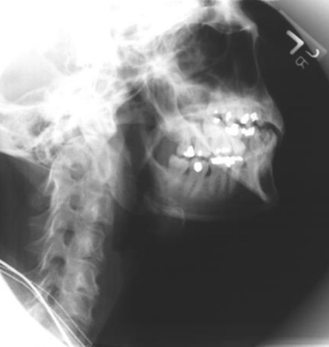
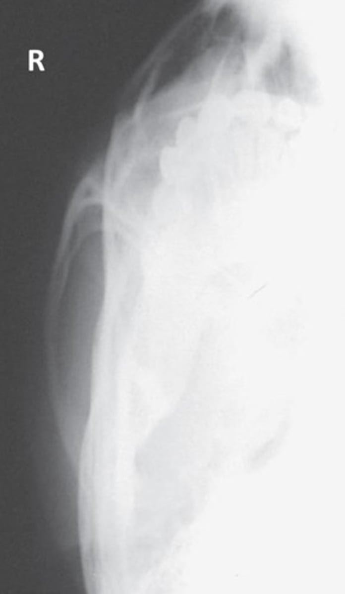
Lateral Facial Bones
Name
PA Mandible
Name
PA Axial Mandible
Name
Axiolateral Mandible for Ramus
Name
Axiolateral Oblique Mandible for Body
Name
Axiolateral Oblique Mandible for Symphysis
Name
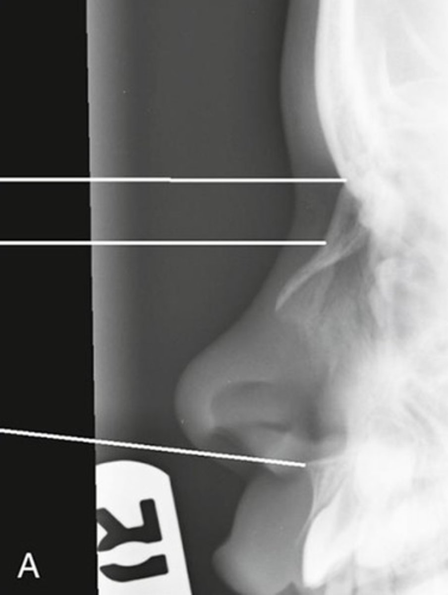
Lateral Nasal Bones
Name
SMV for Zygomatic Arches
Name
Tangential Zygomatic Arch
Name
AP Axial Modified Towne
For a lateral projection of the facial bones, the image receptor is centered to the:
zygomatic bone
When the CR is not perpendicular to the IR what will occur?
Elongation
When the body part is not parallel to the IR what will occur?
Foreshortening
Which projection results in a lateral image of the ramus of the mandible?
Axiolateral mandible
Select the changes that need to be made to an SMV of the zygomatic arches from the SMV for the skull:
- Change centering from 3/4" anterior to the EAM to 1" posterior to the outer canthus
- Use less technique on the SMV for zygomatic arches
- Use more technique on the SMV for zygomatic arches
- Do not use a grid on the SMV for the zygomatic arches
- Centering should be the same on both
- Change centering from 3/4" anterior to the EAM to 1" posterior to the outer canthus
- Use less technique on the SMV for zygomatic arches
- Do not use a grid on the SMV for the zygomatic arches
What baseline should be parallel with the transverse axis of the IR for lateral nasal bones?
IOML
What is the advantage of doing a PA mandible instead of an AP mandible?
- Less magnification
- More magnification
- There is no advantage
- A PA is easier for the patient
- Less magnification
Since skull or facial bone positioning depends on the relationship of the baseline to the CR, using the glabellomeatal line instead of the orbitomeatal line would require a tube adjustment of:
8 degrees
Since skull or facial bone positioning depends on the relationship of the baseline to the CR, using the infraorbitomeatal line instead of the orbitomeatal line would require a tube adjustment of:
7 degrees
For a lateral projection of the facial bones, the image receptor is centered to the:
zygomatic bone
For the Waters method for the facial bones, the orbitomeatal line is placed at what angle to the IR?
37 degrees
For the tangential projection of the zygomatic arch, the MSP of the head is rotated how many degrees?
15 degrees toward the side examined
The axiolateral oblique projection is used to demonstrate the mandible. How is the head positioned to demonstrate the body of the mandible?
True lateral
30 degrees toward the IR
The axiolateral oblique projection is used to demonstrate the mandible. How is the head positioned to demonstrate the symphysis of the mandible?
True lateral
45 degrees toward the IR