RBC Production, Function, and Morphology & Hemoglobin Function
 Call Kai
 Learn
Learn Practice Test
 Spaced Repetition
 Match
Match Flashcards
 Knowt Play
1/43

There's no tags or description
Looks like no tags are added yet.
Name | Mastery | Learn | Test | Matching | Spaced | Call with Kai |
|---|
No analytics yet
Send a link to your students to track their progress
44 Terms
pluripotent stem cells in bone marrow
erythropoietin (EPO) from the kidneys
What do RBCs originate from and what stimulates their production?
cell size decreases
nuclear cytoplasm ratio decreases
chromatin becomes more condensed
cytoplasm changes color
loss of nucleus (becomes anucleate)
What 5 changes do red blood cells go through in maturation?
pronormoblast
basophilic normoblast
polychromatophilic normoblast
orthochromic normoblast
reticulocyte
mature red blood cell
What are the 6 maturation stages of red blood cells (from most premature to mature)?
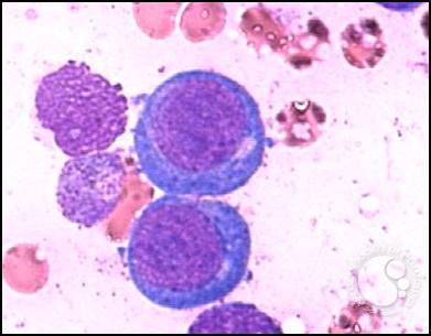
size: 18-20 micrometers
NC ratio: 6:1 (big nucleus)
chromatin: fine texture, deep violet color
Cytoplasm: dark blue, purple
List the size, N:C ratio, and cytoplasm color for pronormoblasts.
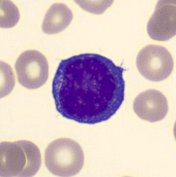
size: 16 micrometers
NC ratio: 6:1 (large nucleus)
cytoplasm: blue, with areas of clearing
List the size, N:C ratio, and cytoplasm color for basophilic normoblasts.
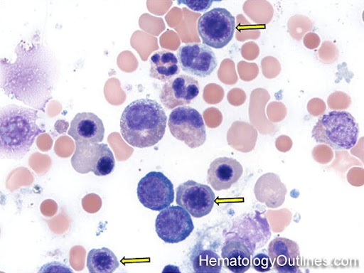
size: 13 micrometers
NC ratio: 4:1
cytoplasm: blue, with tinges of red-orange
List the size, N:C ratio, and cytoplasm color for polychromatophilic normoblast.
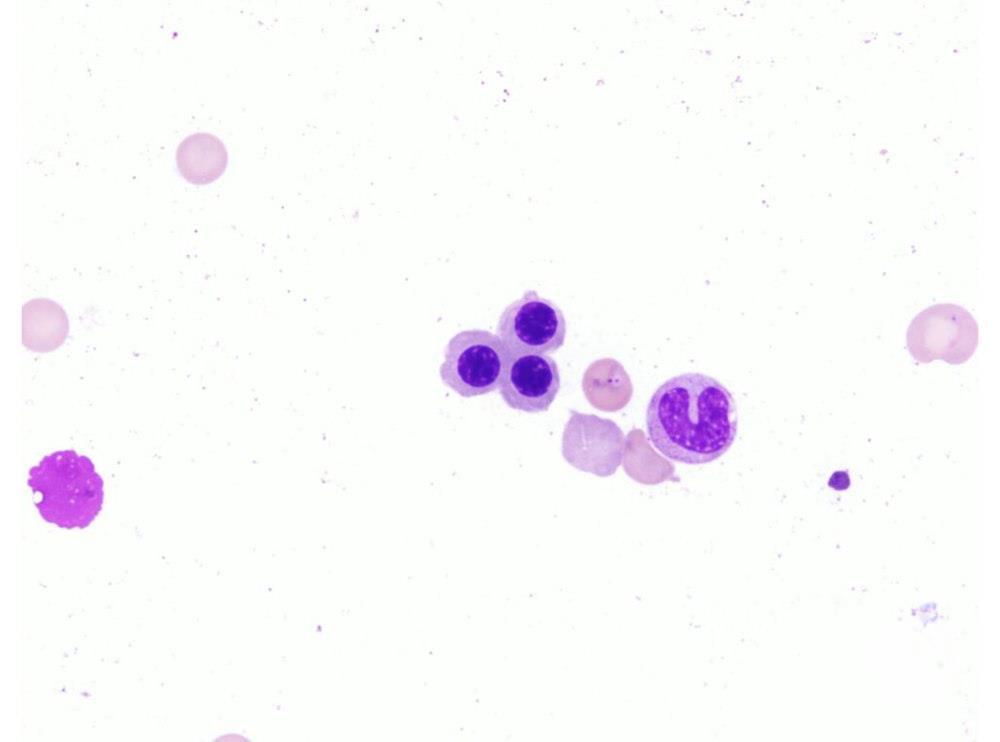
size: 8 micrometers
NC ratio: 1:1
cytoplasm: red-orange tinges with slight blue
chromatin: very round and dense
List the size, N:C ratio, and cytoplasm color for orthochromic normoblasts.
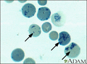
size: 8 micrometers
appearance: large, bluish red cells, reminants of RNA visualized with methylene blue stain
List the size and appearance for reticulocytes (polychromatic macrocyte).
Embden-Meyerhof pathway
produces 90% of ATP and NAD+
Phosphogluconate pathway
provides 5-10% of ATP
Methemoglobin reductase pathway
maintains iron in the ferrous state (Fe2+)
RBC metabolism is anaerobic
What are the 3 pathways of RBC metabolism?
outer layer
made of glycolipids and glycoproteins
middle layer
made of cholesterol and phospholipids
inner layer
cytoskeleton
Describe the 3 layers of the RBC membrane
size: 6-8 micrometers
MCV: 80-100 fL
MCHC: 32-36%
What is the normal size, MCV, and MCHC of red blood cells?
microcytic
less than 6 micrometers
caused by iron deficiency anemia, thalassemia, sideroblastic anemia, and iron overload disorders
macrocytic
larger than 9 micrometers
caused by vitamin B12 deficiency, folic acid deficiency, liver disease, and megaloblastic anemia
What are the two variations in size (anisocytosis) of RBCs?
polychromasia
often seen when the bone marrow responds to anemic stress and releases immature cells into circulation (reticulocytes and orthochromic normoblasts)
appearance: gray-blue and large
hypochromasia
has a larger central pallor
Hgb synthesis is impaired and MCHC is less than 32%
conditions causing: thalassemia, sideroblastic anemia, and IDA (iron def anemia)
What are the two variations in color of RBCs?
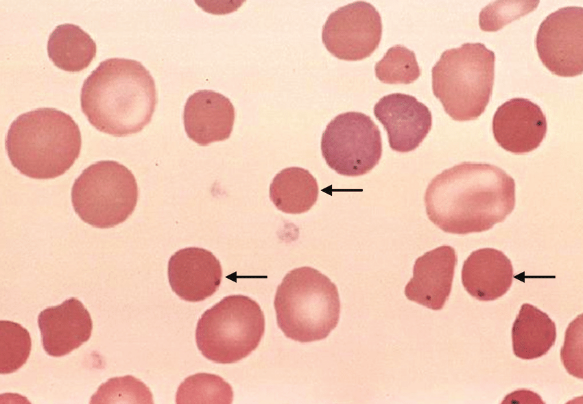
abnormally dark and compacted cells
MCV is nearly normal, but MCHC is greater than 32%
results from hereditary spherocytosis or autoimmune hemolytic anemia
Describe the following abnormal cell shape: spherocytes
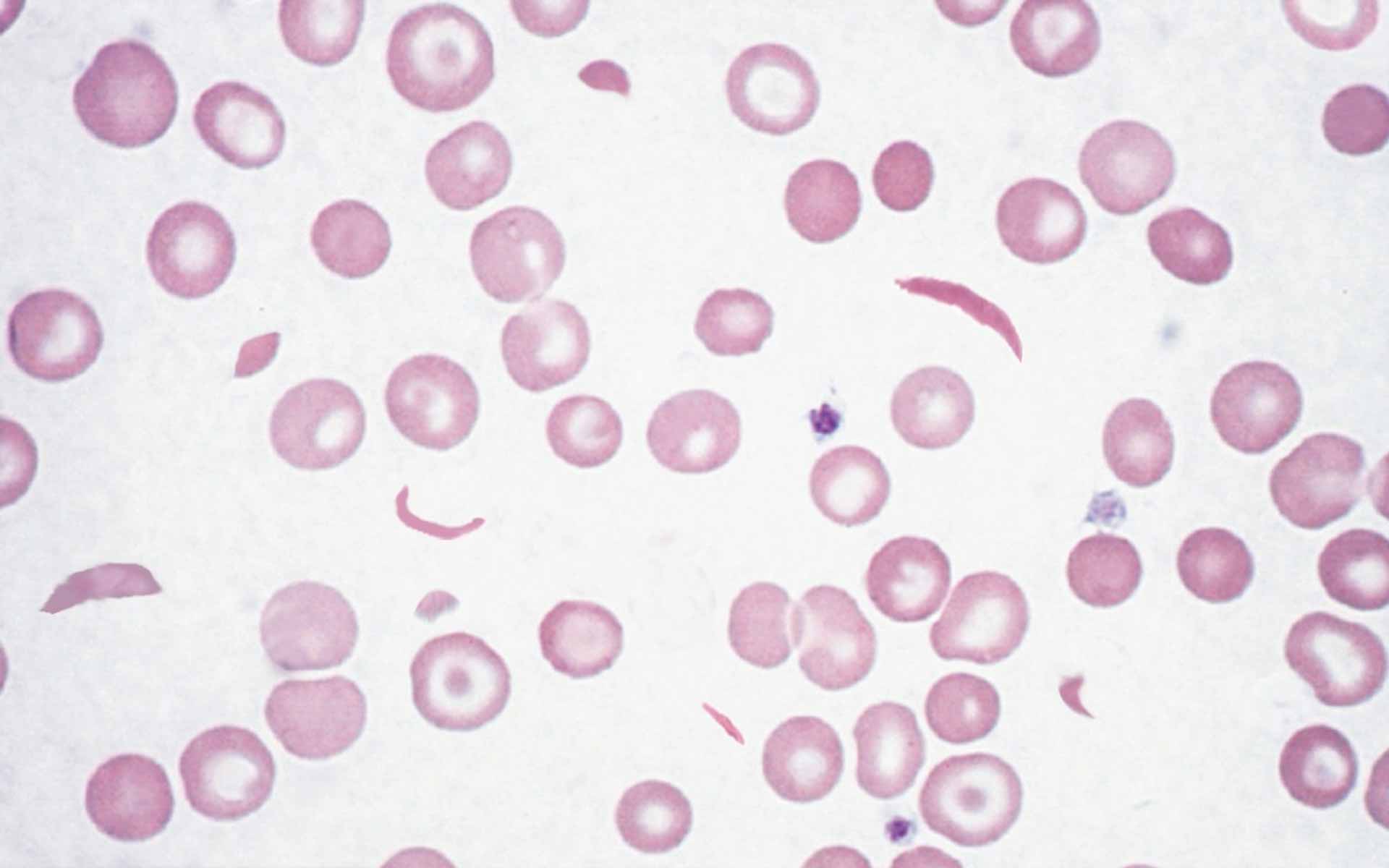
two types:
reversible - rounded, half moon shaped (can revert back to normal RBC)
irreversible - crescent shaped and pointed (cannot revert back)
those with sickle cell have the abnormal hemoglobin S, which forms tactoids (sickles) under hypoxic stress
Describe the following abnormal cell shape: sickle cells
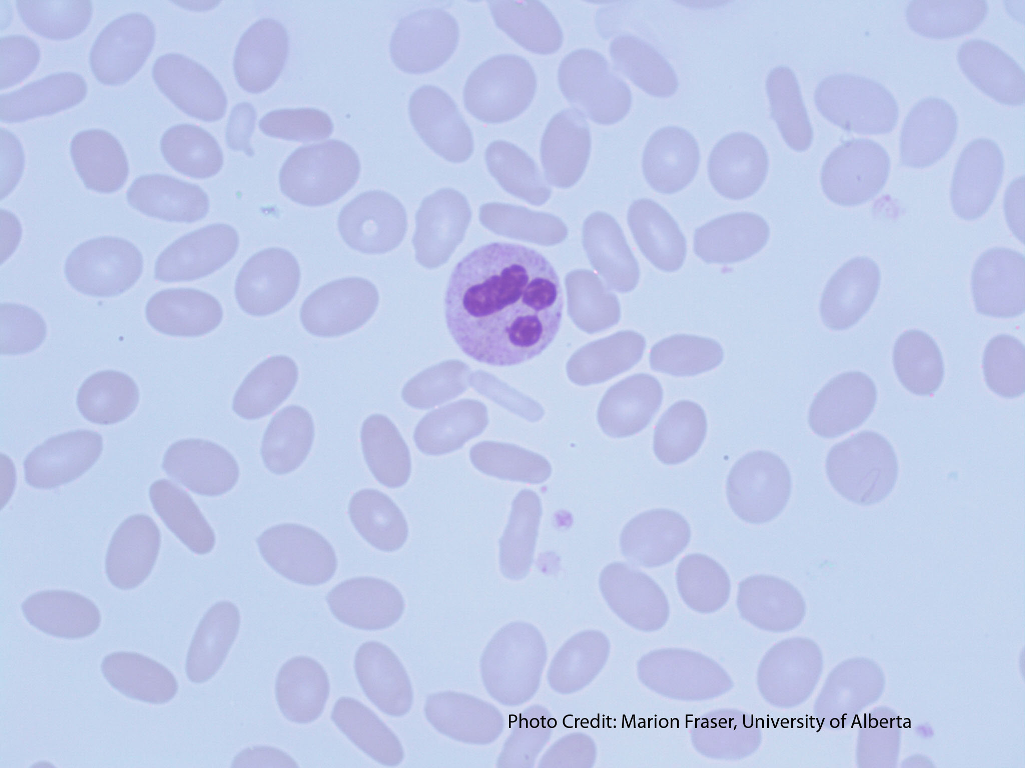
egg-shaped and disc-shaped (respectively)
caused by thalassemia or megaloblastic anemia
elliptocytes stem from abnormal spectrin (an RBC membrane protein), and in serious cases, anemia
Describe the following abnormal cell shape: ovalocytes and elliptocytes
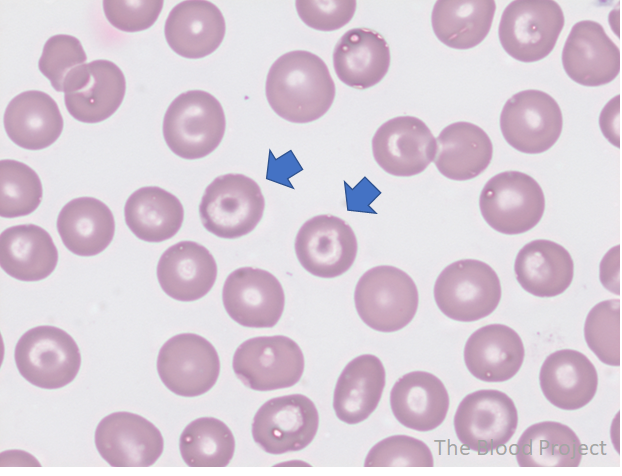
bull’s-eye shaped cells
happens due to an artifact, decreased Hgb, or increased red cell surface membrane
seen in those with IDA, hemoglobin C disease, liver disease, and a splenectomy
Describe the following abnormal cell shape: target cells
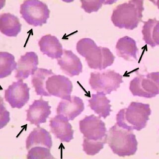
small red cells surrounded by uneven thorn-like spikes
found in those with liver disease, autoimmune hemolytic anemia, and LCAT deficiency
Describe the following abnormal cell shape: acanthocytes
bite cells
burr cells (in dehydrated patients)
schistocytes
List 3 types of fragmented RBCs (pieces of RBC membranes)
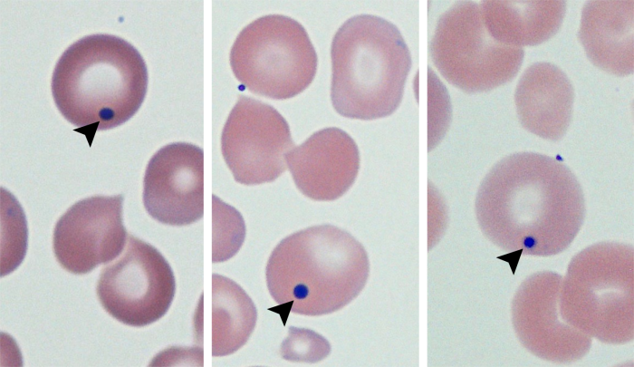
remnants of DNA that look like a dark purple dot inside cells
can be seen in postsplenectomy patients when responding to anemic conditions (erythropoiesis is rushed, no spleen = no removal of abnormal cells)
What are Howell-Jolly Bodies inclusions?
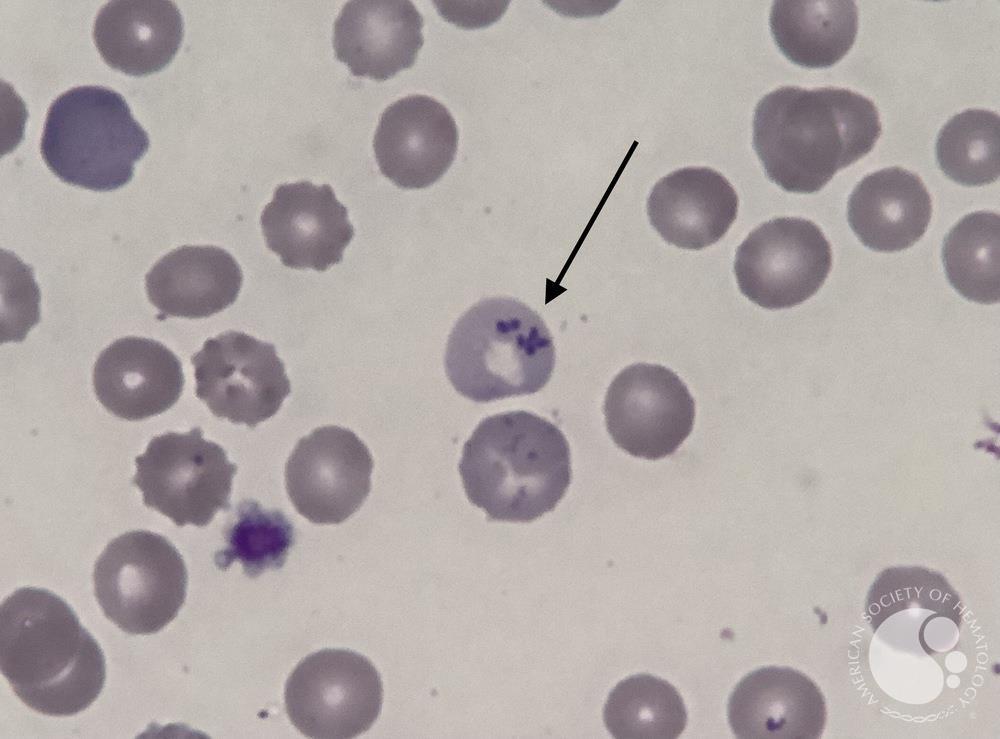
small, light purple beaded inclusions
seen in iron loading anemias (excessive iron)
What are Pappenheimer bodies inclusions?
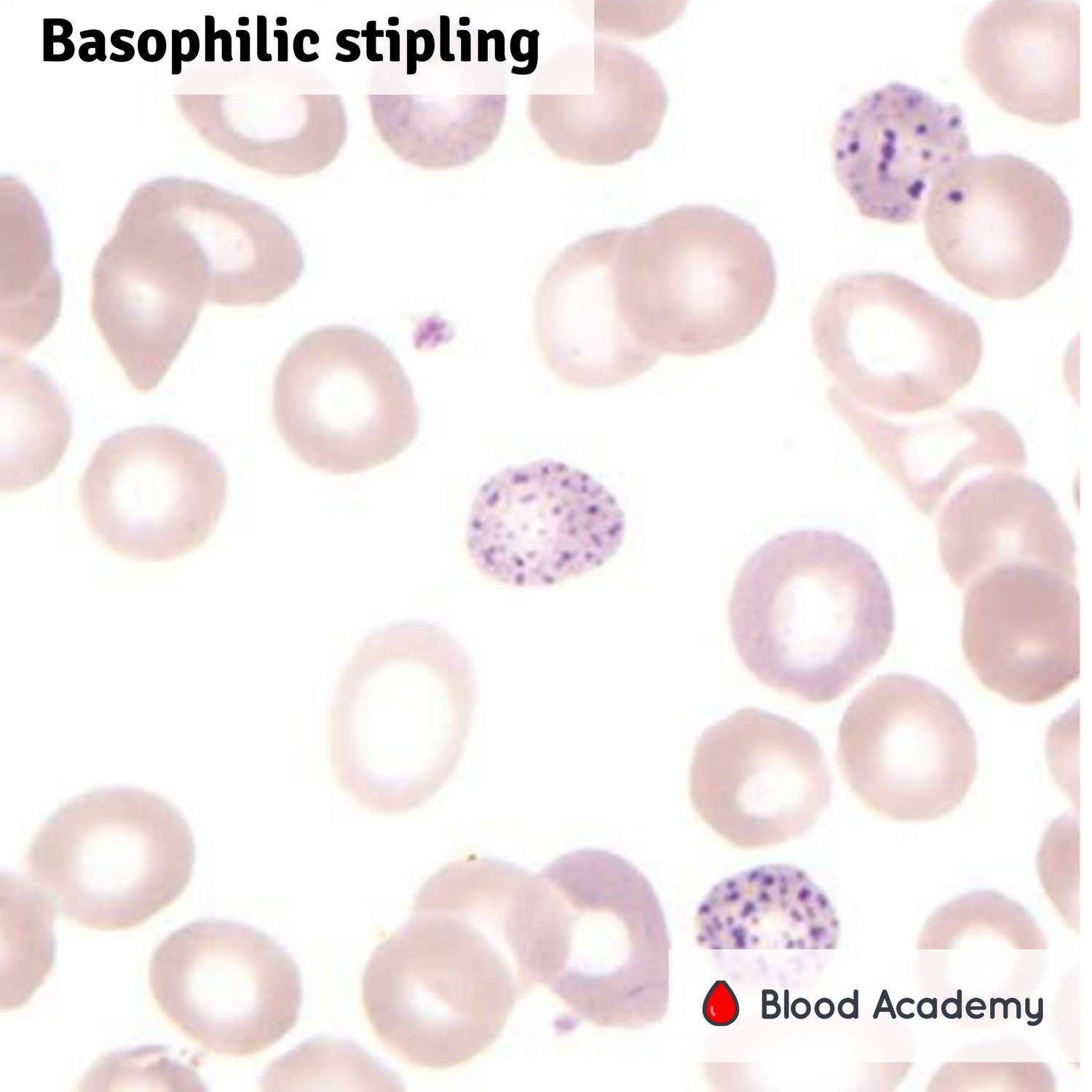
composed of RNA and mitochondrial remnants
course-looking in appearance, granular
caused by lead intoxication or thalassemia
What are basophilic stippling inclusions?
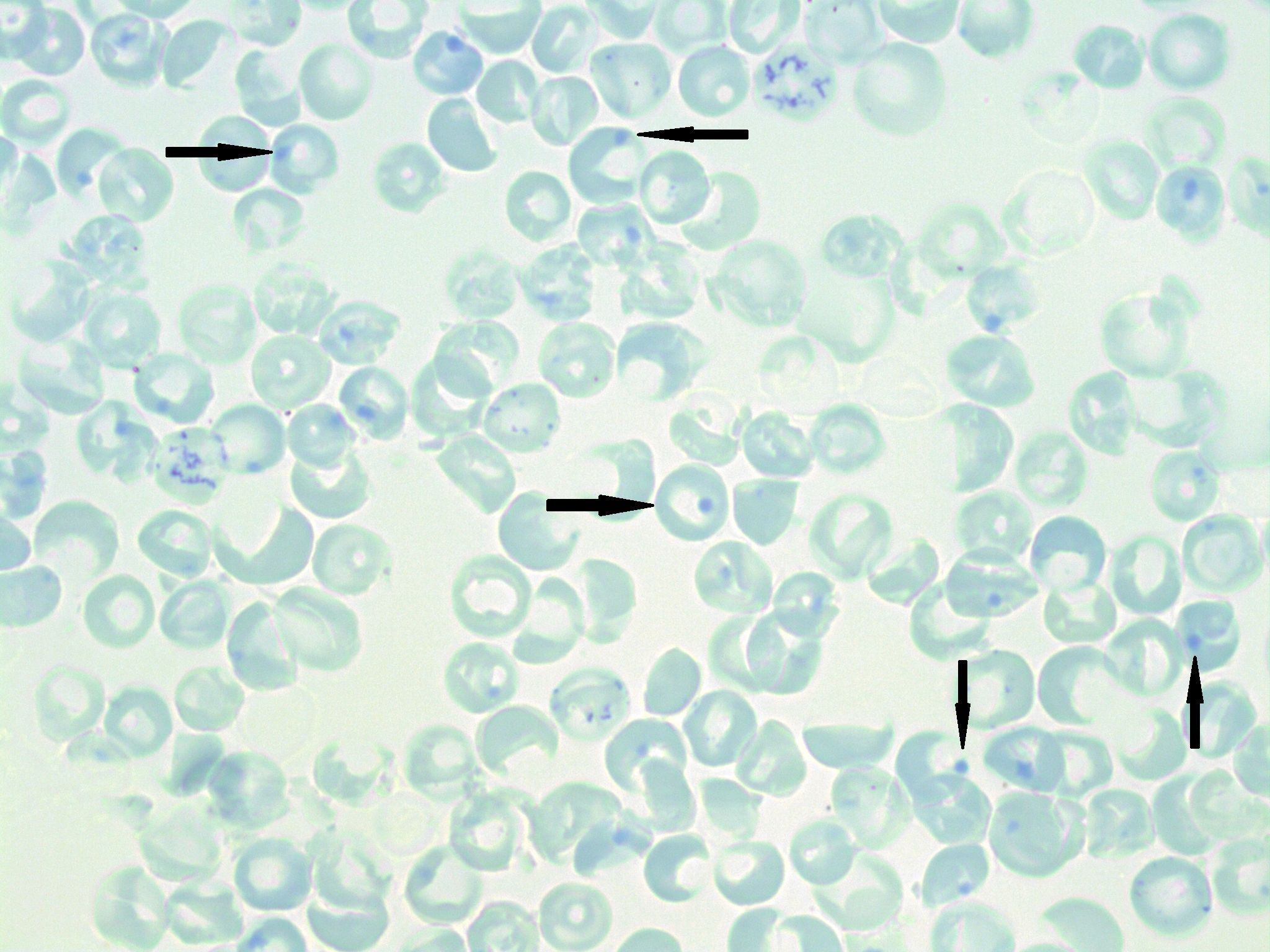
large cells appear bright blue due to staining
a dot is visible on the edge of the cell
results from denatured hemoglobin (Hgb has become unstable)
can only be visualized with supravital stains (such as brilliant cresyl blue)
mostly seen in G6PD deficiency
What are Heinz bodies inclusions?
best way of assessing bone marrow function, RBC generation, and response to anemia
reference range:
0.5% - 2.0% in adults, 2.0% - 6.0% in newborns
What is the purpose of reticulocyte counts and what is the normal reference range?
reticulocytosis
elevated reticulocyte count
body’s response to anemic stress, accompanied by erythroid hyperplasia
reticulocytopenia
decreased reticulocyte count
aplastic conditions
DECREASED RBC PRODUCTION
What is reticulocytosis and reticulocytopenia?
target cells
Which RBC morphology may form as a result of excess cholesterol on the cell’s membrane?
a decrease in hemoglobin content in red blood cells
Hypochromasia is used to define:
heme
4 atoms of iron
globin
2 pairs of globin chains made up of amino acids
What two things make up hemoglobin?
embryonic Hgb
contains zeta and epsilon chains
Hemoglobin Gower One, Hemoglobin Gower 2, and Hemoglobin Portland
fetal Hgb
3 months of fetal development
hemoglobin F (contains alpha and gamma chains)
adult Hgb
contains alpha and beta chains
adult Hgb is 95%-98% Hemoglobin A, 3%-5% Hemoglobin A2, and <2% Hemoglobin F
What are the three types of hemoglobin synthesized in RBC production?
carry oxygen (known as oxyhemoglobin) to tissues
pull CO2 away from tissues
keep a balanced blood pH
What are the three functions of hemoglobin?
Hgb A, Hgb A2, and Hgb F
What are the three normal adult hemoglobins?
a substance produced via the Embden-Meyerhof pathway of RBC metabolism
helps with oxygen affinity of hemoglobin
What is 2,3-DPG?
synthesis begins in the polychromatic normoblast stage of RBC development
What is the origin of hemoglobin?
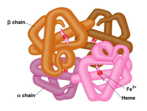
four heme molecules with one iron inside
the heme is lodged in the two globin chains
Describe the structure of a hemoglobin molecule
the Hgb loses its gamma chains and instead, the amount of beta chains increase
adult hemoglobin has a lot of alpha and beta chains
What changes occur when hemoglobin shifts from fetal Hgb to adult Hgb?
4
one oxygen for each heme
How many molecules of oxygen can one hemoglobin carry?
in the lungs, Hgb has a high affinity for oxygen, so they become saturated (4 oxygen per Hgb)
Hgb become oxyhemoglobin
Hgb carries oxygen to the tissues and unloads
in areas where there is low concentration of oxygen, Hgb’s affinity for oxygen decreases, and increases for carbon dioxide
Hgb picks up CO2 and transports it to the lungs to be eliminated
Hgb can change shape depending on if it’s oxygenated or not: tense Hgb is not oxygenated, relaxed Hgb is oxygenated
How does hemoglobin deliver oxygen to tissues and eliminate carbon dioxide?
right
Hgb has less attraction to oxygen, releases oxygen more easily
left
Hgb holds on to oxygen, does not release easily
What does right shift and left shift mean in the oxygen dissociation curve?
anemia, acidosis, or fever
right shift causes RBCs to act more efficiently in delivering oxygen to tissues (compensatory mechanism)
What conditions cause a right shift on the OD curve?
abnormal hemoglobin, alkalosis, or decreased body temp
What conditions cause a left shift in an OD curve?
methemoglobin
iron can no longer bind oxygen
can be causes by drugs or Hgb M (an amino acid substitution)
carboxyhemoglobin
Hgb has a higher affinity for CO2
can lead to carbon monoxide poisoning (no oxygen to tissues)
increased in smokers and industrial workers
can be reversed
sulfhemoglobins
affinity for oxygen is 100 times lower than normal Hgb
caused by exposure to sulfa-containing drugs or sulfonamides
can be toxic at low levels
What are the three abnormal hemoglobins?
the premature lysis of RBCs
two types: intravascular and extravascular
What is hemolysis?
intravascular
occurs when RBCs are lysed directly in blood vessels
results in low Hgb, Hct, and RBC count
high bilirubin, low serum haptoglobin, hemoglobinemia (free hemoglobin), hemoglobinuria, high reticulocytes and LDH, and schistocytes
extravascular
RBCs are destroyed and their contents are phagocytized in the spleen, liver, lymph nodes, and bone marrow
low Hgb, Hct, and RBC count, increased reticulocyte count, polychromasia, increased serum bilirubin, low haptoglobin, high LDH, and spherocytes
What is intravascular and extravascular hemolysis?
PB PORE
Pronormoblast
Basophilic normoblast
Polychromatophilic normoblast
Orthochromic normoblast
Reticulocyte
Erythrocyte
mnemonic to remember RBC maturation stages