Cardiovascular System
1/45
There's no tags or description
Looks like no tags are added yet.
Name | Mastery | Learn | Test | Matching | Spaced | Call with Kai |
|---|
No analytics yet
Send a link to your students to track their progress
46 Terms
What are the major components of the cardiovascular system?
The heart, blood vessels (arteries, veins, capillaries), blood, and lymphatic system.
What are the main components of blood?
Plasma, red blood cells (RBCs), white blood cells (WBCs), and platelets.
What are the functions of plasma?
Carries nutrients, hormones, and waste; helps maintain blood pressure and volume.
Compare and contrast arteries and veins.
Arteries carry blood away from the heart, have thick walls, and elastic fibers; veins carry blood to the heart, have thinner walls, less elastic tissue, and one-way valves.
Describe the structure and function of capillaries.
Thin-walled (one cell thick) vessels that allow gas, nutrient, and heat exchange between blood and tissues.
Explain the difference between single and double circulation.
Single: Blood passes through the heart once per cycle (heart → gills → body → heart). Double: Blood passes through the heart twice (heart → lungs → heart → body → heart).
Describe the flow of blood through a fish heart.
Sinus venosus → atrium → ventricle → conus/bulbus arteriosus → gills → body → sinus venosus. (1 atrium + 1 ventricle = 2 chambers)
What are the specializations of the fish heart in hagfish and lamprey?
They have accessory hearts (cardinal, caudal, and portal hearts) that are contractile but lack cardiac muscle.
How is the lungfish heart specialized for breathing air?
It has partial interatrial and interventricular septa and a spiral valve that reduce mixing and direct blood based on oxygen needs.
What is the purpose of the ductus arteriosus in lungfish?
Connects the pulmonary artery to the systemic circulation, allowing blood to bypass the gills when not in use.
Describe the structure of the amphibian heart.
Three chambers—two atria and one ventricle—with a spiral valve in the conus arteriosus.
How does the spiral valve in the amphibian heart function?
It directs oxygenated blood to systemic circulation and deoxygenated blood to pulmocutaneous circulation, limiting mixing.
Compare the hearts of chelonians/squamates and crocodilians.
Chelonians/squamates: 3 chambers (2 atria, 1 ventricle divided into cavum venosum, cavum pulmonale, and cavum arteriosum). Crocodilians: 4 chambers (2 atria, 2 ventricles) with a Foramen of Panizza connecting systemic arches.
What is the Foramen of Panizza?
An opening between the left and right systemic aortas in crocodilians that allows blood shunting during diving.
How do reptiles shunt blood during diving?
Pulmonary resistance increases, causing a right-to-left shunt that bypasses the lungs; this helps conserve oxygen and regulate temperature, acid-base balance, and digestion.
Describe the structure of the avian and mammalian heart.
Four chambers—right atrium, right ventricle, left atrium, left ventricle—with complete separation of oxygenated and deoxygenated blood.
What are the four chambers of the avian/mammalian heart and their functions?
Right atrium: Receives deoxygenated blood from the body. Right ventricle: Pumps blood to the lungs. Left atrium: Receives oxygenated blood from the lungs. Left ventricle: Pumps blood to the body.
Describe the flow of blood through the avian/mammalian heart.
Vena cava → right atrium → tricuspid valve → right ventricle → pulmonic valve → pulmonary artery → lungs → pulmonary vein → left atrium → mitral valve → left ventricle → aortic valve → aorta → body.
List the four valves of the avian/mammalian heart and their locations.
Tricuspid: Between right atrium and right ventricle. Pulmonic (semilunar): Between right ventricle and pulmonary artery. Mitral (bicuspid): Between left atrium and left ventricle. Aortic (semilunar): Between left ventricle and aorta.
What are the major arteries and their destinations?
Aorta: From heart to body. Pulmonary artery: To lungs. Hepatic artery: To liver. Renal artery: To kidneys. Carotid artery: To head.
What are the major veins and their sources?
Cranial/caudal vena cavae: Return blood from body to heart. Pulmonary vein: From lungs to heart. Hepatic vein: From liver. Renal vein: From kidneys. Jugular vein: From head.
Which artery carries deoxygenated blood?
Pulmonary artery.
Which vein carries oxygenated blood?
Pulmonary vein.
Describe the hepatic portal system and its function.
Drains the stomach, spleen, pancreas, and intestines into the liver for nutrient processing before systemic circulation.
In general, fish have a single | double | triple | figure-of-8 circulatory loop system.
single
List the number of normal heart chambers for each of these species:
Blank 1: Fish (non-lungfish): ___
Blank 2: Reptiles (crocodilians): ___
Blank 3: Amphibians: ___
Blank 4: Reptiles (chelonians/squamates): ___
Blank 5: Birds: ___
Blank 6: Mammals: ___
#1: 2
#2: 4
#3: 3
#4: 3
#5: 4
#6: 4
Lungfish have a _______ _______ located within the conus arteriosus that facilitates separation of oxygenated and deoxygenated blood
spiral value
In which of these would you also find a spiral valve?
shark intestine
As you auscult, he has questions. Thinking of the "lub dub" sound that makes a dog's heartbeat…which of these best describes what makes the sounds?
Closing of tricuspid/bicuspid valves and closing of aortic/pulmonic valves
Blood in the right atrium is oxygenated | non-oxygenated | mixed.
Non-oxygenated
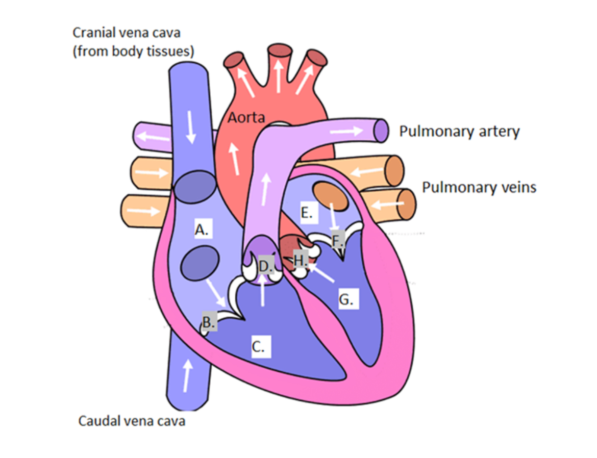
Using the graphic above, fill in the appropriate heart chamber or valve. (Don't forget to indicate left or right where appropriate).
Right atrium, tricuspid value, right ventricle, pulmonary valve, left atrium, mitral valve, left ventricle, aortic value
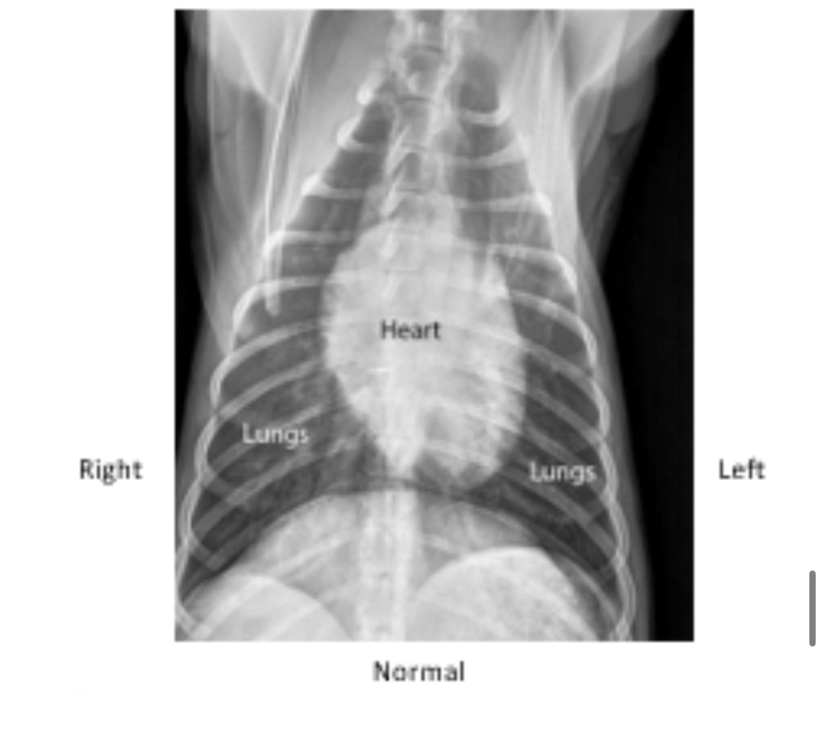
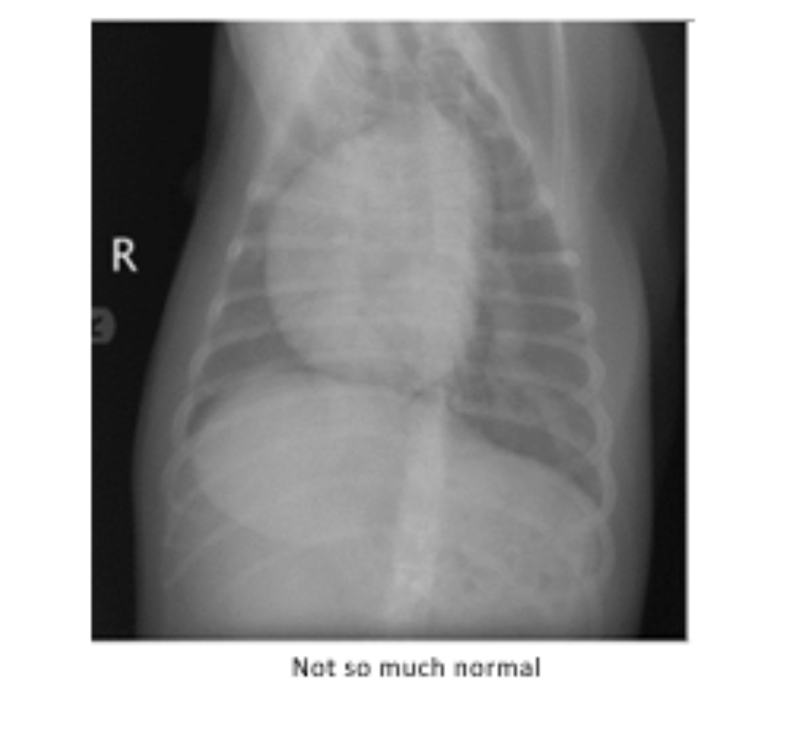
Examine this normal canine chest radiograph compared to Midnight's abnormal one. Which side of the heart appears enlarged?
Right
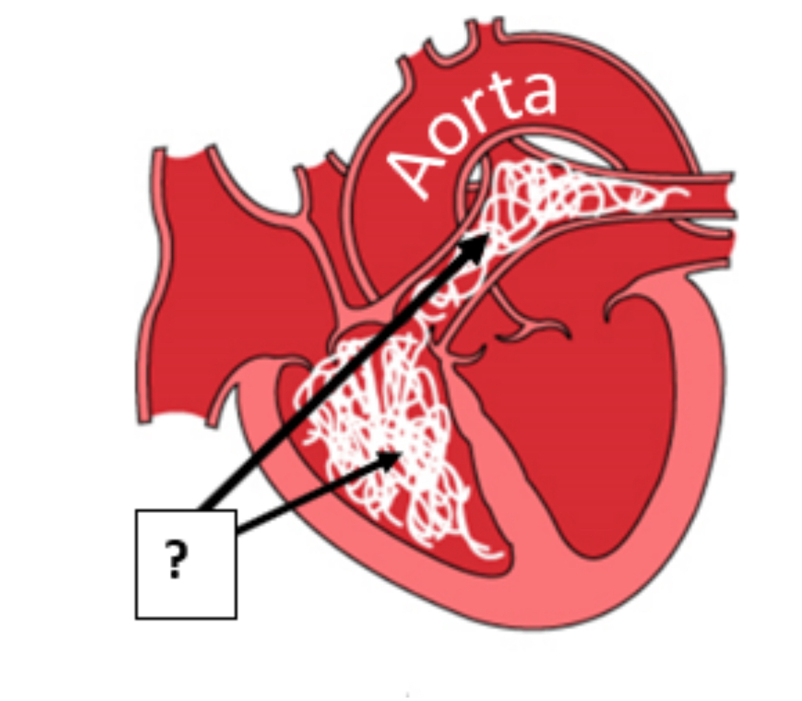
This diagram illustrates what's happening in the abnormal radiograph. In dogs, adult heartworms take up residence in which chamber of the heart and which blood vessel (as indicated)?
Chamber: __________ (Blank 1)
Blood vessel: ___________ (Blank 2)
#1: right ventricle
#2: pulmonary artery
This infliction may lead to pulmonic stenosis (narrowing) of the outflow of the blood vessel listed above/in the previous question.
Using the graphic we discussed in class (in the PowerPoint), define WHERE would be the best place to auscult (listen) for pulmonic stenosis:
Between ribs _____ and _____ near the ________ _______ junction, on the _____ side.
B#1: 2
B#2: four
B#3: costochondral
B#4: left
The doctor asks you to get a heart rate for the iguana by placing a Doppler probe over a blood vessel. What is responsible for the sound you hear?
Movement of blood through the blood vessel, propelled by contraction
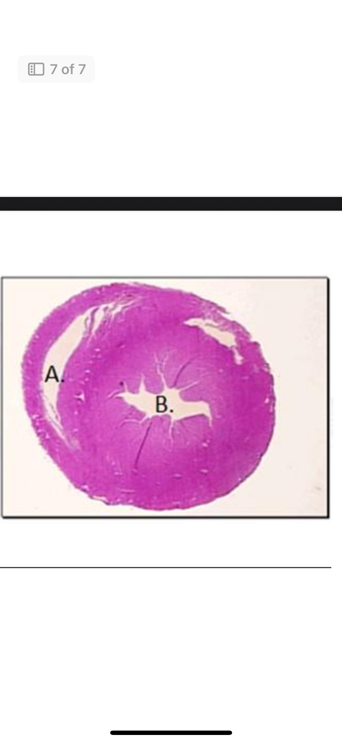
Lastly, examine this transverse section of an avian heart. You are looking at the inside (lumen) of the left and right ventricle. Identify which is which:
A. is the right ventricle
B. is the left ventricle
Match the major blood vessels with the appropriate name: Pulmonary artery
Carries blood to lungs
Match the major blood vessels with the appropriate name: Renal artery
Carries blood to kidneys
Match the major blood vessels with the appropriate name: Afferent artery
Carries blood to the gills
Match the major blood vessels with the appropriate name: Hepatic portal vein
Carries blood from stomach, pancreas, spleen, to liver
Carotid artery
Carries blood to the head
Match the major blood vessels with the appropriate name: Renal vein
Carries blood from kidneys toward heart
Match the major blood vessels with the appropriate name: Hepatic vein
Carries blood from liver toward heart
Match the major blood vessels with the appropriate name: Jugular vein
Drains blood from head
Match the major blood vessels with the appropriate name: Vena cava
Carries blood to right atrium
Match the major blood vessels with the appropriate name: Iliac artery
Carries blood to hind limbs