Radiation Saftey and The Machine
1/240
There's no tags or description
Looks like no tags are added yet.
Name | Mastery | Learn | Test | Matching | Spaced | Call with Kai |
|---|
No analytics yet
Send a link to your students to track their progress
241 Terms
what is it (Intro to Diagnostic Imaging)
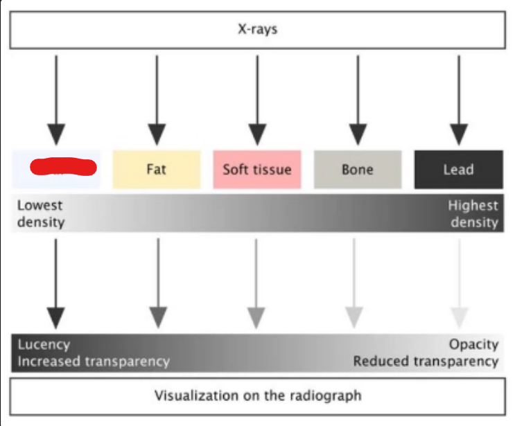
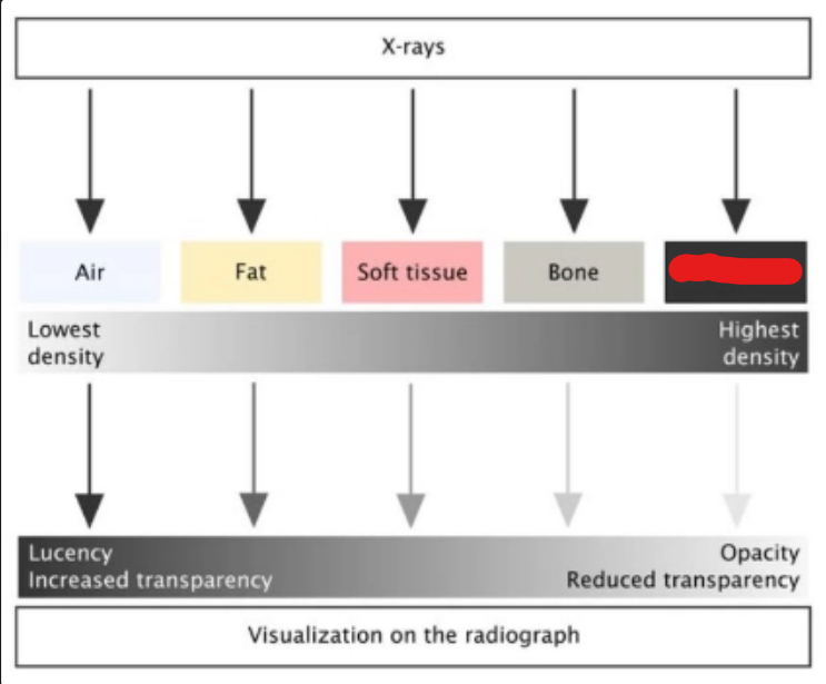
air
what is it (Intro to Diagnostic Imaging)
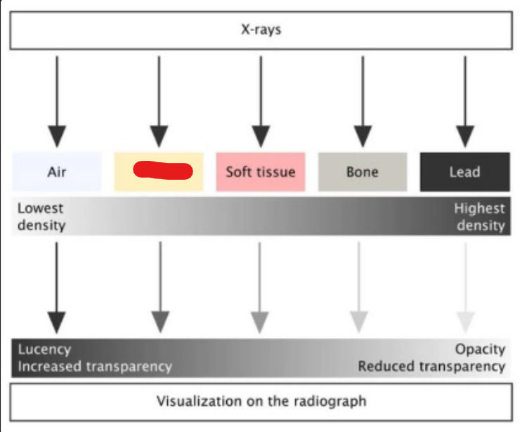
fat
what is it (Intro to Diagnostic Imaging)
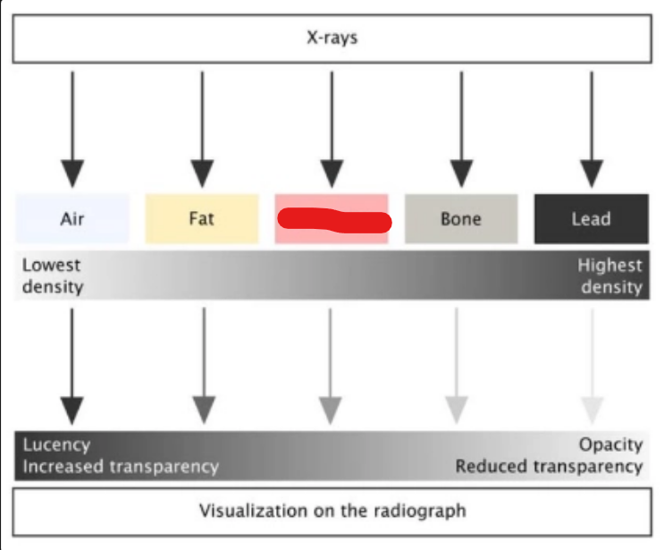
soft tissue
what is it (Intro to Diagnostic Imaging)
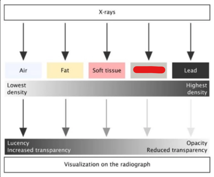
bone
what is it (Intro to Diagnostic Imaging)
lead
Diagnostic value is related to the … (Intro to Diagnostic Imaging)
quality of the image
High quality images = (Intro to Diagnostic Imaging)
High diagnostic value
Low quality images = (result + consequence) (Intro to Diagnostic Imaging)
Low diagnostic value/Misdiagnosis
photons have — energy to pass through tissues (Intro to Diagnostic Imaging)
high
X Ray machines turn electrical current into — — — (Intro to Diagnostic Imaging)
high energy photons
Electrical current flows through (Intro to Diagnostic Imaging)
cathode
the cathode is —- charged (Intro to Diagnostic Imaging)
negativly
the anode is — charged (Intro to Diagnostic Imaging)
positively
the anode attracts — (Intro to Diagnostic Imaging)
electrons
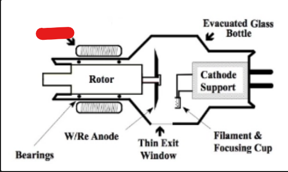
what is it (Intro to Diagnostic Imaging)
stator
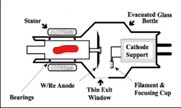
what is it (Intro to Diagnostic Imaging)
rotor
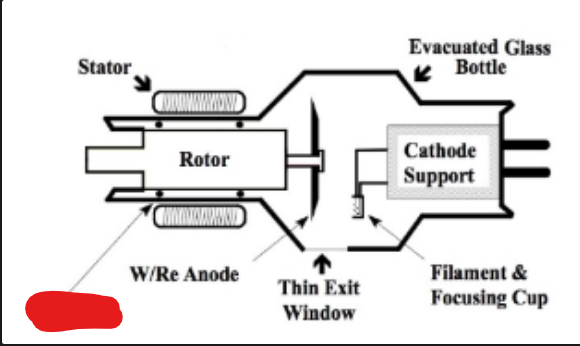
what is it (Intro to Diagnostic Imaging)
bearings
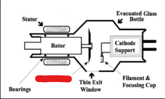
what is it (Intro to Diagnostic Imaging)
W/Re anode
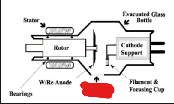
what is it (Intro to Diagnostic Imaging)
thin exit window
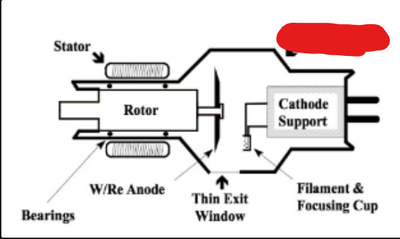
what is it (Intro to Diagnostic Imaging)
evacuated glass bottle
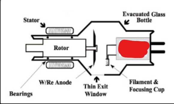
what is it (Intro to Diagnostic Imaging)
cathode support
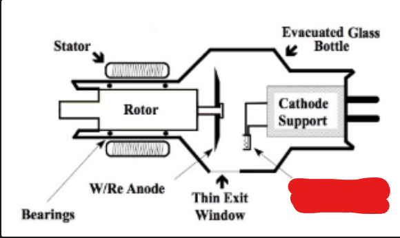
what is it (2) (Intro to Diagnostic Imaging)
filament and focusing cup
We can control 3 parameters to alter the nature of the X Ray beam produced and so optimize our radiographic image (Intro to Diagnostic Imaging)
kVp
mA
time
what does kVp stand for (Intro to Diagnostic Imaging)
kilovolts
what does mA stand for (Intro to Diagnostic Imaging)
milliamps
kVp is the — with which the photons move (Intro to Diagnostic Imaging)
Power
Higher kVp gives the photons … (Intro to Diagnostic Imaging)
more power to penetrate tissues.
Increasing kVp will — contrast (Intro to Diagnostic Imaging)
decrease
mA is the — — of photons in the beam (Intro to Diagnostic Imaging)
total number
Higher mA increases the total number of photons and —- the image overall (Intro to Diagnostic Imaging)
darkens
Higher mA increases the total number of photons and — contrast (Intro to Diagnostic Imaging)
reduces
long contrast is — kVp, — mAs (Intro to Diagnostic Imaging)
high, low
short contrast is — kVp, — mAs (Intro to Diagnostic Imaging)
low, high
X-Rays are a type of — radiation (Radiation Safety)
ionizing
X Rays are highly penetrating but have low (Radiation Safety)
LET
what does LET stand for (Radiation Safety)
linear energy transfer
Risks are — and increased based on —- —- —- to radiation (Radiation Safety)
CUMULATIVE, TOTAL LIFETIME EXPOSURE
define Inverse square law of radiation intensity (Radiation Safety)
The beam intensity at the image receptor is directly proportional to the exposure parameters mAs
what ppe should be worn (5) (Radiation Safety)
0.5mm lead gown
thyroid protector
gloves
glasses
dosimeter
what is the dosimeter for (Radiation Safety)
to allow monitoring of excessive exposure
PPE only protects from (Radiation Safety)
scatter radiation
NO PART OF THE TECHNICIANS ANATOMY SHOULD BE PLACED IN … (Radiation Safety)
THE PRIMARY BEAM
who discovered x-rays (The Basics of Atoms and Electricity)
Wilhelm Conrad Roentgen
when were x-rays discovered (The Basics of Atoms and Electricity)
1895
what are the12 unique properties of x-rays (The Basics of Atoms and Electricity)
Are invisible
Are electrically neutral
Have no mass
Travel at the speed of light in a vacuum
Cannot be focused by a lens
Form a polyenergetic (heterogeneous) beam
Can be produced in a range of energies (kV)
Travel in straight lines
Cause fluorescence in certain substances
Can cause chemical changes to occur in radiographic and photographic film
Can be absorbed or scattered by tissues in the body; can produce scattered and secondary radiation
Can cause chemical and biological damage to living tissue
define element (The Basics of Atoms and Electricity)
Smallest particle of a substance
define atom (The Basics of Atoms and Electricity)
Smallest particle of an element
the nucleus of atoms contains (2) (The Basics of Atoms and Electricity)
protons
neutrons
— circle the nucleus in rings (The Basics of Atoms and Electricity)
electrons
define thermionic emission (The Basics of Atoms and Electricity)
heat is applied to the cathode of the x-ray tube, electrons are “boiled off”
how are x-rays made (4) (The Basics of Atoms and Electricity)
boiling electrons off
exposure switch is closed
“photons” or energy packets are drawn across from the cathode to the anode
interacts with the anode
what charge do protons have (The Basics of Atoms and Electricity)
positive
what charge do neutrons have (The Basics of Atoms and Electricity)
none
none
what charge are electrons (The Basics of Atoms and Electricity)
negative
X-rays created from converting — into — (The Basics of Atoms and Electricity)
matter, energy
Principle characteristic of “matter” (2) (The Basics of Atoms and Electricity)
Matter occupies space
Mass or weight
Energy’s principle characteristic (2) (The Basics of Atoms and Electricity)
Movement or motion
The ability to do work
what kind of energy is used in radiography (The Basics of Atoms and Electricity)
electromagnetic
amplitude is the —- of waves (The Basics of Atoms and Electricity)
height
frequency is the —- between waves (The Basics of Atoms and Electricity)
wavelength
frequency is connected to the kilovoltage used to … (2) (The Basics of Atoms and Electricity)
penetrate the tissue of the patient
the time that is set to produce the x-rays.
Shorter wavelengths with higher frequencies penetrate the tissue —- effectively than long wavelengths with low frequency (The Basics of Atoms and Electricity)
more
A photon may be pictured as a … (The Basics of Atoms and Electricity)
small bundle of energy.
As energy increases, frequency —- (The Basics of Atoms and Electricity)
increases
heat dissapation is —-% heat/—-% x-rays (Diagnostic X-Ray Production)
99, 1
what are the 4 x-ray generator settings (Diagnostic X-Ray Production)
voltage
current
time
resistance
what are the units for voltage (Diagnostic X-Ray Production)
V
define voltage (Diagnostic X-Ray Production)
the speed with which the electrons in the electric current transfer energy along the circuit.
what are the units for high voltage (Diagnostic X-Ray Production)
kV
high voltage produces (3) x-rays (Diagnostic X-Ray Production)
short-wavelength
high frequency
highly penetrating
current is also known as (Diagnostic X-Ray Production)
amerage
what is the unit for current (Diagnostic X-Ray Production)
upper case ‘i’
define current (Diagnostic X-Ray Production)
to measure the electric current that activates the x-ray tube.
the x-ray unit consists of a closed circuit with 5 main criteria: (Diagnostic X-Ray Production)
It must have enough power to eventually produce x-rays.
It must have selections where the power can be increased or decreased as necessary.
The electric current must travel in the same direction through the x-ray tube.
There must be a way to produce free electrons with enough energy to produce x-rays.
There must be an efficient way to dissipate the heat that results in the interaction of the photons and the anode.
circuit breakers acts as a (Diagnostic X-Ray Production)
fuse
how do circuit breakers act as fuses (Diagnostic X-Ray Production)
If the voltage or current is set too high, or if a component malfunctions, the circuit breaker will cut the power to the unit
define fuse (Diagnostic X-Ray Production)
safety devices that will mechanically interrupt the flow of electricity if a problem arises that would overheat the circuit
define ground wire (Diagnostic X-Ray Production)
Redirects current flow of excess electrons if circuit is broken
what color is the ground wire usually (Diagnostic X-Ray Production)
green
operating console is aka (Diagnostic X-Ray Production)
generator
voltage controls — —- (Diagnostic X-Ray Production)
penetrating power
current controls (2) (Diagnostic X-Ray Production)
number of photons
density of image
power (WATT) equation (Diagnostic X-Ray Production)
Power (WATT) = voltage (V) X Current (A)
purpose of line voltage compensator (Diagnostic X-Ray Production)
Stabilizes incoming power line
define direct current (Diagnostic X-Ray Production)
Source of power near the end user
define Alternating Current (Diagnostic X-Ray Production)
One positive pulse and then one matching negative pulse
how many cycles are in alternating current (Diagnostic X-Ray Production)
120 cycles/sec
define transformers (Diagnostic X-Ray Production)
Transformers step up (increase) the power at one end of the journey and then step down (decrease) the power at the destination
define rectifiers (Diagnostic X-Ray Production)
Allow current to keep moving in one direction within x-ray unit
What is the problem with single-phase circuits (Diagnostic X-Ray Production)
The problem that arose was the long exposure time due to the low power of these units and the loss of power as the voltage dropped during the exposure with the completion of each pulse
what are 3-phase circuits used for (Diagnostic X-Ray Production)
large animal chest and abdominals
why is 3-phase circuits used for large animal (Diagnostic X-Ray Production)
high milliamperes (mA) combined with short times (seconds) are necessary to image large-animal chests and abdomen
what are the 3 kinds of circuits (Diagnostic X-Ray Production)
single-phase
three-phase
high-frequency
what are the 3 components of the x-ray unit (Diagnostic X-Ray Production)
High-voltage transformer / high-tension transformer
X-ray generator
X-ray tube
define High-voltage transformer / high-tension transformer (Diagnostic X-Ray Production)
It is connected to the hospital power lines and uses the power supplied to the hospital by the outside power lines.
define X-ray generator (Diagnostic X-Ray Production)
receives its power from the highvoltage transformer and sends it through the circuit to the rectifiers and then to the x-ray tube
define x-ray tube (Diagnostic X-Ray Production)
it is here that the x-rays are produced
what is in the x-ray tube (7) (Diagnostic X-Ray Production)
Cathode
anode
Near vacuum
Power must enter thru the cathode
Heat will exit through stem of the anode
Stem is mounted on ball bearings which are coated in light oil
Every x-ray tube is shielded with a metal covering, which restricts most off-focus radiation from exiting the tube other than at the tube port.
the x-ray tube process to produce x-rays (5) (Diagnostic X-Ray Production)
the exposure switch is closed
the electrons are drawn across to the anode by electromagnetic force
they are stopped very suddenly by the density of the metal of the anode.
They react immediately by losing speed
converting the forward motion to energy, which results in heat and x-rays (99% heat and 1% x-rays).
define thermionic emission (Diagnostic X-Ray Production)
emission due to heating