Chapter 26- The Heart .
 Call Kai
 Learn
Learn Practice Test
 Spaced Repetition
 Match
Match Flashcards
 Knowt Play
1/132

There's no tags or description
Looks like no tags are added yet.
Name | Mastery | Learn | Test | Matching | Spaced | Call with Kai |
|---|
No analytics yet
Send a link to your students to track their progress
133 Terms
what is the cardiovascular system also called?
circulatory system
what is the cardiovascular/circulatory system concerned with?
heart & blood vessels
what is the heart & what is it composed of?
-heart is a muscle
-composed of striated muscle, cardiac in type
what is the function of the heart?
pumping blood through vessels of the cardiovascular system
where is the heart located?
in the thorax, between the lungs, resting on the diaprhagm
how many internal cavities/chambers does the heart have?
4
what are the 4 chambers of the heart?
right atrium
left atrium
right ventricle
left ventricle
how is the heart positioned?
in such a way that the atria lie posterior to the ventricles
in what direction does blood pass through the atria to the ventricles?
horizontally
what structure protects the heart anteriorly?
sternum
what structure protects the heart laterally?
ribs, intercostal muscles, and lungs
what structure protects the heart posteriorly?
thoracic vertebrae
what is the heart enclosed in (that further protects it)?
pericardium (a tough connective tissue sac)
what is the pericardium?
fibro-serous sac that encloses the heart
what does the outer layer of the pericardium blend with inferiorly?
central tendon of the diaphragm
what does the fibrous layer of the pericardium blend with superiorly & posteriorly?
major blood vessels of the heart: vena cava, aorta, and pulmonary trunk
what is cardiac muscle referred to as?
myocardium
what is the endocardium?
inner epithelial lining of the chamber of the heart
what is the external anatomy of the heart composed of?
apex, base, and three surfaces
what are the three surfaces of the external heart?
sternocostal surface
diaphragmatic surface
pulmonary surface
where is the sternocostal surface?
adjacent to the sternum (sterno) and costal cartilages (costal)
where is the diaphragmatic surface?
adjacent to the diaphragm
where is the pulmonary surface?
adjacent to the medial surface of the left lung
what is the apex of the heart?
rounded projection that points inferiorly and to the left
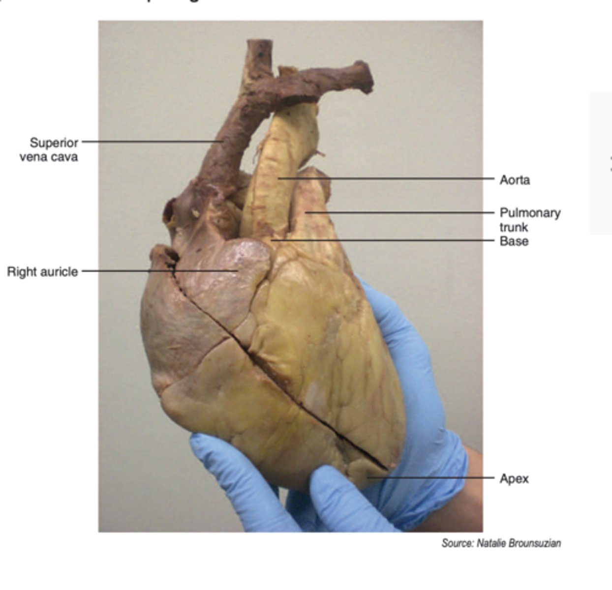
where does the apex of the heart usually lie?
level of the left sixth costal cartilage
what is the base of the heart?
area where the great vessels (aorta, pulmonary trunk, and superior vena cava) attach
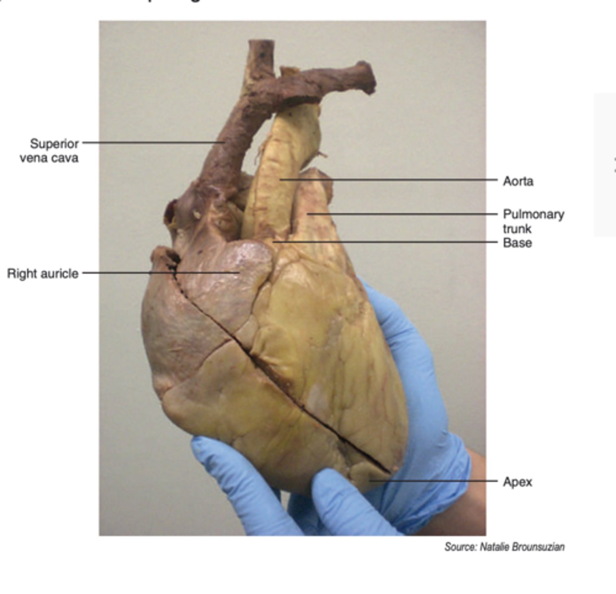
what do BOTH the left and right atria have attached to their lateral surface?
auricles (also referred to as appendages)
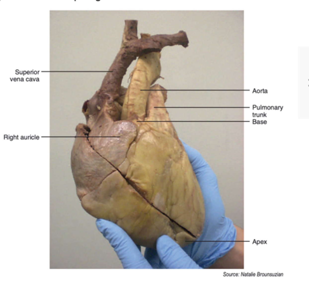
what does the auricle/appendage allow for?
increases the capacity of the atrium
what does the internal anatomy of the right heart consist of?
right atrium
tricuspid valve (right atrioventricular valve)
right ventricle
what is the crista terminalis?
vertical, muscular ridge on the posterior wall of the right atrium
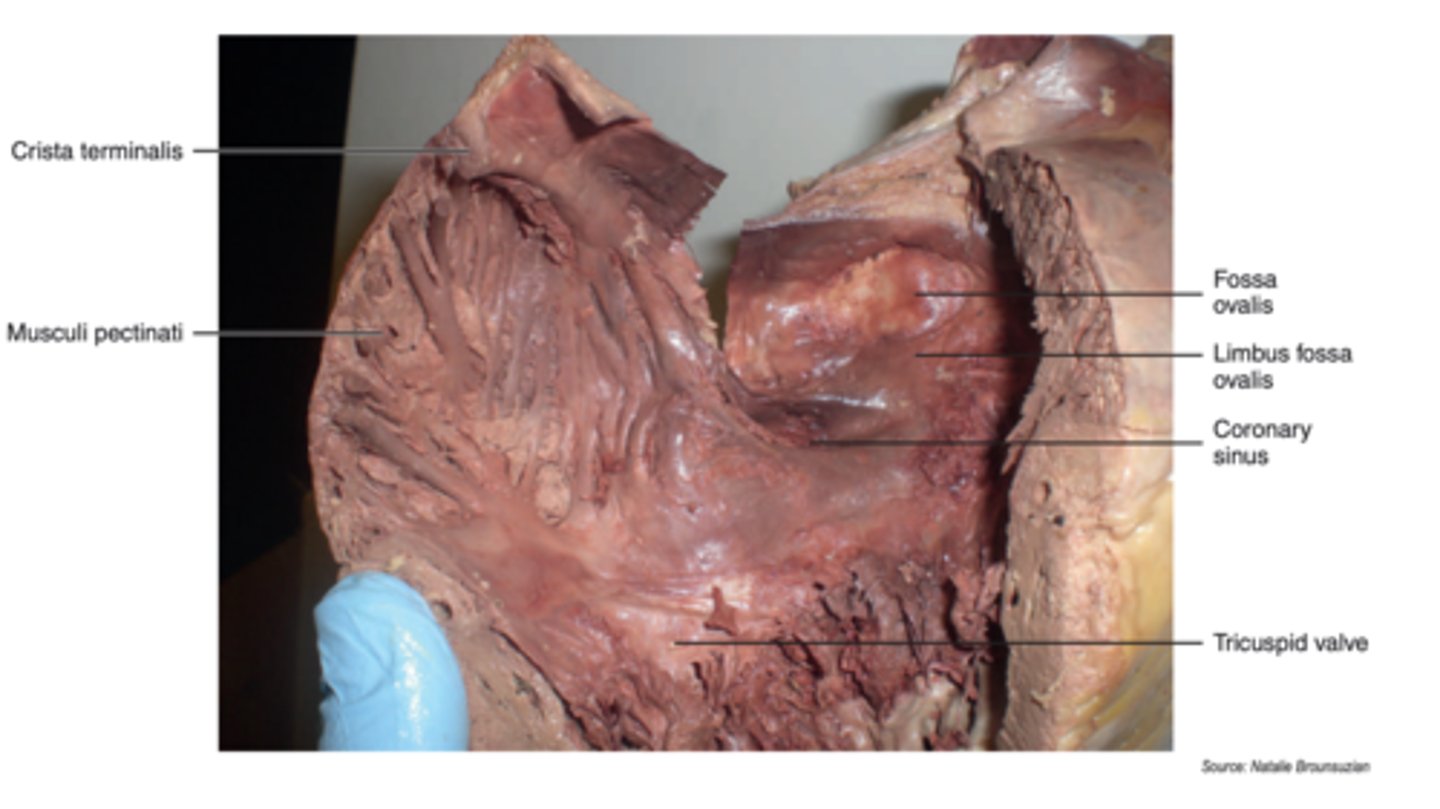
what are musculi pectinati?
muscular, ridge like extensions from the crista terminalis
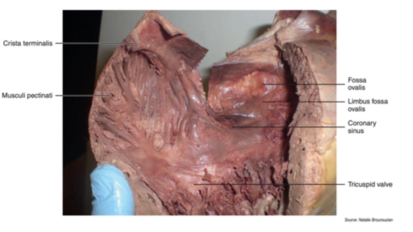
where else can musculi pectinati be found?
right atrium
right auricle
left auricle
what are the three major openings that enter into the right atrium?
superior vena caval opening
inferior vena caval opening
opening of the coronary sinus
what is the superior vena caval opening for?
the main vein returning blood to the RIGHT atrium from the upper half of the body
what is the inferior vena caval opening for?
the main vein returning blood to the RIGHT atrium from the lower half of the body
what is the inferior vena caval opening marked by?
non-fucntional valve of the inferior vena cava (small flap of tissue)
what does the opening of the coronary sinus lead to?
a venous chamber located on the posterior surface of the heart
what does the coronary sinus receive?
receives ALL THREE CARDIAC veins that drain the heart muscle
what does the coronary sinus open into?
opens into the right atrium just anterior and superior to the inferior vena cava
what is the partition between right and left atrium?
interatrial septum
what is the fossa ovalis?
- oval depression in the RA
- remnant of foramen ovale of fetal heart (that existed between the right and left atrium) that closed at birth
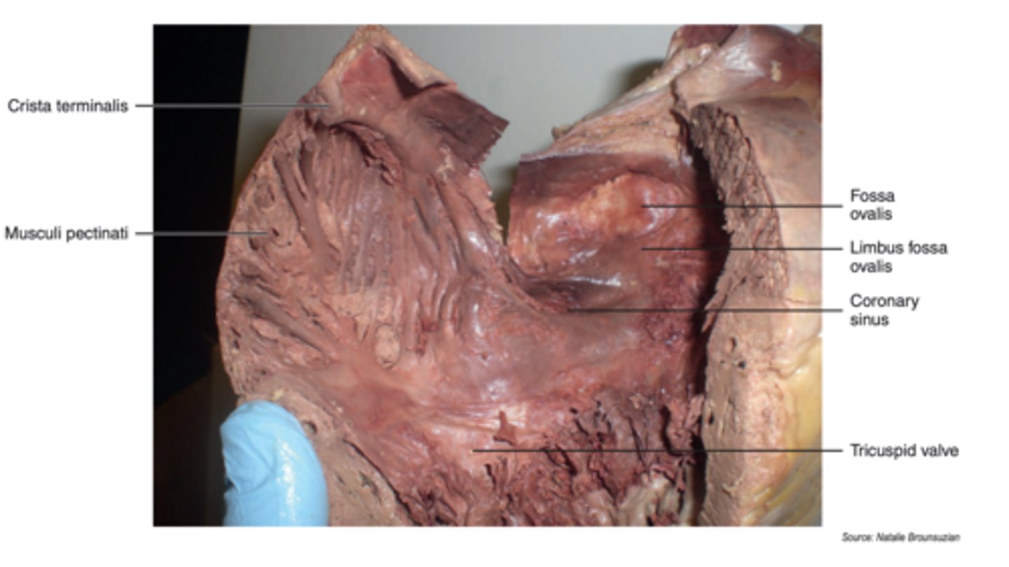
where is the fossa ovalis located?
right atrial interatrial septum
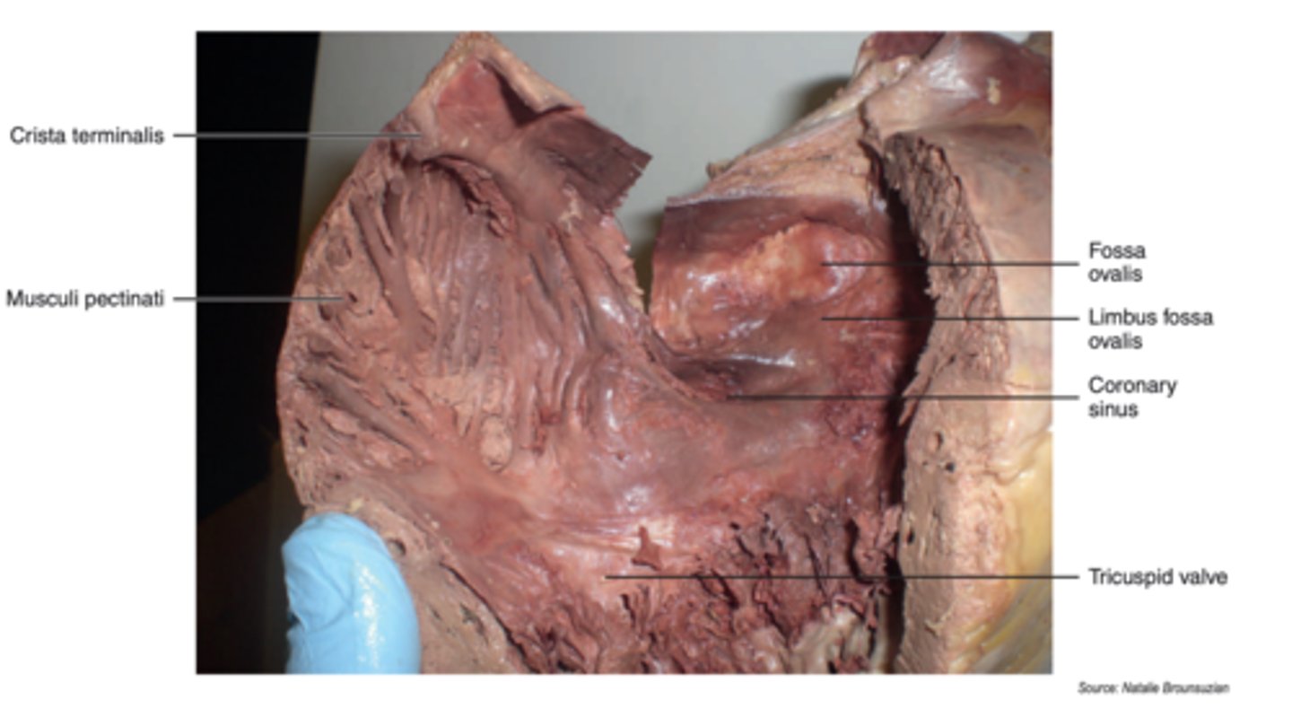
how does the right atria open to the right ventricle anteriorly?
right atrioventricular opening
what is the right atrioventricular opening guarded by?
the tricuspid valve (aka right AV valve)
what is a valve?
mechanical device that allows flow of blood in one direction preventing retrograde flow (flow in the wrong direction)
what types of valves compose the heart?
atrioventricular (AV) valves and semilunar vavles
what is an AV valve composed of?
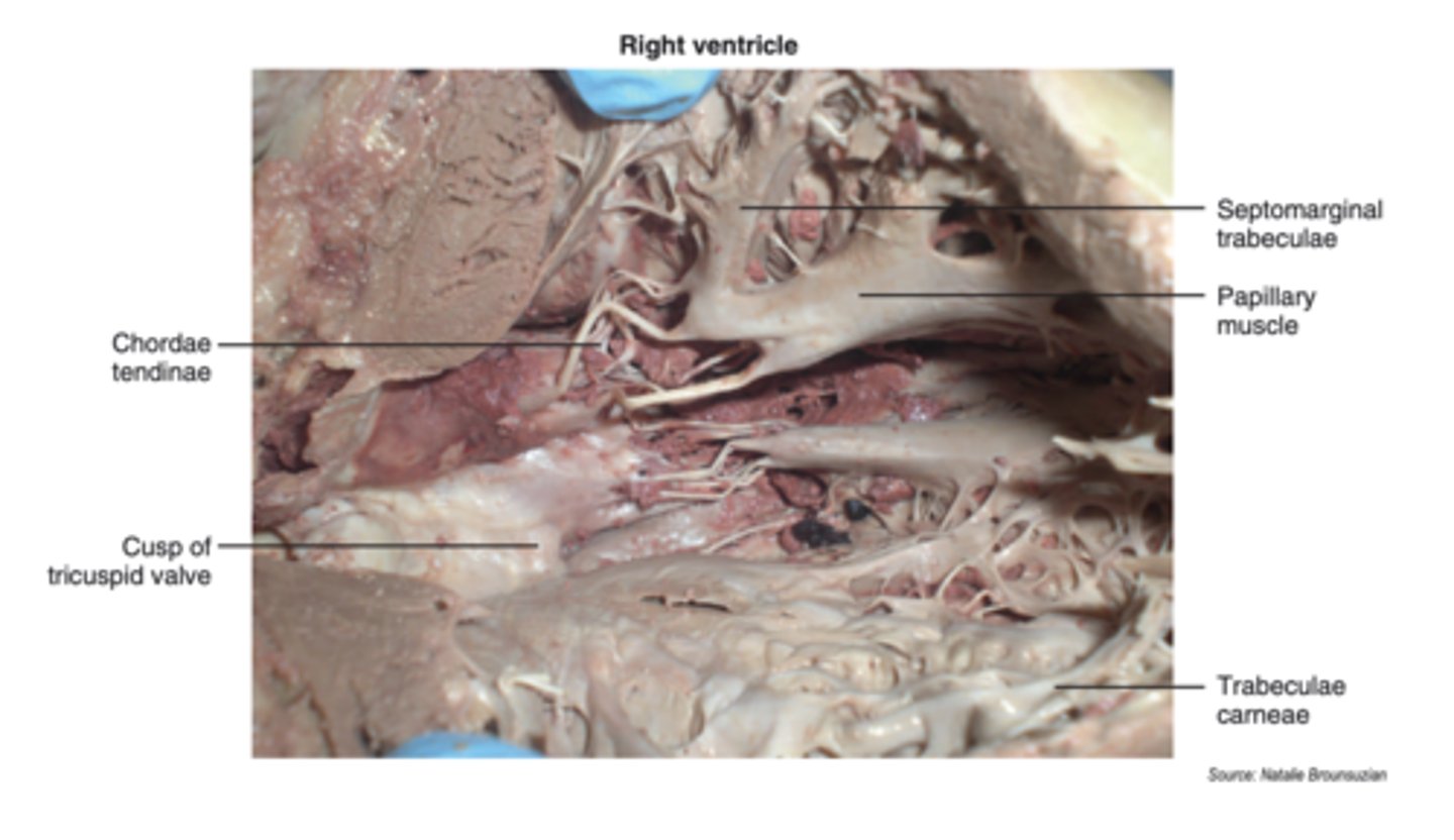
cusps, chordae tendinae, and papillary muscle
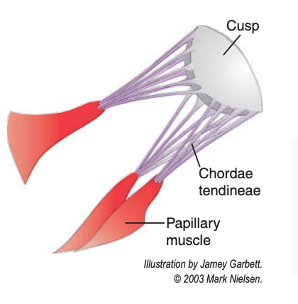
what are cusps?
flaps of connective tissue
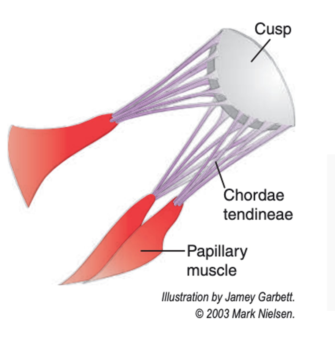
what are chordae tendinae?
connective tissue cords that attach to the lower surface of the cusps & support the cusp
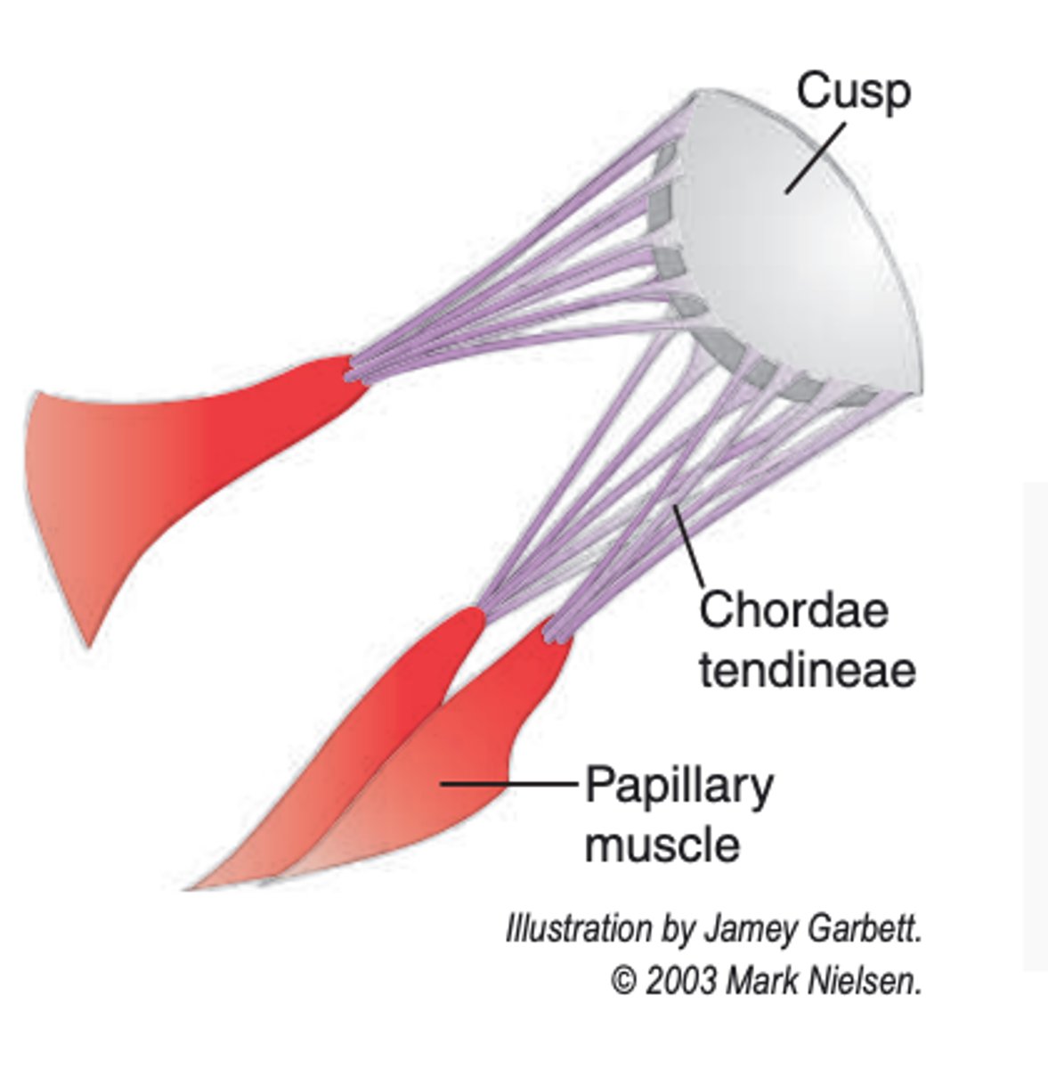
what does the papillary muscle consist of?
internal folds of ventricular muscle that give attachment to the chordae tendineae
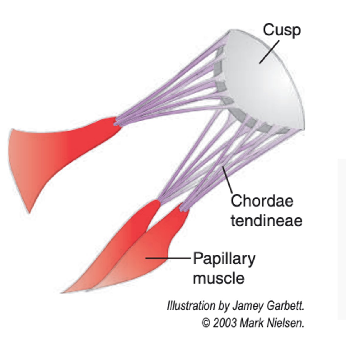
how are the 3 cusps of the tricuspid (right AV) valve named?
according to their anatomical positions
how are the papillary muscles named?
similarly to the cusps - according to the anatomical positions
what happens to the tricuspid valve during contraction of the ventricles?
the tricuspid valve closes - preventing backflow of blood from the right ventricle to the right atrium
how is the tricuspid valve held in a closed position?
via the chordae tendinae which are attached to the lower surface of cusps (and thus not everted into the right atrium)
what does the internal surface of the right ventricle consist of?
trabeculae carneae
what is trabeculae carneae?
irregular, ridge-like projections of muscle on the internal surface of the right ventricle
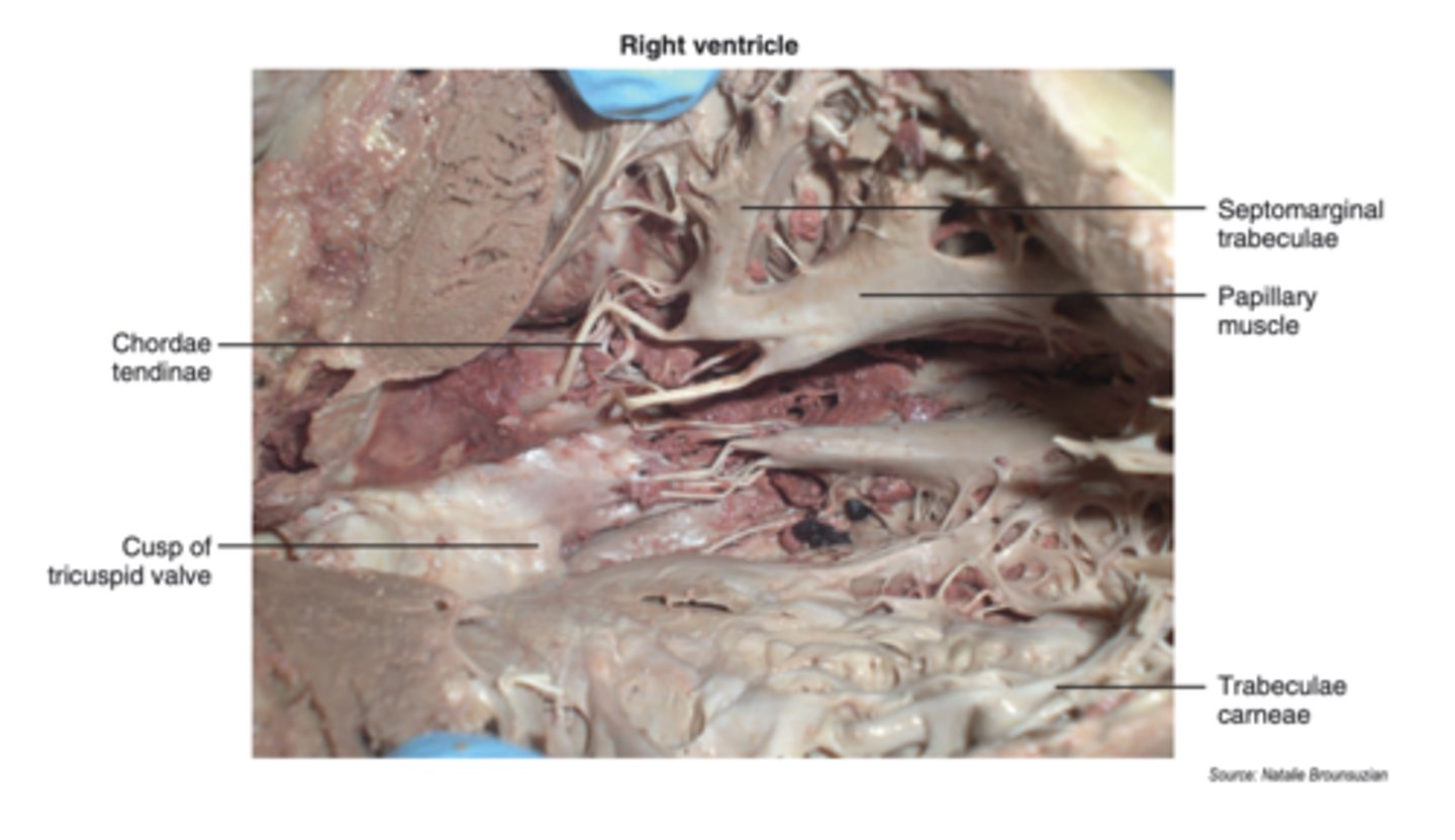
what are some types of trabeculae carneae?
-papillary muscles
-moderator band/septomarginal trabeculae (special type)
what does the moderator band/septomarginal trabeculae attach to?
attaches to the interventricular septum and to the anterior papillary muscle
what does the moderator band/septomarginal trabeculae contain?
contains the right branch of the AV bundle (which is part of the collection of fibers making up the conduction system of the heart)
what are the only openings into the right ventricle?
-right atrioventriclar opening (guarded by the tricuspid valve)
-pulmonary trunk opening (guarded by the pulmonary semilunar valve)
what passes through the pulmonary trunk opening?
blood leaving the right ventricle to enter the pulmonary trunk
what is the pulmonary trunk opening guarded by?
pulmonary semilunar valve
does a semilunar valve possess papillary muscles and chordae tendinae?
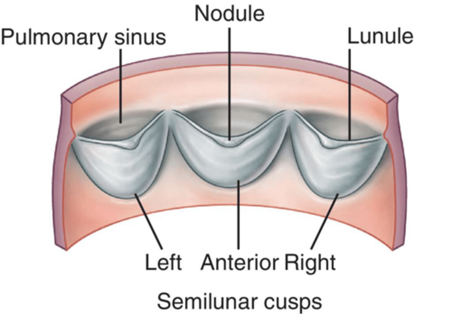
no - it exists merely as pocket-like flaps surrounding an opening
how many flaps/cusps for each semilunar valve?
3
what is the nodule (of the cusp)?
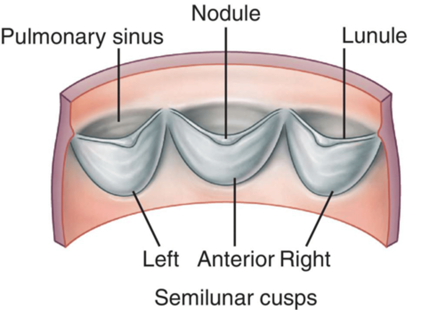
free-edge of each cusp that has a small, central thickening of fibrous tissue
what do the nodules do when the three flaps close?
fill the small gap left (at the closure of the three flaps)
what is the lunula?
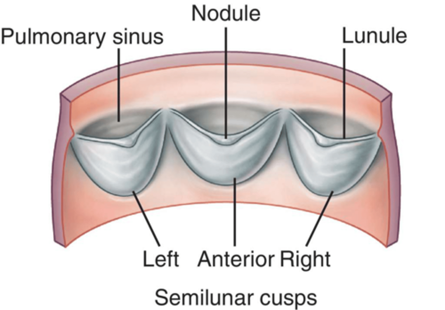
narrow, thin lip extending from each side of the nodule
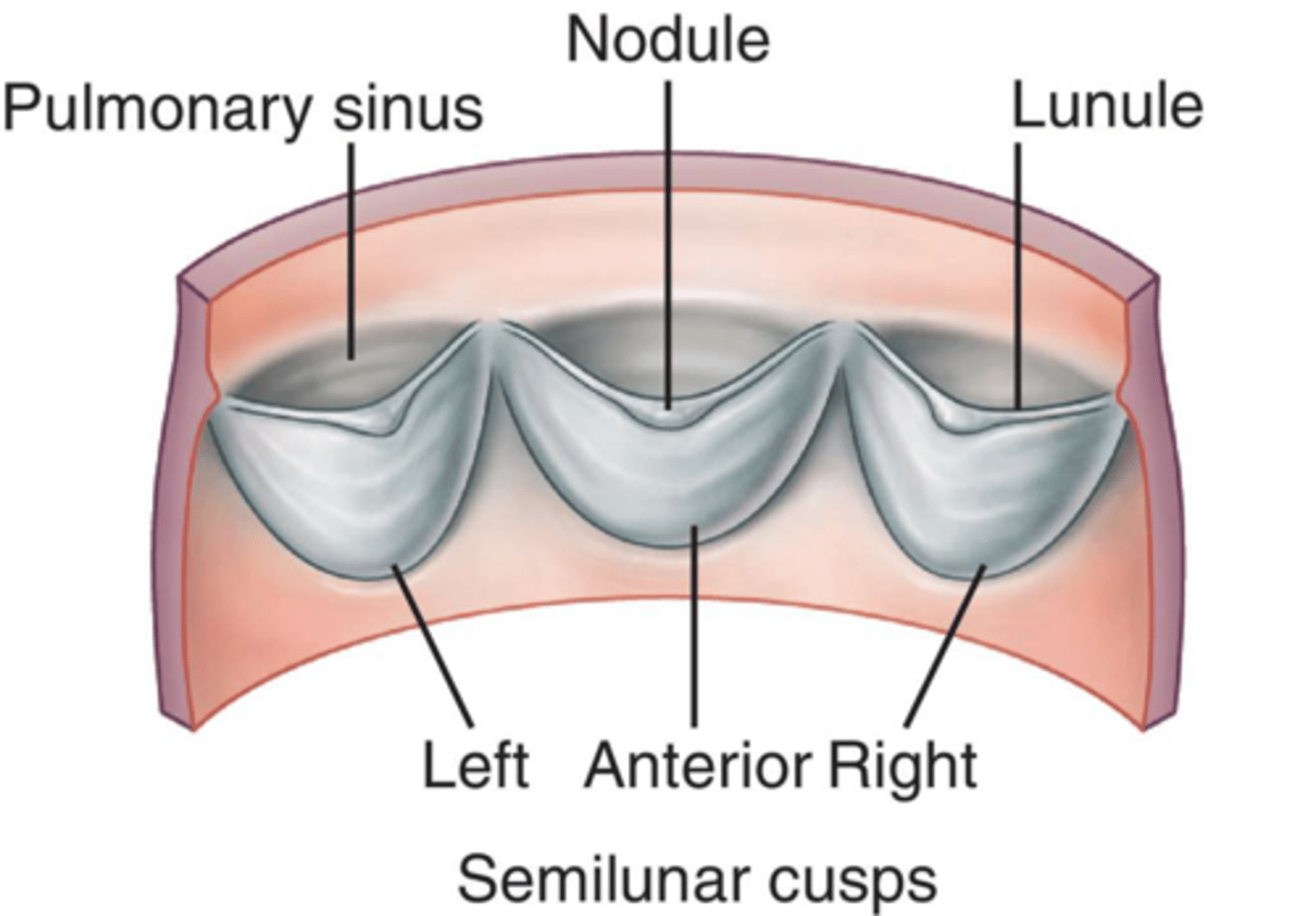
what are the sinuses of the semilunar valve?
spaces between the cusps and wall of the vessels (each named for its corresponding cusp)
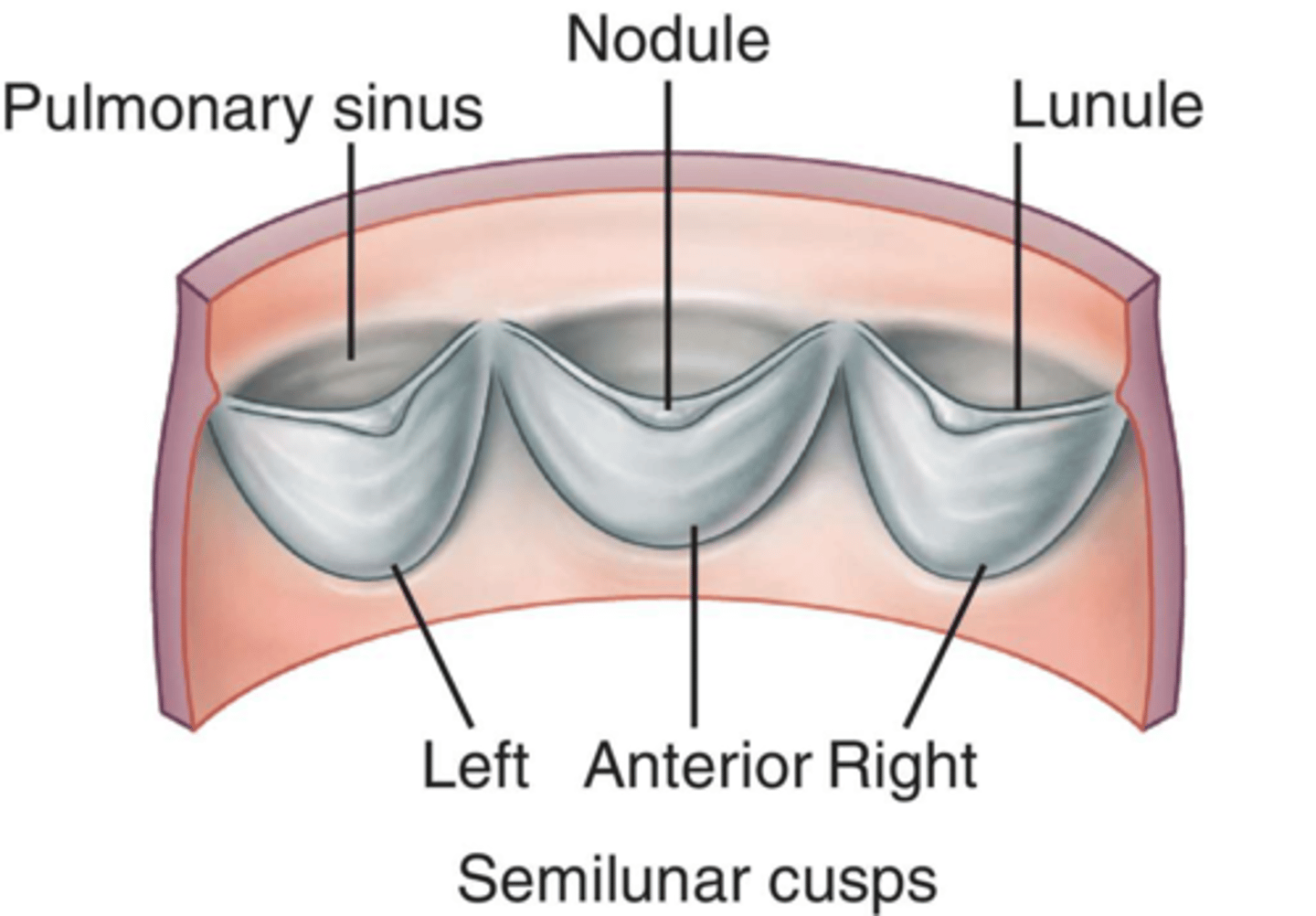
how many flaps in the pulmonary semilunar valve?
3
how many sinuses between the cusps and wall of the pulmonary trunk?
3
when does the pulmonary semilunar valve open?
when blood is flowing from the right ventricle into the pulmonary trunk
when does the pulmonary semilunar valve close?
if the flow (between the right ventricle and pulmonary trunk) is RETROGRADE
what is the pulmonary circulation?
blood flows through the pulmonary trunk --> right and left pulmonary arteries --> right and left lungs
what happens to blood in the lungs?
blood gives off carbon dioxide and receives oxygen
how does oxygenated blood return to the heart?
through four pulmonary veins (2 from the right lung and 2 from the left lung)
what do the four pulmonary veins do?
carry the oxygenated blood into the left atrium
where is the musculi pectinati located within the left heart?
left auricle ONLY
what is the main part of the (internal) left atrium taken up by?
four separate openings for the pulmonary veins
what is the only other opening (besides the openings for the pulm veins) of the left atrium?
left atrioventricular opening
what is the AV valve of the left heart?
mitraL (bicuspid valve)
what is the left atrioventricular opening guarded by?
the left AV valve (aka mitraL aka bicuspid)
because the mitral valve is an AV valve, what does it contain?
cusps (2 - bicuspid), chordae tendinae, and papillary muscles
what does the chordae tendinae do?
connects supporting papillary muscle to the cusps
how does blood flow from the left atrium into the left ventricle?
through the bicuspid (mitral) valve
what does the left ventricle contain?
like the right ventricle, the left ventricle contains trabeculae carneae (internal muscular ridge-like folds)
what is a type of trabeculae carneae in the left ventricle?
papillary muscles of the bicuspid valve
what is one major difference between the right and left ventricles?
THICKNESS of the wall
which ventricle wall is thicker & why?
left ventricle wall is usually more than twice as thick
- RV only pumps blood to ONE organ--> the lungs
- LV pumps to every other organ in the body--> thus it performs more work and is larger
what are the only openings of the left ventricle?
-left atrioventricular opening
-opening of the aorta
what happens when the left ventricle contracts?
blood leaves & enters the aorta
what is the opening of the aorta guarded by?
aortic semilunar valve
where is the aortic semilunar valve located?
opening of the aorta
what does the aortic semilunar valve possess?
3 cusps; these cusps possess nodules and lunulae (like the pulmonary semilunars)
what are the aortic sinuses?
spaces between the cusps and wall of the aorta
how does the aortic sinus differ from the pulmonary sinus?
right aortic sinus is the site of origin for the right coronary artery and the left aortic sinus is the site of origin for the left coronary artery
what happens when blood leaves the left ventricle?
it enters the aorta and then passes to all parts of the body
in addition to the fossa ovalis of the RA, what else is a remnant of fetal circulation?
ligamentum arteriosum
what is the ligamentum arteriosum?
-remnant of fetal circulation
-chord-like band connecting the pulmonary trunk and aortic arch
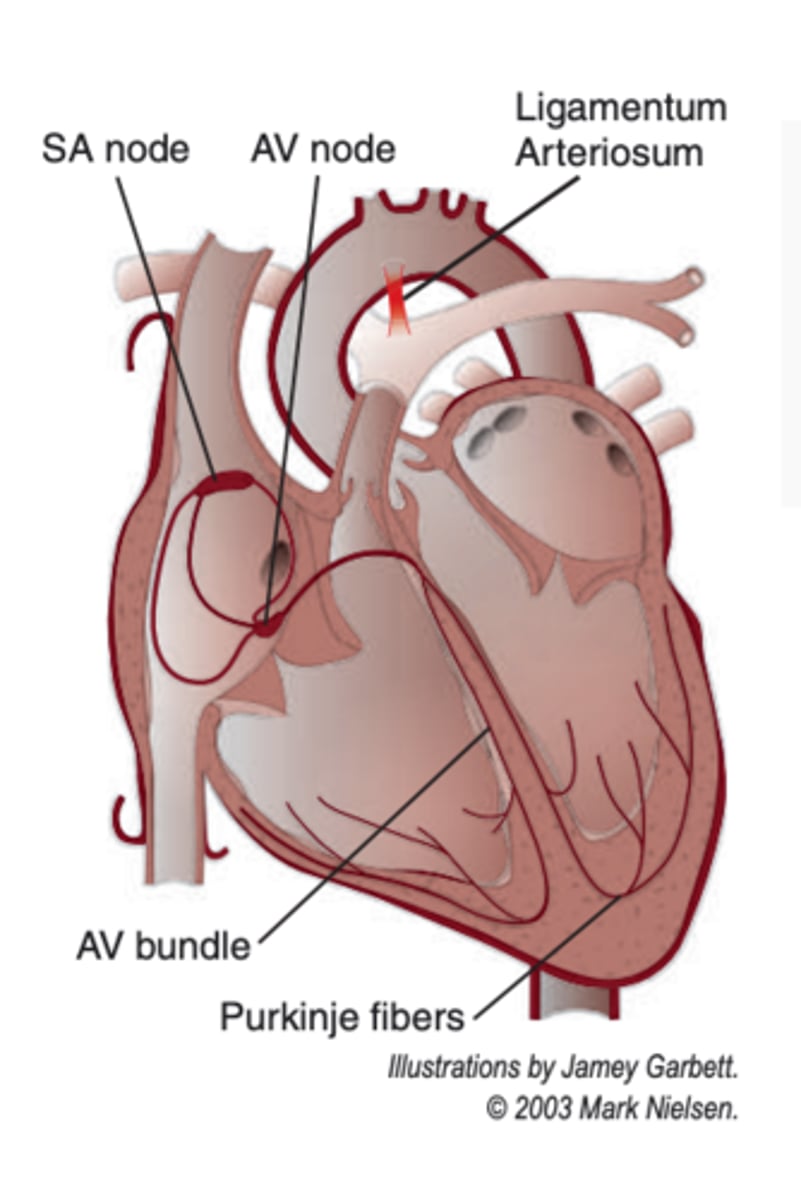
what is the conducting system of the heart?
system of the heart that is made of specialized muscle fibers that conduct impulses
how does the heart work efficiently?
when the conduction system carries out certain events of the heart in an orderly sequence