(L5) IMED2002 - Premature Red Blood Cell Destruction
1/36
There's no tags or description
Looks like no tags are added yet.
Name | Mastery | Learn | Test | Matching | Spaced | Call with Kai |
|---|
No analytics yet
Send a link to your students to track their progress
37 Terms
Iron is absorbed in the ___
Deficiency in adults is most commonly due to ___
Ferritin is the best test to measure
Iron is transported in the plasma by
B12 needs
- upper small bowel (duodenum and jejunum)
- blood loss or gastrointestinal lesion
- iron stores
- transferrin
- intrinsic factor to be absorbed (iron does not)
Physiology of RBC Destruction
- red cells destroyed by macrophages
DEGREDATION OF RED CELL COMPONENTS:
- globin degraded to amino acids
- Haem binds to haptoglobin and is degraded to protoporphyrin and metabolised to bilirubin
- Iron is released and recycled
.
- if red cells are destroyed in vessels (intravascular): Hb present in plasma of vessel
.
- someone with haemolytic anaemia may have elevated bilirubin
- extravascular means outside blood vessel
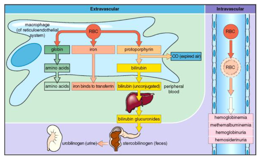
Biochemistry of Haemolysis
- Extravascular and Intravascular

Haemolytic Anaemia
- anaemia due to shortened RBC lifespan
Haemolysis: RBC survival is <120 days
- Usually bone marrow responds and increases erythropoiesis (driven by EPO stimulus)
- Red cell production can increase 6-8 times
.
Anaemia occurs when:
- Red cell survival is <15 days
- Haematinic deficiency (esp. folate)
- bone marrow disease
Red cell destruction
- increased bilirubin (haem breakdown)
- increased LDH
- reduced haptoglobins (Hb-haptoglobin complex)
.
- levels of haptoglobins are reduced because they are busy binding to Hb thats free from lysed cells
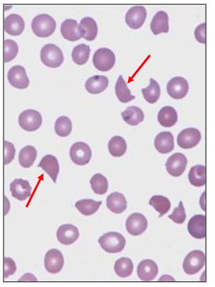
Evidence of damaged red cells
- spherocytes
- fragmented red cells (schistocytes)
.
- schistocytes look like a knife has cut the cell

Evidence of increased RBC production
- reticulocytosis (polychromasia)
- nucleated red blood cells

Laboratory Features of Haemolytic Anaemia - Extravascular
- Red cell lysis occurs outside the blood vessels
- Blood film: polychromasia; altered RBC shape
- Lactate dehydrogenase (LDH) elevated
- Bilirubin elevated

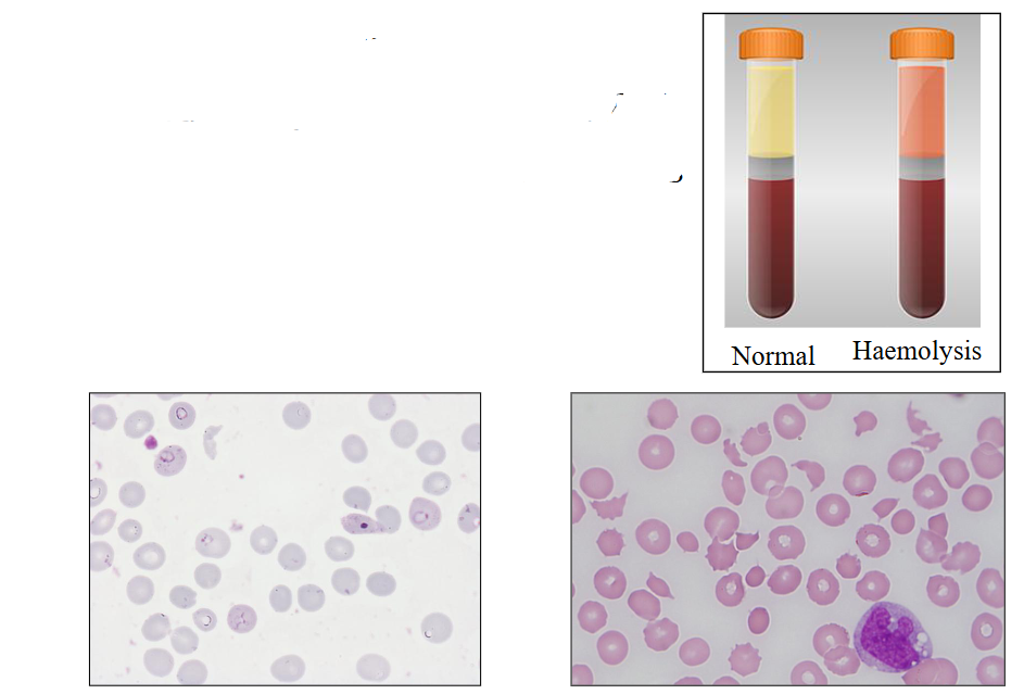
Laboratory Features of Haemolytic Anaemia - Intravascular
- Red cell lysis occurs inside BV (blood vessel)
- Blood film: fragmented red cells
- Haptoglobin low
- Plasma Hb
- Haemosiderin in urine
.
- plasma looks red because there is Hb in the plasma (which is red)
Haemosiderin (or hemosiderin) is an iron-storage complex formed when red blood cells break down, releasing hemoglobin which macrophages then convert into insoluble, iron-rich pigments
Clinical Features of Haemolytic Anaemia
- anaemia
- Jaundice: bilirubin in plasma
- Pigment gallstones (composed primarily of bilirubin) may occur
- Splenomegaly (common)
- Ankle ulcers: sickle cell anaemia
- Expanded bone marrow
- aplastic crises: parvovirus
- Megaloblastic anaemia: folate deficiency
Causes of Haemolytic Anaemia (Hereditary and Acquired)
HEREDITARY:
a) Membrane defect: Hereditary spherocytosis
b) Enzyme defect: G6PD and PK defeciencies
c) Globin chain defects: haemoglobinopathies
.
ACQUIRED:
a) Immune haemolytic anaemia
b) Fragmentation haemolysis
c) Oxidative haemolysis
d) Other: including;
- Liver disease (spur cell anaemia)
- Infections, renal disease
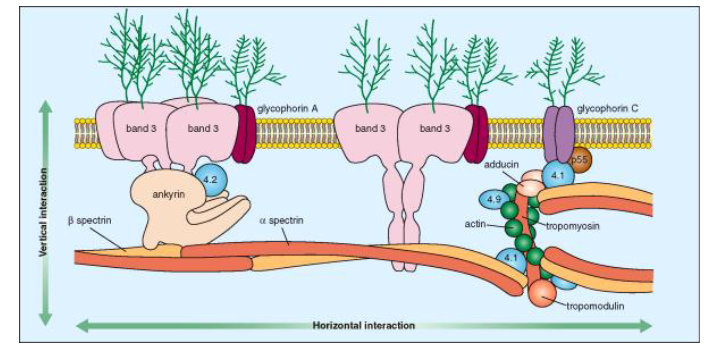
a) Red Blood Cell Membrane
- must be deformable and stabile
- Phospholipid bilayer with embedded proteins
Membrane proteins (52%):
- peripheral: membrane elasticity (spectrin, actin, protein 4.1)
- integral: embedded with lipids (Band 3, glycophorin)

Red Cell Membrane Defects
- Abnormal RBC membrane deformabilty
- Reduced 120-day life span
- Determined by phospholipid and membrane proteins
- Mutations in genes of membrane skeletal proteins
INHERITED DISORDERS:
- Hereditary spherocytosis
- Hereditary elliptocytosis
- Hereditary ovalocytosis
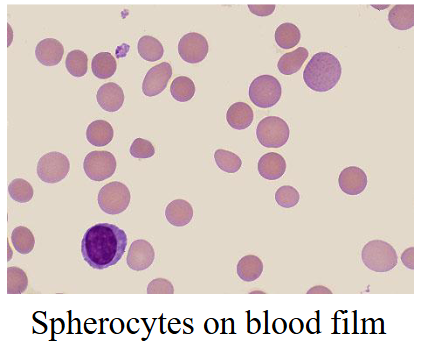
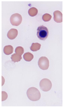
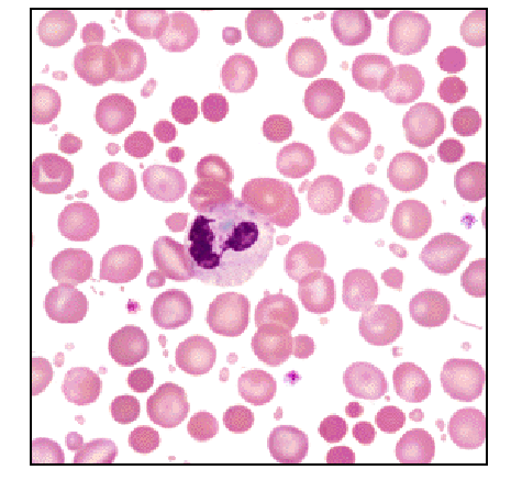
Hereditary Spherocytosis
- Most common inherited haemolytic anaemia
- Autosomal dominant with variable severity
- Defect in a structural RBC membrane protein: Spectrin; ankyrin; band 3
- RBC less deformable and lose membrane when passing through spleen
- Red cells become spherical, rigid and then destroyed
.
- in this case there was no central pallor
- Central pallor refers to the pale-staining, white center of a normal red blood cell (erythrocyte) seen on a blood smear, occupying about one-third of its diameter

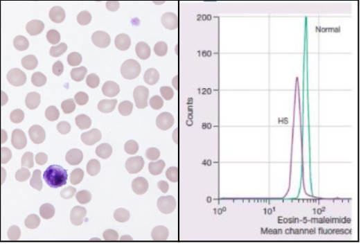
Hereditary Spherocytosis Lab Features and Treatment
- Fluctuating anaemia and jaundice
- Splenomegaly and (pigment) gall stones
LABORATORY FEATURES:
- Spherocytes on blood film
- Polychromasia due to increased reticulocytes
- Negative DAT
- Positive EMA (band-3)
.
TREATMENT:
- splenectomy
- Folic acid
- Cholecystectomy

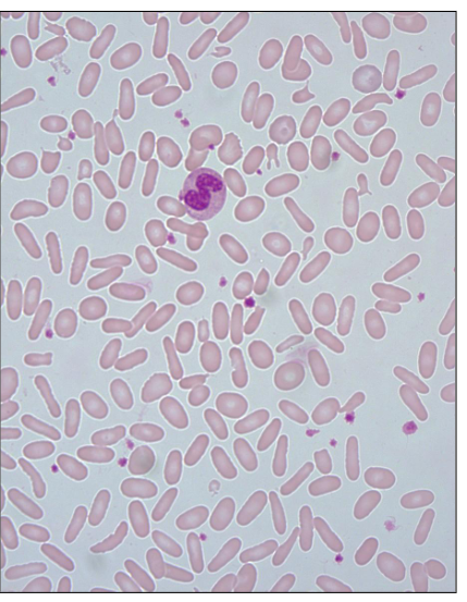
Hereditary Elliptocytosis
- autosomal dominant
- milder then Hereditary spherocytosis
- Many asymptomatic
- Mutations in spectrin
- 10% have haemolysis
- Elliptical shaped red cells
VARIANTS:
- Hereditary pyro-poikilocytosis
- South-east asian ovalocytosis


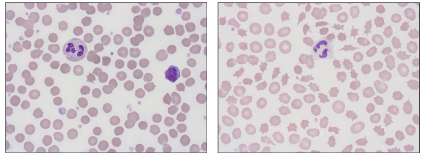
Red Cell Enzyme Defects
- Inherited genetic defects in red cell enzymes
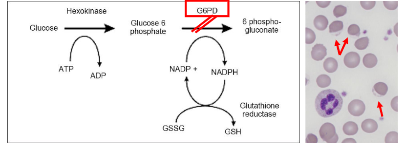
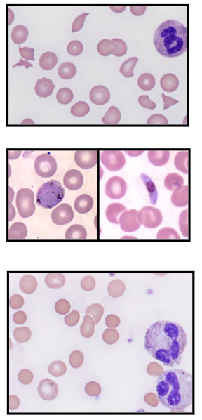
Most common: Glucose-6-phosphate dehydrogenase
- Enzyme in the hexose monophosphate shunt which generates reducing power as NADPH
- G6PD deficiency: most common red cell enzyme disorder worldwide (>200 million affected)
- Gene on X chromosome (X-linked): males affected
.
- on the right diagram, there are a couple of cells with no staining on the bottom (bite cells)
- thats because when the cell was placed in oxidative stress (ROS)(Free radicals), the haemoglobin becomes oxidised and moves there, hence it has that cap

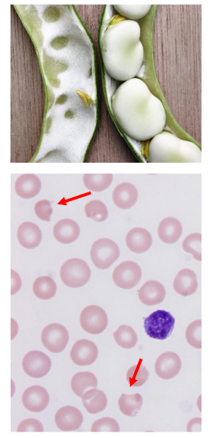
G6PD Deficiency
- X-linked inheritance; 400 million affected
- RBC susceptible to oxidant stress
- Triggers of oxidant haemolysis: drugs, fava (broad) beans, infection, hypoxia
- Abrupt: jaundice, dark urine; lumbar pain
- Intravascular haemolysis (self-limiting)
- Oxidised Hb removed; "bite"/blister cells
- Rx: Remove/stop/treat offending agent; treat infection; transfuse if necessary
- Normal blood count between crises
- Relative resistance to malaria
.
- in the diagram, the Hb part of the cell has been bittne (because spleen doesnt let it pass), hence it looks weird
- before its bitten its called blister cell
- after being bitten its called bite cells
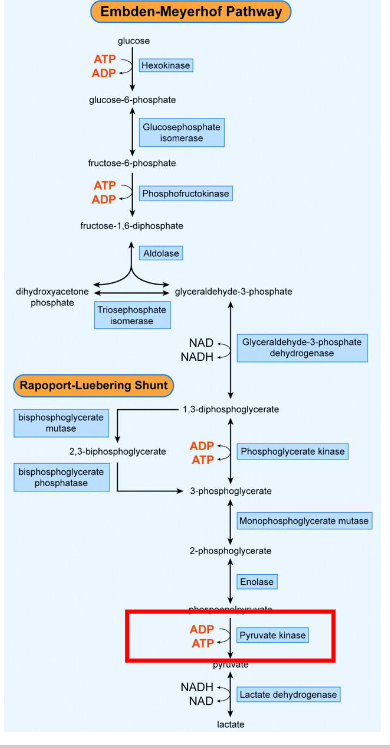
Pyruvate Kinase
- Glucose is metabolised in RBCs by anaerobic glycolysis
- Embden-Meyerhof Pathway
- Pyruvate kinase is the enzyme that catalyses the final step of glycolysis and is required to make ATP
.
PYRUVATE KINASE DEFICIENCY:
- Inherited enzyme defect
- Lack of PK: insufficient ATP made
- Rigid cell membrane and cell death
- Haemolytic anaemia
- Splenectomy partly improves anaemia

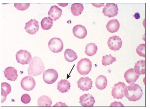
Pyruvate Kinase Deficiency
- autosomal recessive inheritance
Variable clinical presentation
- Mild: ocassional compensated haemolysis
- Severe: can present in neonatal period with failure to thrive, splenomegaly
.
- prickle-shaped RBC'
DIAGNOSIS:
- Blood film
- Pyruvate Kinase Assay

Immune Haemolytic Anaemia
- haemolysis due to antibodies directed at RBC
.
AUTO-IMMUNE HAEMOLYTIC ANAEMIA:
- Auto-antibody directed at own red blood cells (self)
- Causes: idiopathic/unknown (60%); B cell lymphoma
.
ALLO-IMMUNE HAEMOLYTIC ANAEMIA
- antibody made by one individual reacts with the RBC of another individual (transfusion; newborns)
.
DRUGS:
- Antibody against a drug-RBC complex; antibiotics

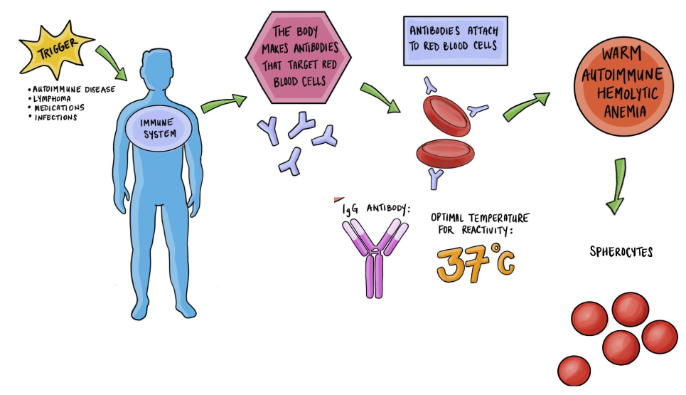
Immune Haemolytic Anaemia
- there is some sort of trigger
- trigger activates immune system - body makes antibodies
- antibodies target red cells
- antibody coated red cells will go to spleen
- spleen doesnt allow it through, binds to the FC receptor on the antibody and the red cells are either removed totally or partially damaged, resulting in the formation of spherocytes
.
- we can identify antibody coated red cells using the direct anti-globulin test, and this would be positive in immune haemolytic anaemia where its negative in patients with hereditary spherocytosis
Auto-immune Haemolytic Anaemia
- an antibody directed against its own red blood cells
- Body destroys its own red blood cells ("auto")
- Causes: unknown, leukaemia; auto-immune syndromes
- Blood film: spherocytes and polychromasia
- Positive Direct Antiglobulin Test (DAT)
- Bone marrow: erythroid hyperplasia (response)
.
TREATMENT:
- Suppress the immune system (corticosteroids)
- Treat the cause (e.g leukaemia)
- Remove the spleen (site of cell destruction)
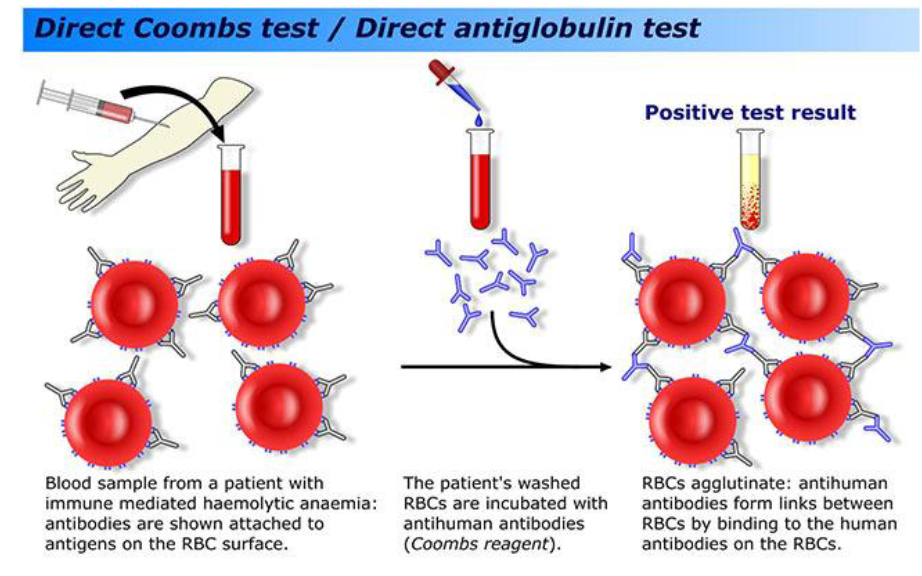
Direct Antiglobulin Test (DAT)
- test for antibodies coating red blood cells

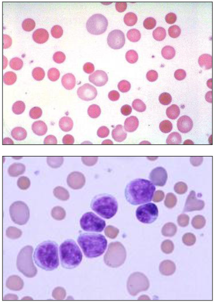
Autoimmune Haemolytic Anaemia
Haematology of AIHA
- Spherocytes: no central pallor due to loss of membrane; small and dark Hb
- Polychromasia (increased reticulocytes)
- Nucleated red cells: (BM response to anaemia)
- Underlying disease (e.g leukaemia)

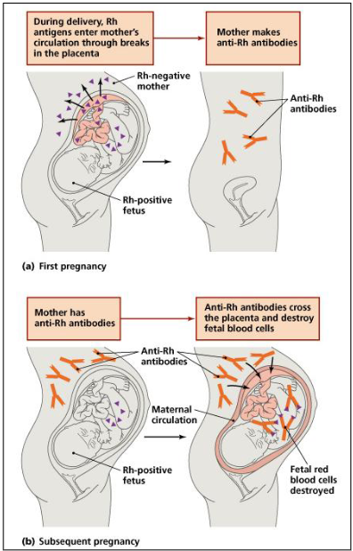
Haemolytic Disease of the Newborn
ALLO-IMMUNE HAEMOLYTIC ANAEMIA:
- Transplacental passage of maternal RBC antibodies that bind to fetal RBC
- Maternal antibodies destroy fetal RBC
.
Usually Rh(D) negative mother and Rh(D) positive fetus
- Anti-D made by mother in 1st pregnancy when exposed to D antigen
- Subsequent pregnancy affected
- Maternal anti-D crosses the placenta
- Anti-D binds to and destroys fetal Rh(D)-positive RBC
.
- D represnts a specific blood type. basically it menas that the mother naturally has anti something (e.g Anti A). If the fetus is A, then these Anti A travel through placenta and destory fetal blood cells

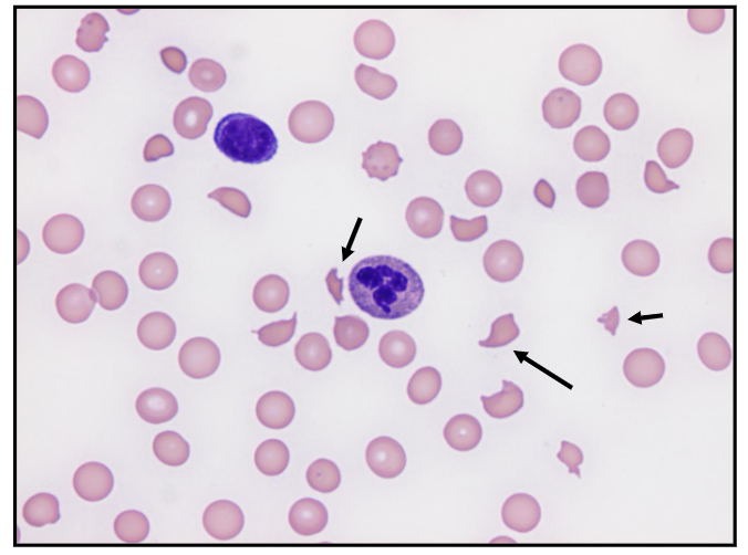
Red Cell Fragmentation
- mechanical red cell damage: red cell fragments; anaemia
.
- "Microangiopathic haemolytic anaemia"
- haemolytic anaemia means haemolysis of blood, premature red cell destruction
- micro means small, angio is vessel, pathic means pathology
- term doesnt really make sense but thats what we term fragmentation haemolysis
- there are a number of entities that can cause it
.
RED CELLS EXPOSED TO AN ABNORMAL SURFACE:
- Heart valve; pinhole lesion in heart
- Fibrin stands in the blood vessels
.
RED CELL FRAGMENTATION "SCHISTOCYTES"
Intravascular Haemolysis (Inside Blood vessel)
- Disseminated intravascular coagulation (DIC)
- Thrombotic Thrombocytopenic purpura
- Haemolytic uraemic syndrome
- Malignant hypertension
- Drugs; graft-versus-host disease

Sepsis can cause haemolytic anaemia
- Fragmentation haemolytic anaemia: E. coli
- haemolytic uraemic syndrome
- haemolytic means haemolysis
- uraemic means kidney failure

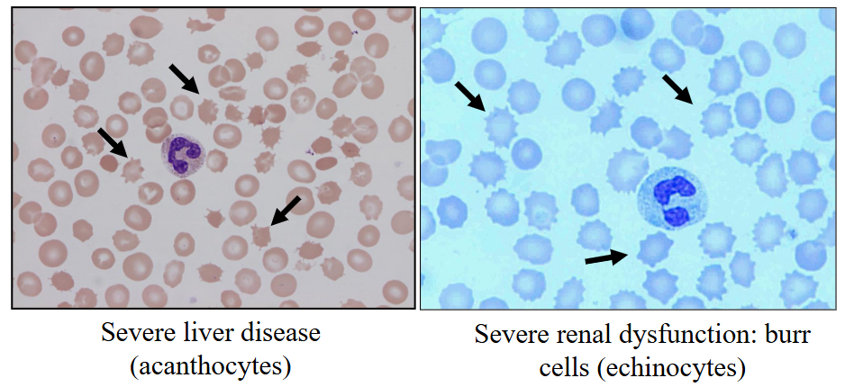
Other Causes of Haemolytic Anaemia
- Liver disease ("spur cell anaemia") and renal failure alter the RBC membrane
- Infections: can be inside RBC (e.g malaria)


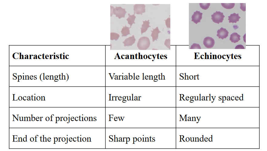
Alcohol and Red Cells
- alcoholic liver disease causes "acanthocytes"
- Renal failure causes "echinocytes"
Severe Burns and Haemolytic Anaemia
- Burns: small, contracted red blood cells
- Heat-damage to the red blood cell membrane

Sepsis
Sepsis is a life-threatening medical emergency occurring when the body's immune response to an infection triggers an extreme, cascading chain reaction, damaging its own tissues and organs
Infections and Haemolytic Anaemia
SEVERE BACTERIAL SEPSIS:
- Dissem. intravascular coagulation; fragmented red cells (MAHA)
.
MALARIA (plasmodium sp.)
- P.vivax, falciparum, ovale, malarie
- Trophozoites; schizonts; gametocytes
.
CLOSTRIDIUM WELCHII:
- microspherocytes in PB film
.
order of images is the order of the subheadings

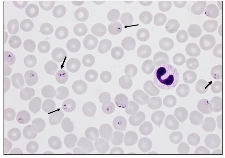
Malaria causes Haemolytic Anaemia
- Malaria (Plasmodium falciparum)
- Parasites inside RBC burst releasing Hb and dark urine


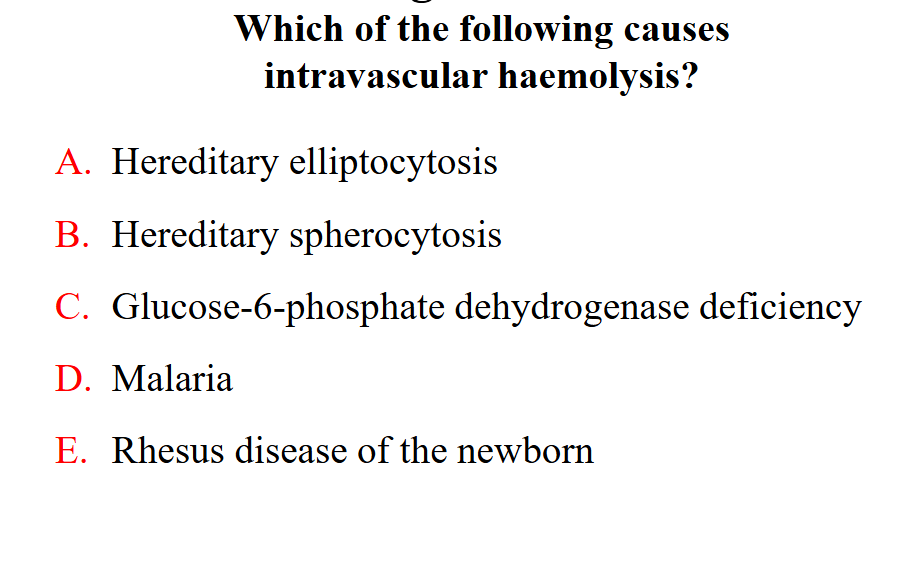
PUT THE QUESTION ON SLIDE 43
- answer is Malaria

Learning Outcomes
DIAGRAM ON SLIDE 44
Summary
DIAGRAM ON SLIDE 45
