locomotor overview
1/134
There's no tags or description
Looks like no tags are added yet.
Name | Mastery | Learn | Test | Matching | Spaced | Call with Kai |
|---|
No analytics yet
Send a link to your students to track their progress
135 Terms
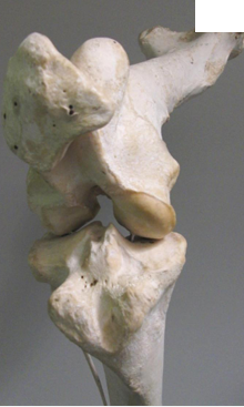
synovial joint
most common
most moveable
articulating bones separated by fluid filled spce
joint space surrounded by synovial membrane which is strengthened bya fibrous joint capsule
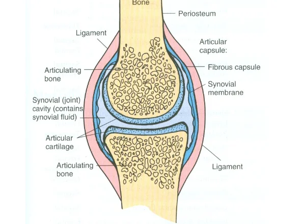
articular (hyaline) cartilage
interface between bones at a synovial joint
lubrication
shock absorption - stiff to compression
load transmission
no nerves or blood vessels
synovial fluid
secreted by synovial membrane
clear/straw coloured viscous fluid
contains hyaluronic acid
lubrication
shock absorption
nutrient and waste transportation

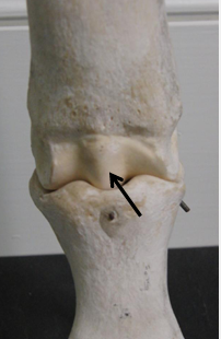
menisci
shape
where are they located
function
medial and lateral are c shaped fibrocartilage rings located within knee joint
deepens articular surface of tibia- increases joint stability
act as shock absorber- increase SA to dissopate force
bursae
structure and what does it contain
function
sac like structure containing small amount synovial fluid
decrease friction between tendon, skin and bones
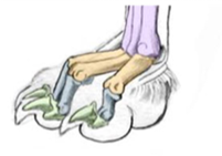
tendon sheath and clinical importance
-like bursae but wrap around tendon where they pass over joints
especially important clinically in horse
blood and nerve supply of
articular cartilage
free sensory fibres from where
what kind of fibres to blood
what kind of fibres from blood vessels
what kind of fibres from joint capsule
articular cartilage is avascular
blood vessels supply epiphysis and joint capsule/ synovial membrane
nerves for pain ,reflex, posture, and locomotion
1. free sensory fibres from joint capsule and synovial membrane
efferent fibres to blood vessles
sensory fibres from blood vessels
proprioceptive fibres from joint capsule
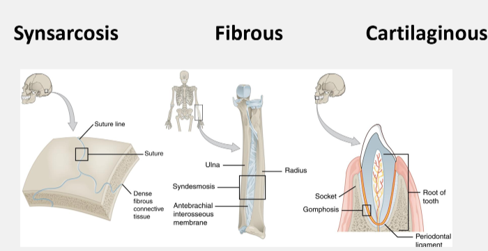
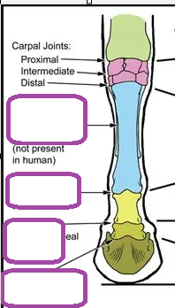
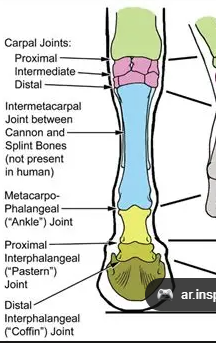
other types of joint
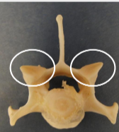
planar
articular surfaces
movement
rotation?
examples
flat or slightly curved articular surfaces
gliding
no rotation
carpal/tarsal
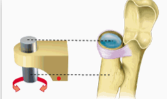
pivot
rounded end of one bone fits into ring of another
rotation
eg proximal radioulnar or antlantoaxial
hinge
slightly rounded eg of one bone fits into slightly hollow of another
one bone moves, the other is stationary
elbow
equine MCP
condylar
oval convex surface fits into hollow
angular movement
biaxial
femoro tibial, radiocarpal
saddle
2 surfaces
convex in one direction
concave in the other at right angles to the first
eg DIP (distal interphalangeal) joint
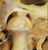
ball and socket
greates range of motion
eg hip

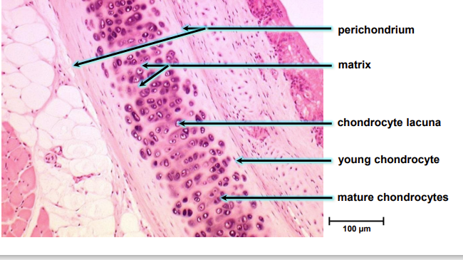
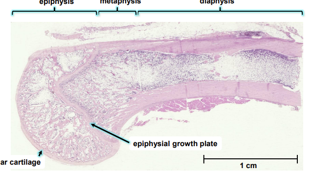
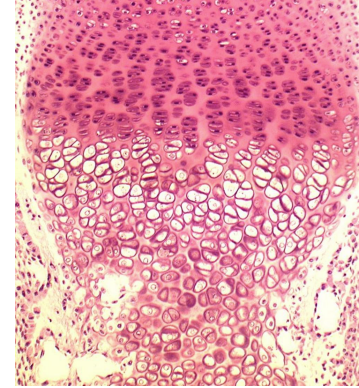
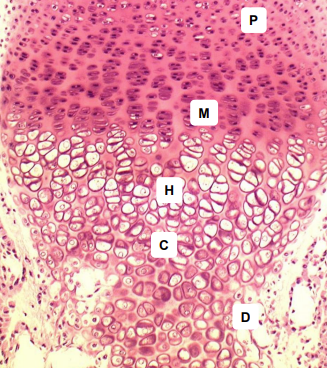
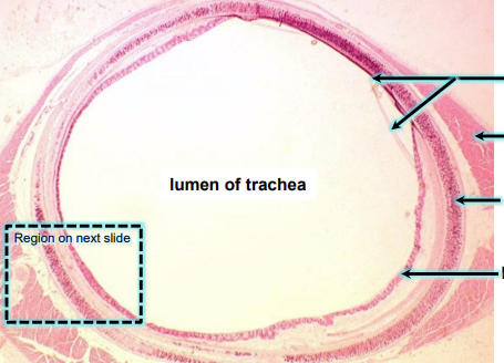
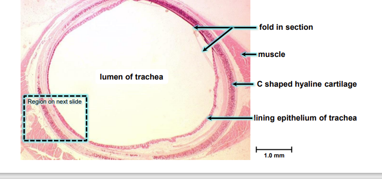
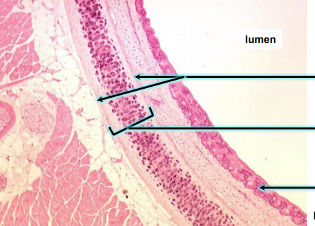
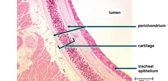
joints
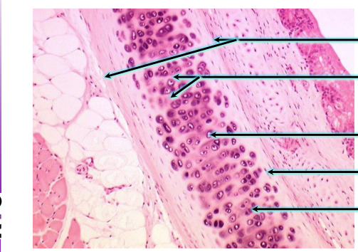
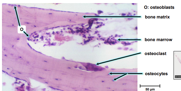
why are osteoprogenitor cells hard to see
they are difficult to distinguish from the surrounding connective tissue
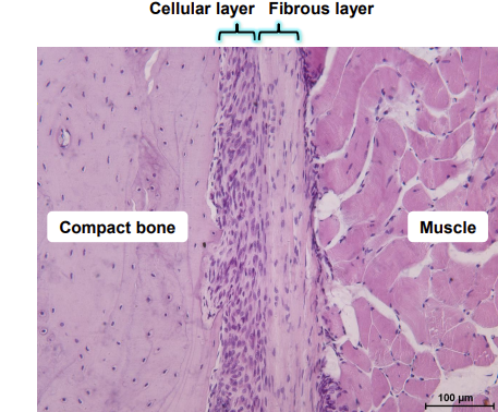
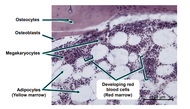
how do oesteoblasts appear
closely arranged in a dense single layer of cells covering the bone surface
where bone formation is active there may be several layers
osteocyte shape
where does it reside
feature of mature osteocyte
star which reside in lacunae
mature osteocyte contains single nucleus
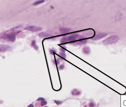
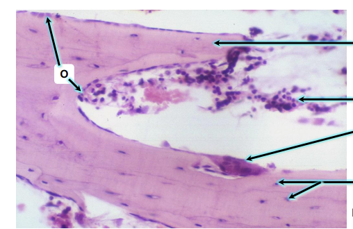
osteoclast shape
very large up to 100um
multi nucleated
attach themselves to the bone matrix and sit within deep indentations of the bone matrix that are formed by their activity:resorption bays or howship lacunae
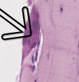
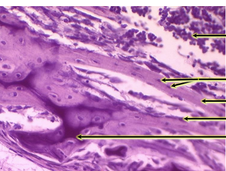
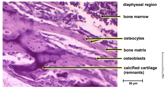
osteoclast
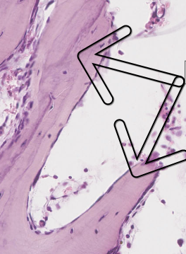
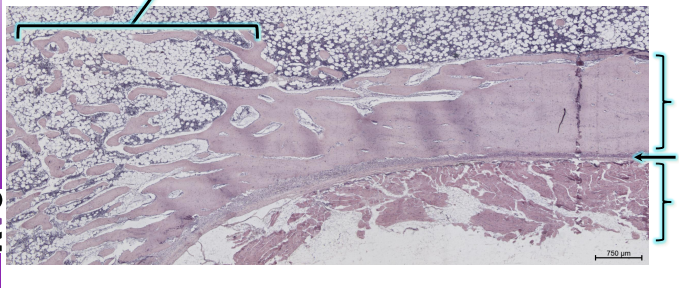
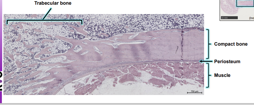
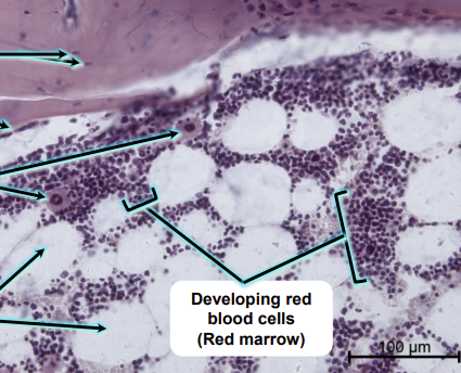
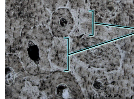
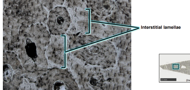
trabeculae
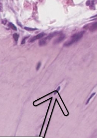
osteocyte in matrix
osteoblast
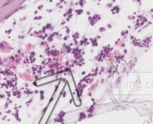
bone marrow
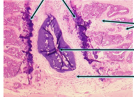
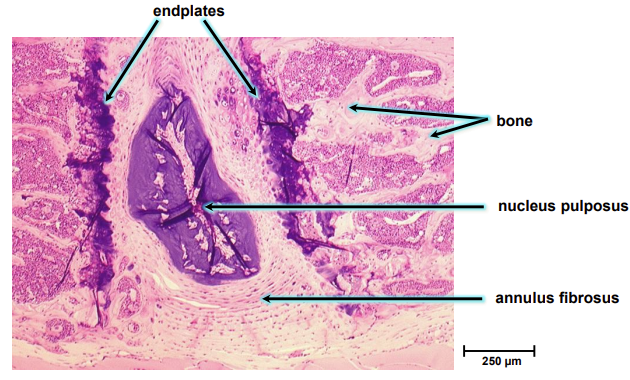
which collagen type predominates the annulus fibrosus
collagen type i
why is the cytoplasm of the osteoblast basophilic
because of the well developed rer
characteristic of cell specialised in the secretion of proteins
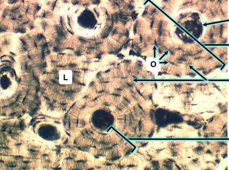
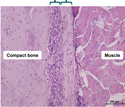
where are the volkmann
where are the bone lining cells found
on quiescent bone surfaces
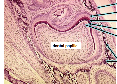
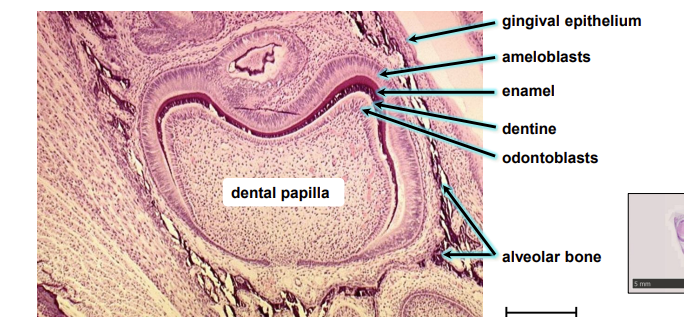
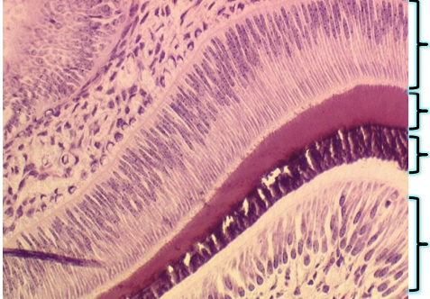
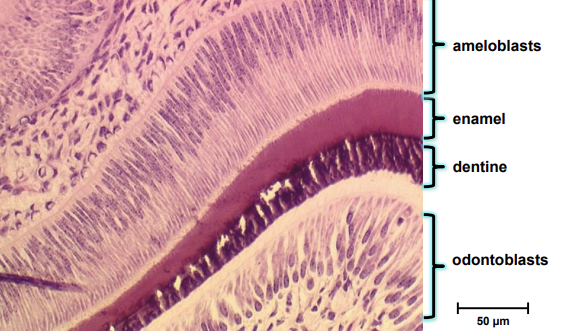
tooth
aperneurosis
flat sheet of dense connective tissue connecting muscle to bones/ fascia
epimysium
dense irregular connective tissue surrounding each muscle protecting it from friction
fasicle
a bundle of muscle fibers
perimysium
connective tissue surrounding a bundle of muscle fibres
endomysium
surrounds individual muscle fibres
muscle belly
fleshy, thickest part of the muscle, is encased in the epimysium
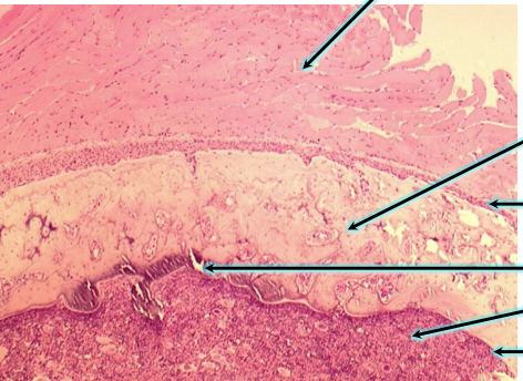
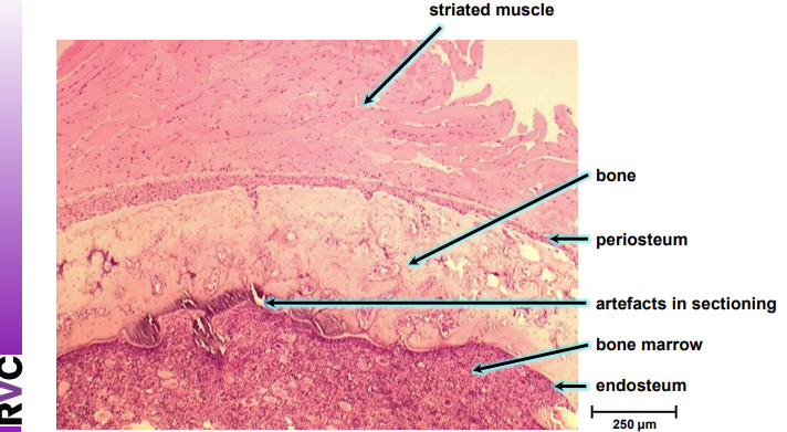
skeletal muscle structure
multiple peripheral nuclei
voluntary
striated
regular parallel bundles
outermost layer surrounded by epimysium
cardiac muscle structure
striated
single central nucleus
involuntary
irregular arrangement
intercalated disks
intercalated disks have extensive gap junctions allowing cell to cell communication
smooth muscle contraction
not striated
single nucleus
involuntary
longer contractions
overlapping sheets of spindle shaped cells
microscopically appear homogenous
connected through end to end junctions called gap junctions creating a watertight seal
function of skeletal muscle
voluntary movement of skeleton controlled by somatic nervous system
maintain body position and posture
stabilise joints
support underlying organs and soft tissue
store nutrient reserves
maintain correc body temperature
smooth muscle function
involuntary contrction controlled by autonomic nervous system
lines inner wall of vasculature, hollow visceral organs, major bodily tracts
regulate blood pressure by altering systemic vascular resistance
peristalsis
regulate bodily secretion
lines respiratory tract
iris controls light entering
hair follicles
what is muscle architecture
the arrangement of muscle fibres relative to the axis of force
maximum force developed by muscle is proportional to the number of sarcoeres hence fibre length
pennate muscle
short fibres at an angle to internal tendon/aperneurosis
increases PCSA
PSCA is directly proportional to force
short fibres mean less contraction distance so it is economical
however trade off with speed
parallel muscles
fibres run parallel to line of pull of muscle
more sarcomeres in series mean more total muscle fibre shortening so more work
work= force x distance
moves joints through a large range of motion
speed= distance/time
roles of tendon
minimise distal limb mass
join muscle to bone
store elastic energy
conserve energy
power amplification: stretched tendons recoil faster than muscle shortens so more power. only a small amount of work is done but in a shorter time so power output is higher
power= rate of doing work
tendon structure
tenoblasts and tenocytes
chondrocytes, synovial cells and vascular cells
tendon collagen fibres are in a crimped pattern
collagen fibrils- collagen fibres- fascicles (surrounded by endotenon)
fascicles are bound together by the endotenon a dense irregular connective tissue sheath to form the tendon
type i
slow oxidative
low myosin ATPase activity
high oxidative capacity
smaller diameter
fatigue resistant
less force production
steady fatigue curve
type iia
fast oxidative glycolytic
high myosin ATPase activity
high oxidative AND glycolytic capacity
type iib
fast glycolytic
high myosin ATPase activity
high glycolytic capacity
larger diameter (stronger) fatigue easily
what can fibre types be influenced by
genetics
training
age
lifestyle
diet
isometric contraction
muscle contracts but does not change length
produces force but it is equal to resistance
eg holding something
concentric contraction
shortens as it generates force
force greater than resistance
movement
eccentric contraction
muscle lengthens under tension
lowering
high force and low energy
eg control or resist flexion of the elbow caused by the ground reaction force during landing impact
lactertus fibrosis
horse
Biceps stretches during stance when carpus locked in extension
– LF stores ELASTIC ENERGY
– When carpus buckles in late stance this rapidly releases the stored energy
– The leg swings forward
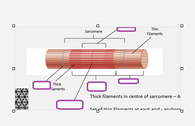
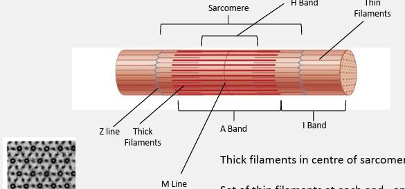
myosin
thick filaments
polypeptide chains
2 globular heads and a long tail
heads are the site of myosin ATP enzyme
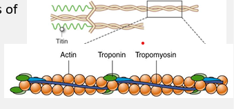
thin filaments
2 intertwned chains of actin molecules plus
tropononin- small globular protein bound to actin and tropomyosin
tropomyosin- rod shaped, located end to end along thin filament
sliding filament theory
tropononin controls position of tropomyosin on the thin filament. myosin cant bind with tropomyosin on its binding site
acetylcholine diffuses from neuron and ca ions are released into sarcoplasm and bind to troponin
calcium acts on tropomyosin and the myosin binding site is exposed
myosin head binds to actin at newly exposed site. pi and adp are released
thin filament moves in the direction of its negative end because the myosin head is firmly attached to the thin filament during its power stroke
the two heads of each myosin molecule work independently. only one head attaches to actin at a given time
myosin head and atp bind. myosin head detaches from the thin filament
atp is hhydrilysed
events during a muscle contraction
resting state- troponin controls the position of tropomyosin on the thin filament- here tropomyosin blocks the myosin binding site on the actin molecules
excitation contraction coupling- calcium ions bind to troponin which changes shape. this moves tropomyosin on the thin filament away from the myosin binding site
myosin heeads bind to actin on the thin filament. causes detachment of adp and phosphate molecules
power stroke: myosin heads move performin a power stroke which drags the thin filament towards the centre of the sarcomere
detachment- ATP binds to myosin causing it to lose affinity for actin and to detach from the actin binding site
atp is hydrolysed into adp and phosphate which reenergises myosin head to return to prevous poition
no calcim- resting state
if calcium is oresent then return to stage 3
what are the 3 functions of ATP in skeletal muscle contraction
energy released from atp hydrolysis re enrges the myosin head providing enery for cross bride movement and force generation
binding of atp to myosin causes the release of the myosin head from actin allowing repeated contractions
in the sacoplasmic reticulum ca-atpase hydrolyses atp in order to take ca ions back to the sr to end a muscle contraction
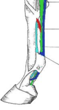
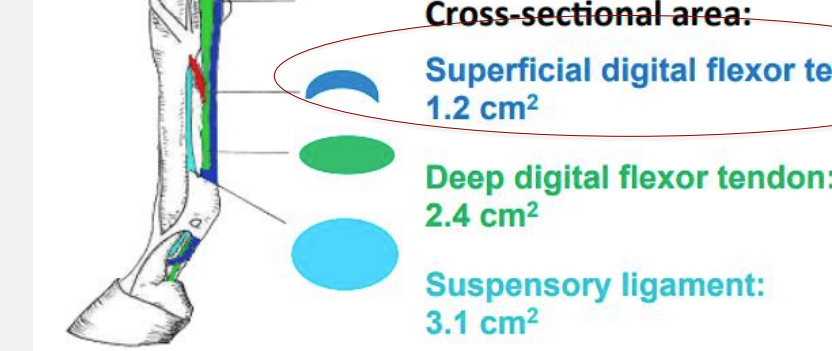
what is the mechanical safety factor
what is it
example
ratio of failure stress to typical stress experienced during locomotion
the superficial digital flexor tendon has a small cross section and experiences relatively high streses operating near its mechanicl limit
this is bcause it plays an important role in elastic energy cycling during locomotion
to fulfill its locomotor function it must operate at a low safety factor
what do extrinsic muscles do
hold scapula to thorax
what flex distal joints
carpal and digital flexors
what happens during stance in forelimb
withstand ground reaction force
stabilise joints
reisst over extension of joints
which muscles elevate and protract limb during swing
cranial extrinsic muscles- protracting limb
dorsal extrinsic- elevating
events during hindlimb stance
withstand ground reaction force
ressit over extension of joints
create propulsion
swing phase
protraction of limb
clearing foot away from ground
preparing limb/foot position for stance
how do GRF change with speed
increase
how do GRF alter during incline
forelimb vertical forces decrease uphill
hind limb peak vertical forces increase uphill
what are gaits based on
footfall patterns
biomechanical properties
PE and KE during
braking
midstance
propulsion
braking and propulsion PE low KE high
other way in midstance
basic structure of the ECM
fibroblasts in most connective tissue
GAG polysaccharide chains (repeating dissacharides)
GAGs which covently bond to proteins- proteoglycans
fibrous proteins such as collagen
proteoglycans form a gel like substance where fibrous proteins are embedded which resists compressive forces, allow diffusion
glucosaminoglycans
structure
branched or unbranched
hydorphobic or hydrophilic
how do they withstand compression
unbranched polysaccharide chains
repeating disaccharide chains
hydrophilic
porous gels (hydrated) filling up most of the extracellular space
attract cations so water in by osmosis- turgor- withstand compression
collagen
how many polypeptide chains
how many forms
triple stranded helical structure
3 polypeptide chains
29 forms
elastin
hydrophobic or hydrophilic
what is the precursor molecule
how does the precursor form elastin
highly hydrophobic protein
precursor is tropoelastin
tropoelastin is secreted into the extracellular space and assembled into elastic fibres close to the plasma membrane, which then cross link
coiled
stretch and recoil
tendon
collagen
proteoglycans
elastin
tenocytes
ligament
fibrocytes
ecm
lower collagen, more proteoglycan
elastin
cartilage
which collagen
which is the predominant proteoglycan
other molecules
how much percent of water
chondrocytes
type ii collagen mostly
proteoglycan predominantly chondrotin sulphate
hyaluoran
68 percent water
regional variation
ecm of bone
what gives rigidity and compressive strength and where is this deposited?
which gives tensile strength and elasticity
hydroxyapatite gives rigidity and compressive strength
this is deposited on collagen fibres
load bearing
inorganic
type i collagen gives tensile strength and flexibility, elasticy, structural organisation, organic
what is mechanical loading
ecm turnover is triggered by mechanical loading
involves cell signalling
loading increases synthesis of new ecm proteins and degrading enzymes
loading can alter the molecular conformation of proteins changing how enzymes bind and degrade (change in collagen type and organisation)
tissue properties influence how they degrade eg stiffness
wolffs law
increase in loading causes architecture of spongy bone to strengthen and cortical layer strengthening whilst decrease causes bones to weaken and bone tissue to be resorbed