A225 - IU - Block 4
1/211
There's no tags or description
Looks like no tags are added yet.
Name | Mastery | Learn | Test | Matching | Spaced | Call with Kai |
|---|
No analytics yet
Send a link to your students to track their progress
212 Terms
Heart
Muscular organ on the left side of the body.
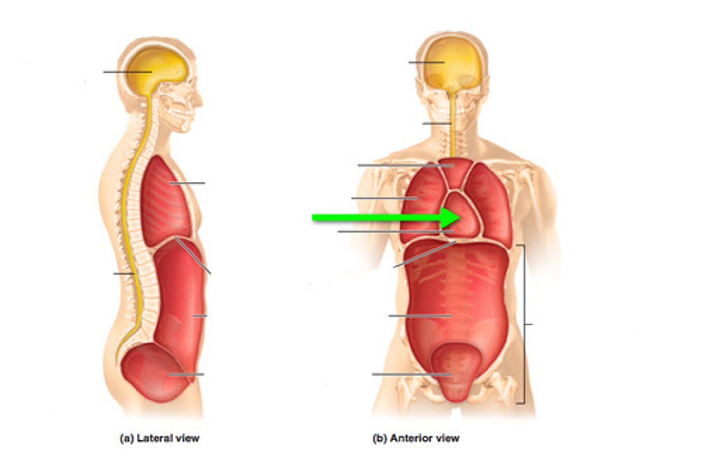
Thoracic cavity
Space housing the heart in mediastinum.
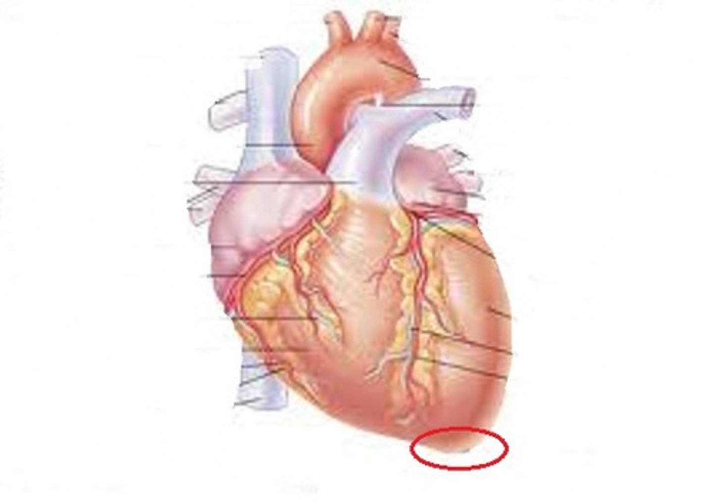
Apex of heart
Pointed tip of heart directing leftward.
Four chambers
Heart consists of two atria and two ventricles.
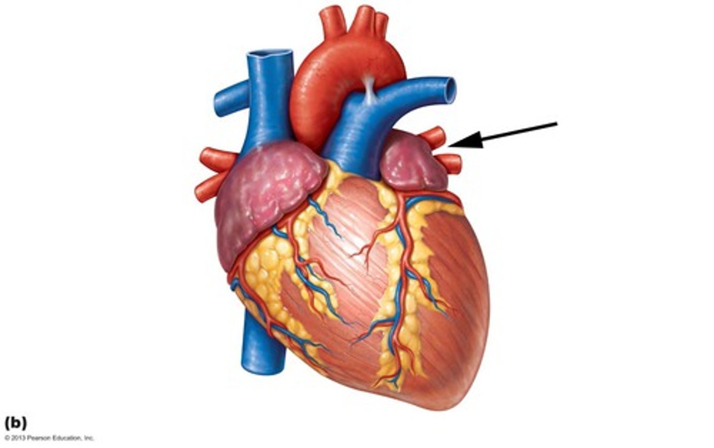
Right atrium
Chamber receiving deoxygenated blood from body.
Left atrium
Chamber receiving oxygenated blood from lungs.
Right ventricle
Pumps deoxygenated blood to the lungs.
Left ventricle
Pumps oxygenated blood to the body.
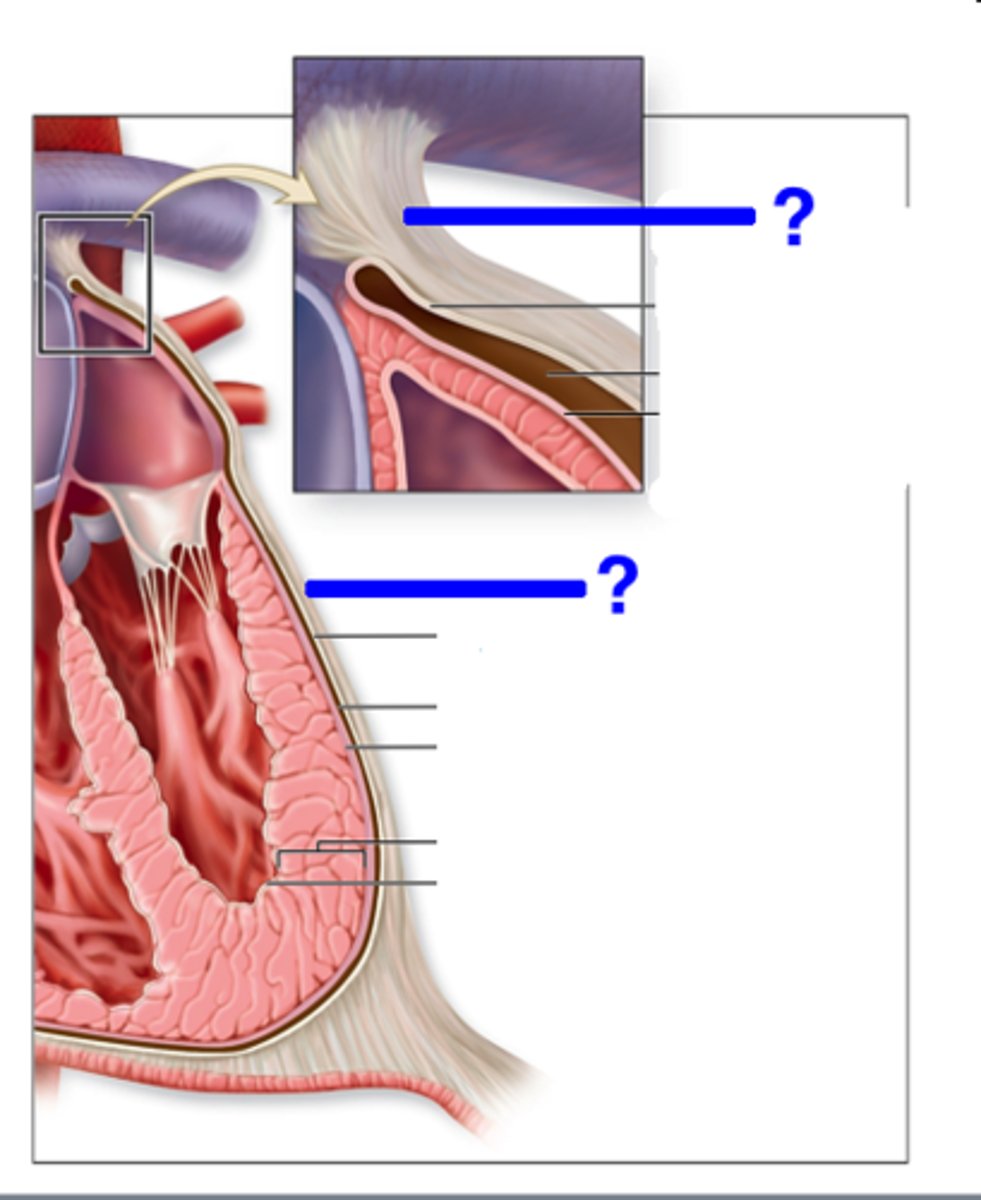
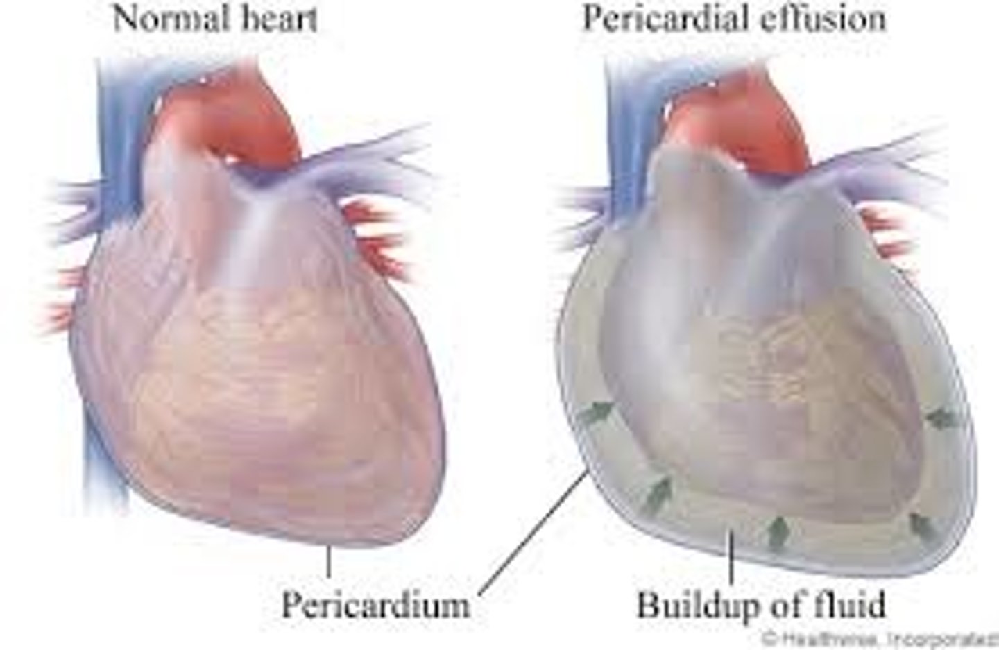
Pericardium
Double-walled sac enclosing the heart.
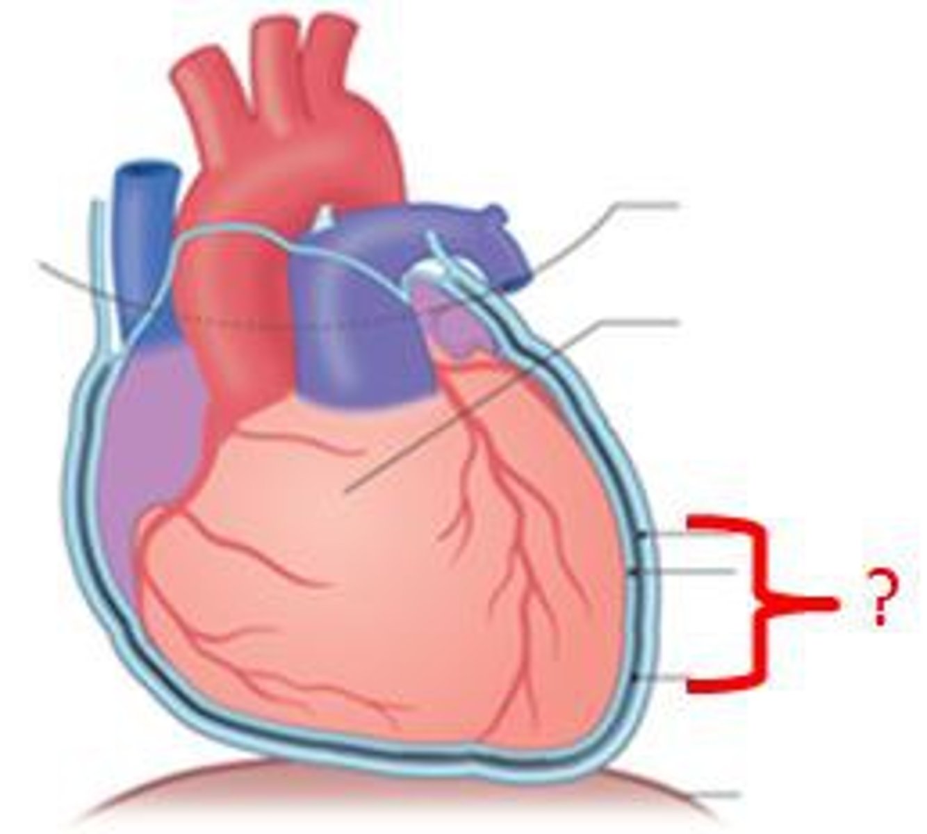
Fibrous pericardium
Outermost layer; prevents heart overfilling.
Serous pericardium
Inner layer with parietal and visceral components.
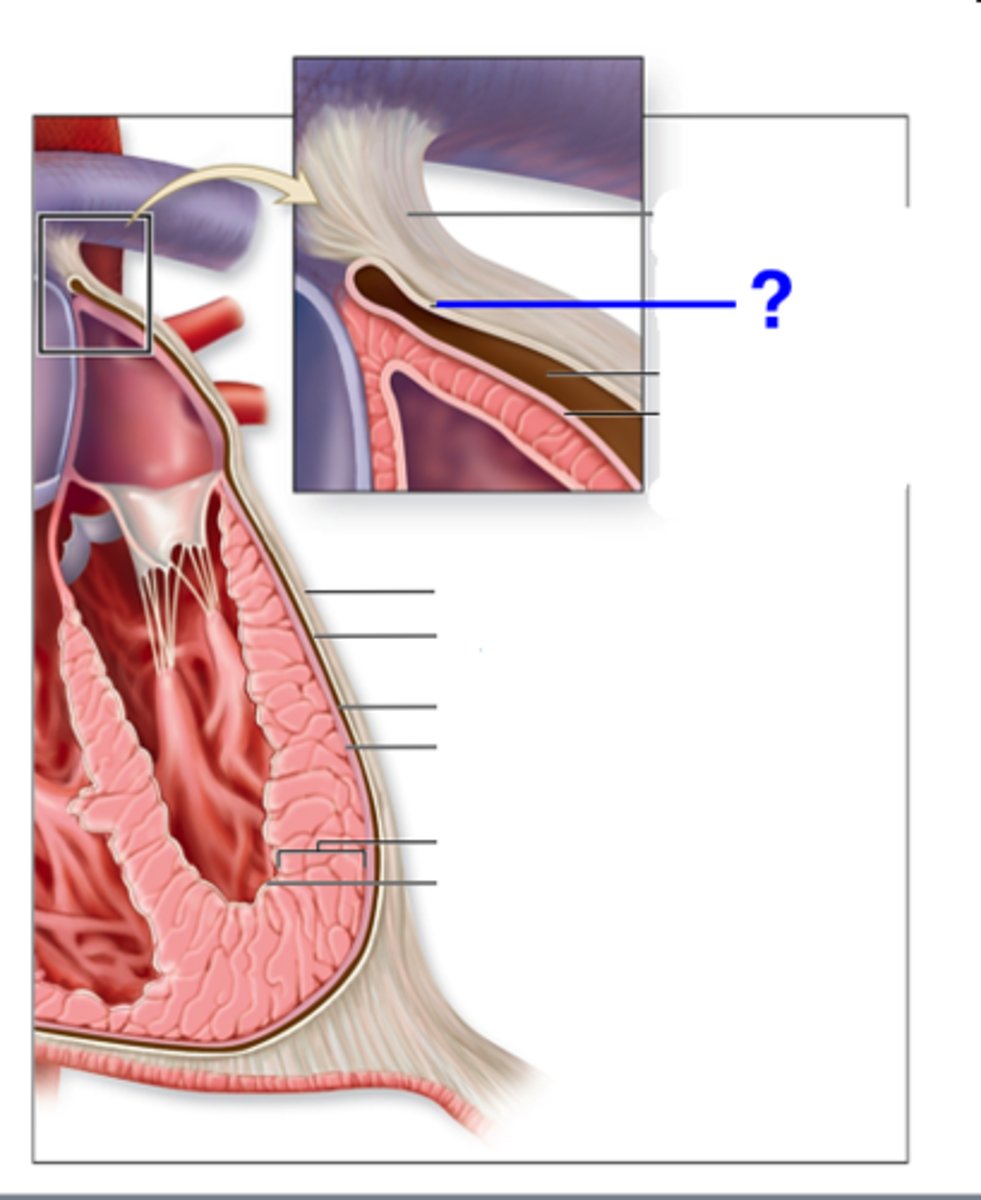
Pericardial cavity
Space with serous fluid reducing friction.
Pericardial effusion
Excess fluid in pericardial cavity causing pressure.
Endocardium
Inner layer of heart; simple squamous epithelium.
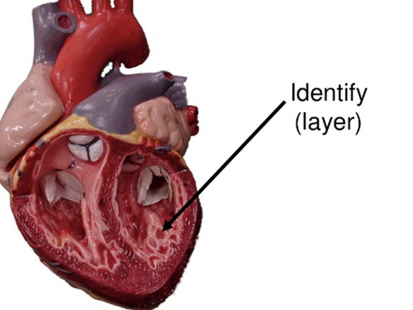
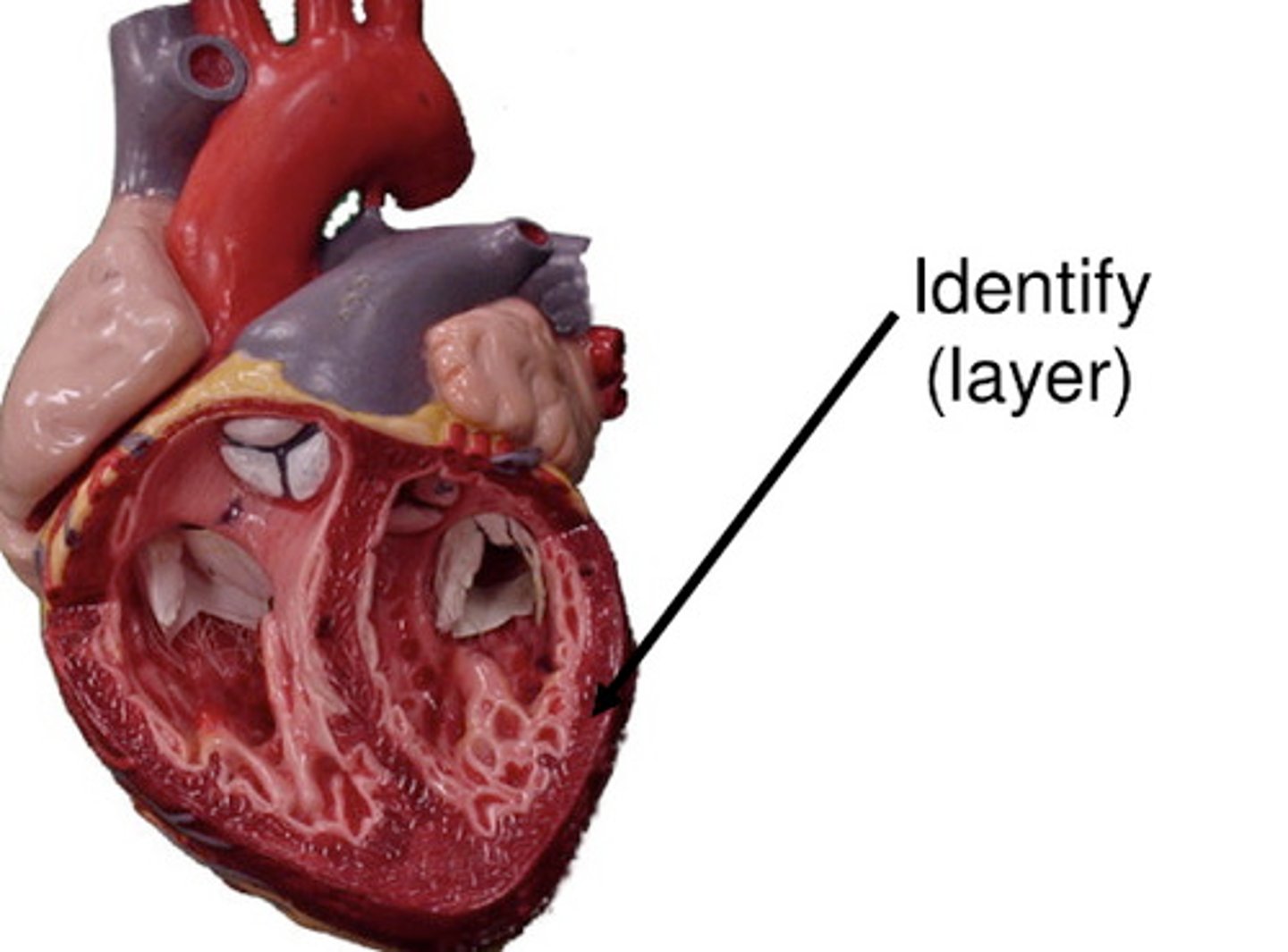
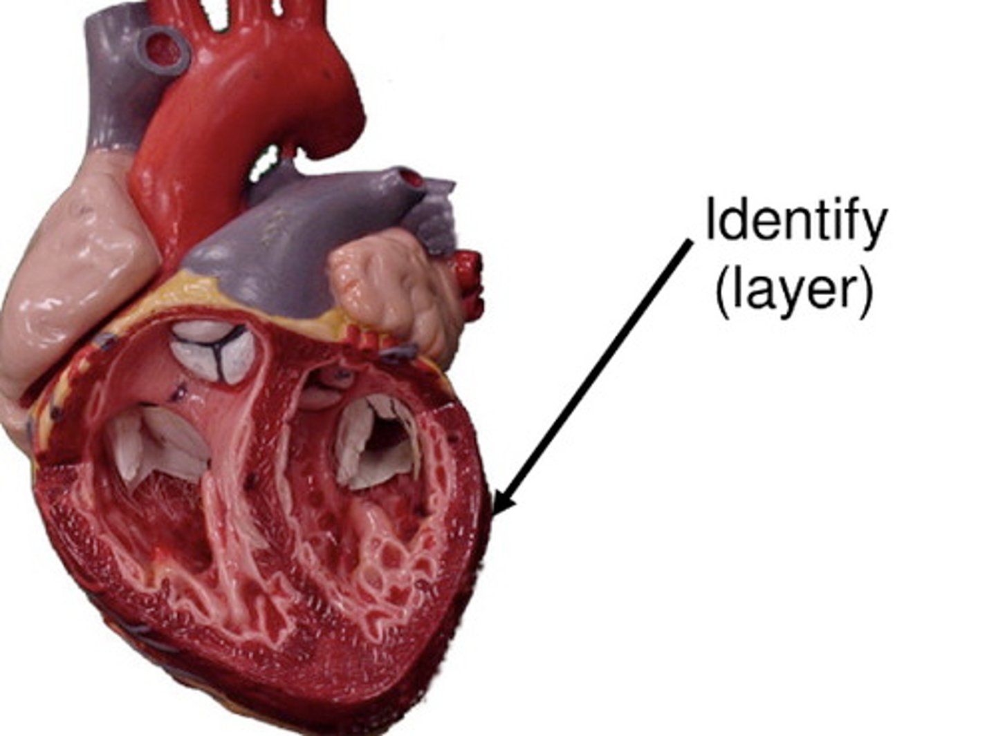
Myocardium
Thick cardiac muscle layer of the heart.
Epicardium
Visceral layer of serous pericardium, thin membrane.
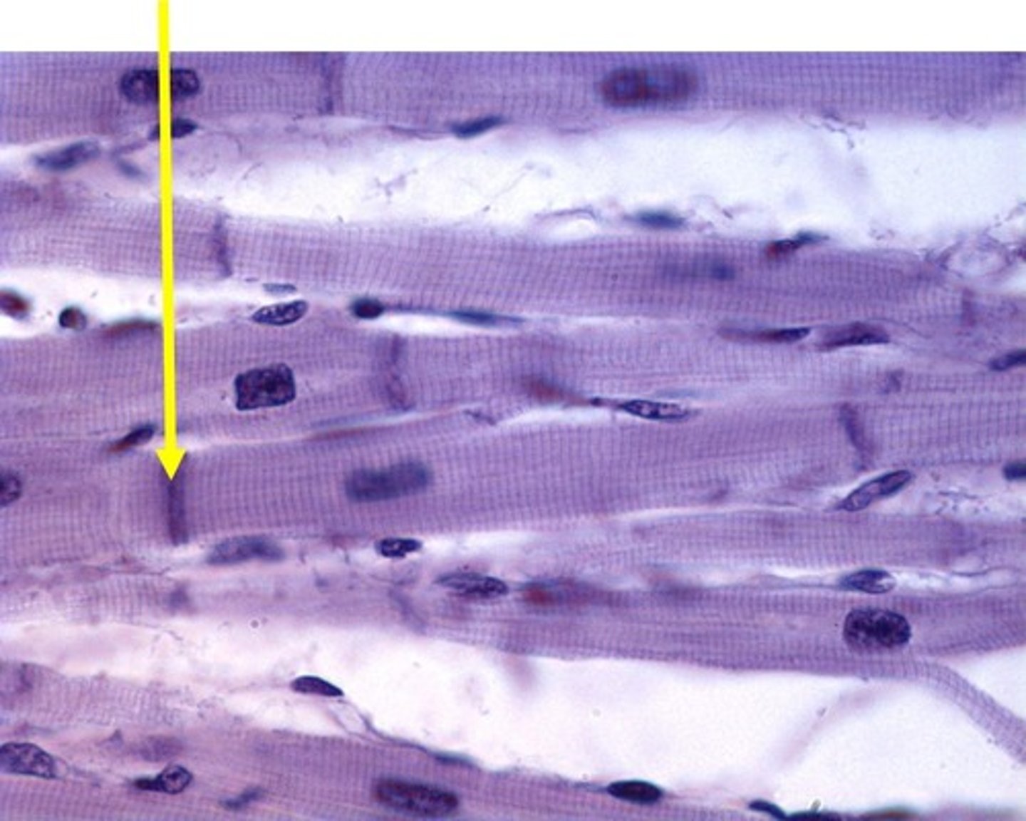
Intercalated discs
Specialized junctions connecting cardiac muscle cells. Transmit nerve impulses
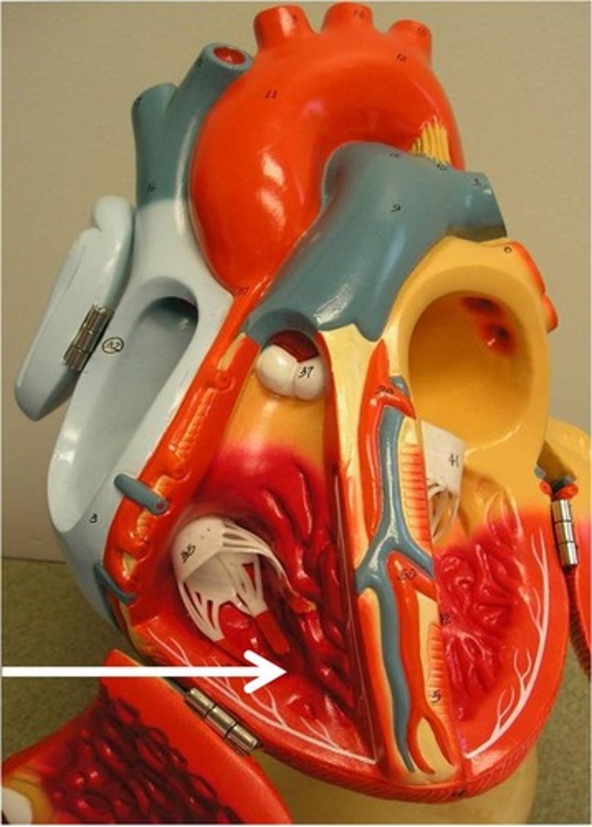
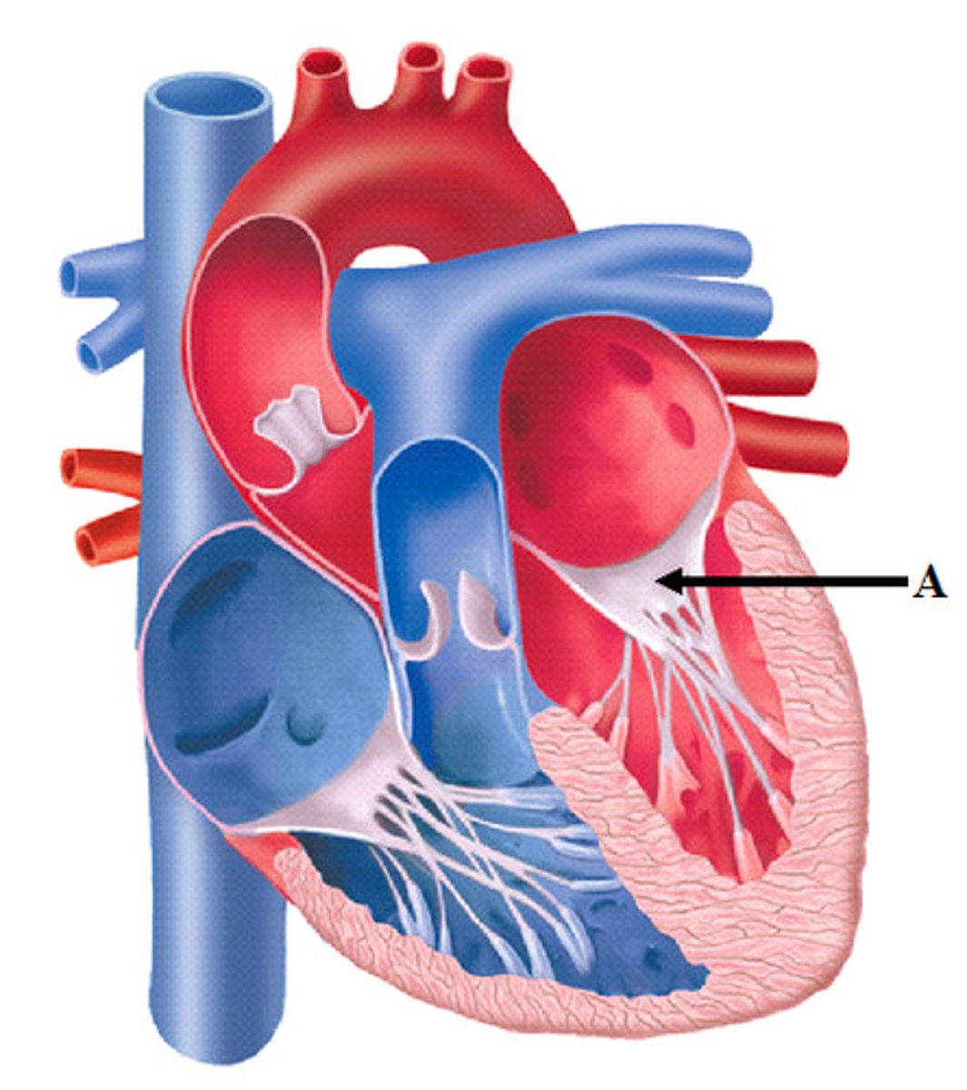
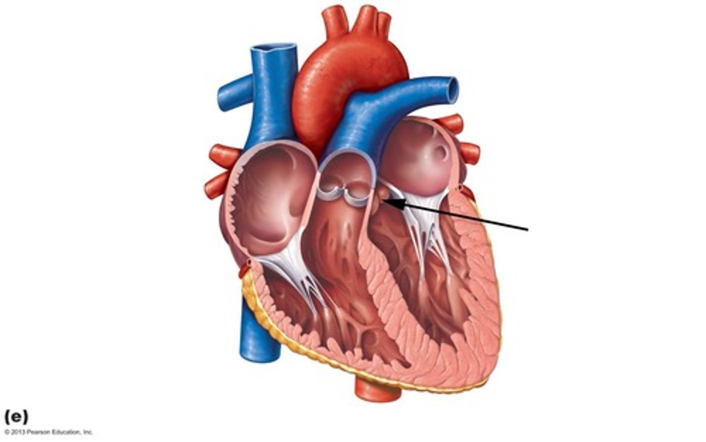
Tricuspid Valve
Valve between right atrium and ventricle.
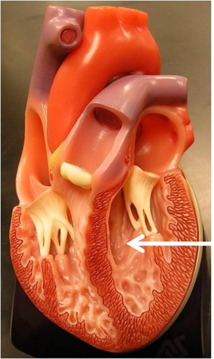
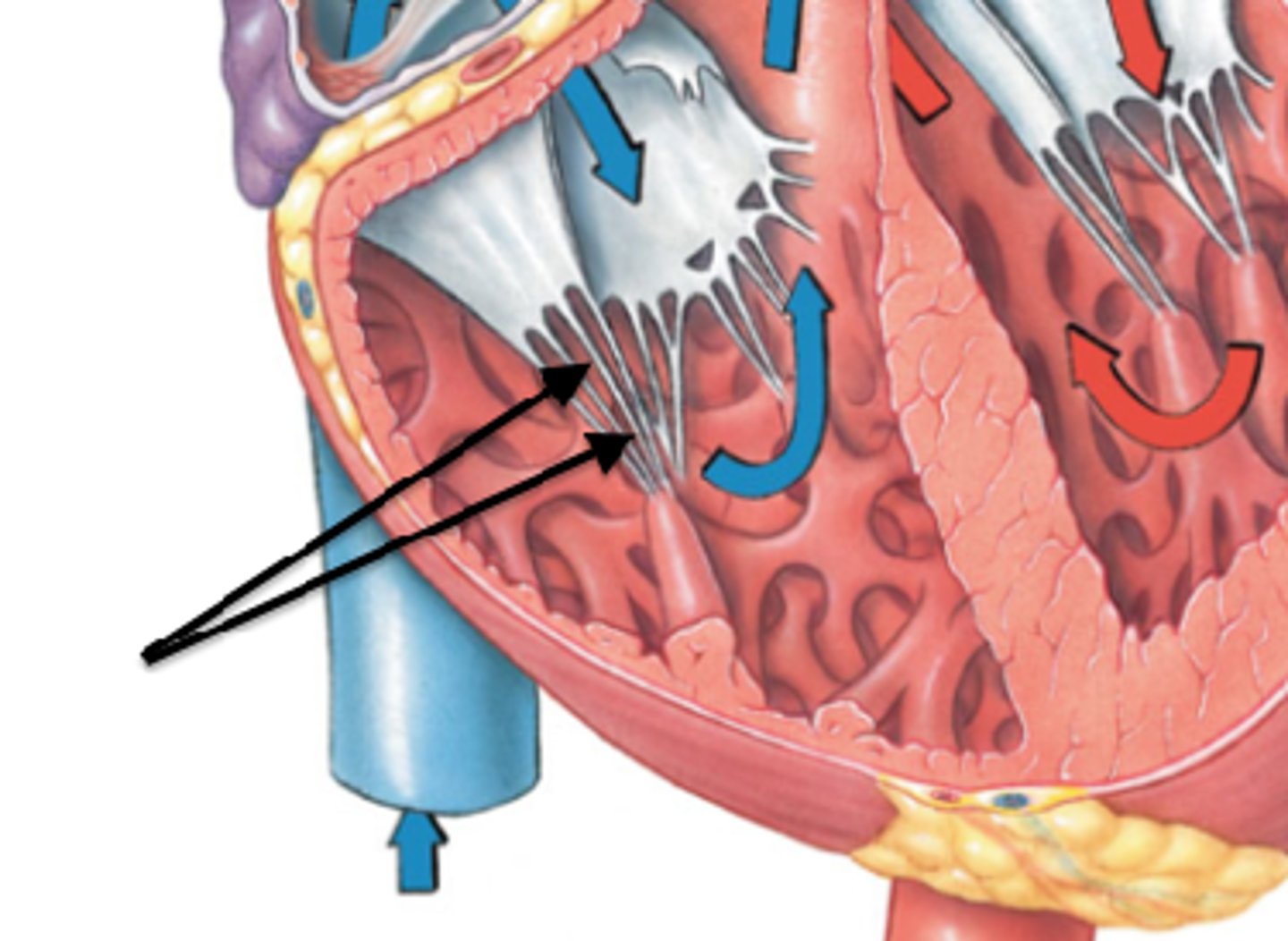
Papillary Muscles
Muscles that anchor heart valves via chordae tendineae.
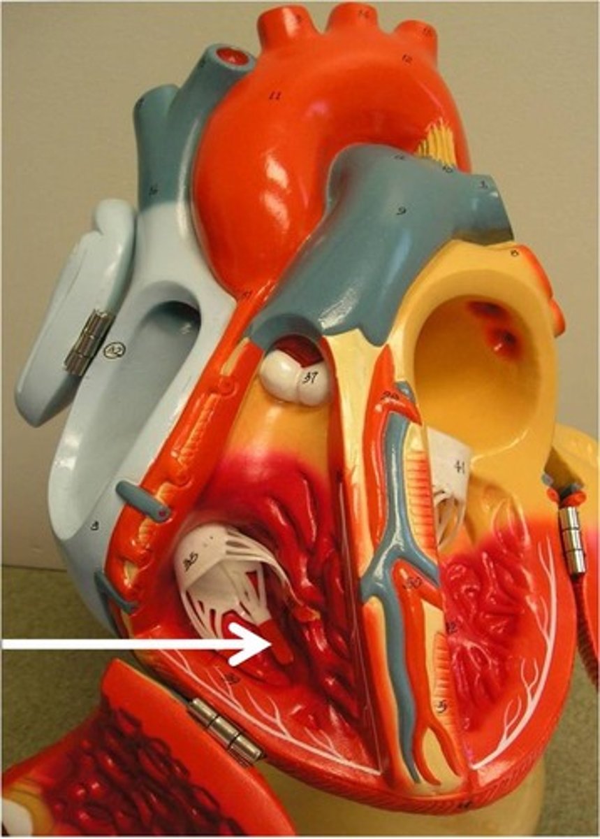
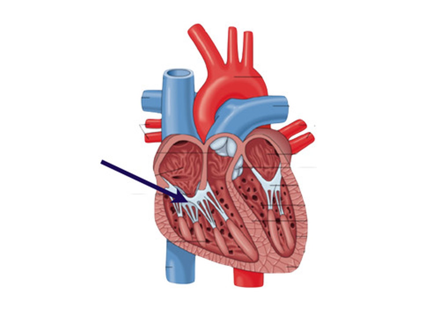
Chordae Tendineae
Tendons preventing valve eversion during contraction.
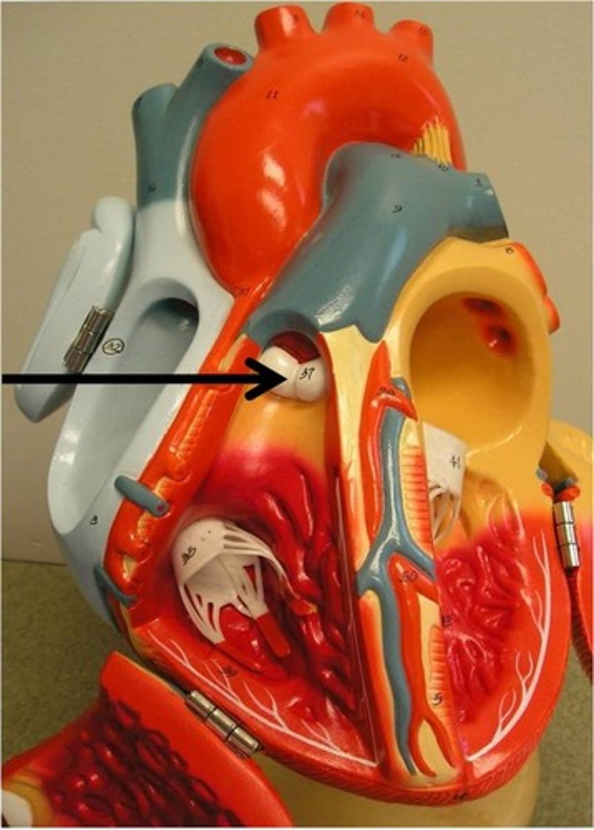
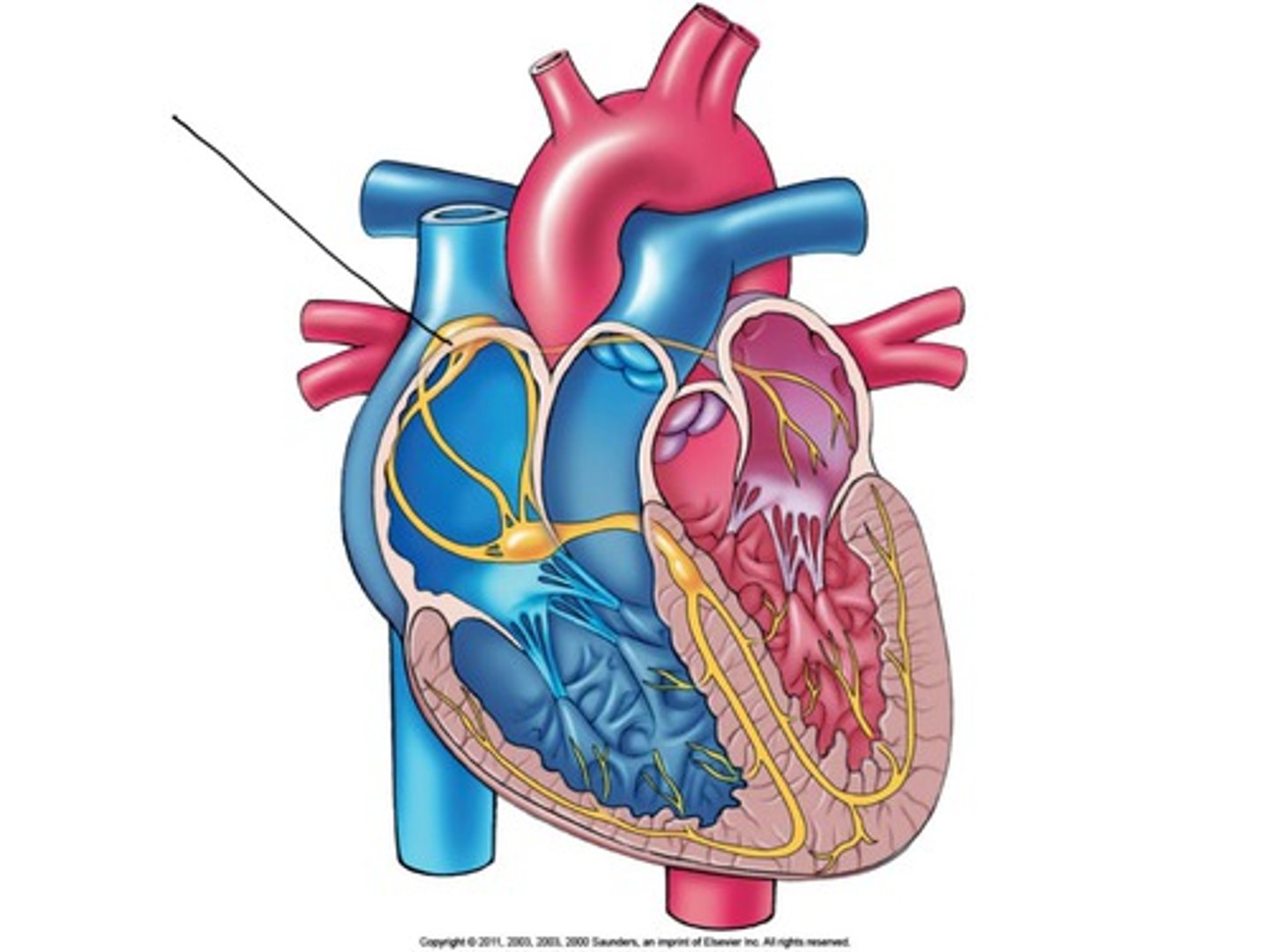
Pulmonary Semilunar Valve
Valve between right ventricle and pulmonary trunk.
left atrioventricular valve (Mitral or Bicuspid)
Valve between left atrium and left ventricle.
Aortic Semilunar Valve
Valve between left ventricle and aorta.
Coronary Sulcus
Groove separating atria from ventricles externally.
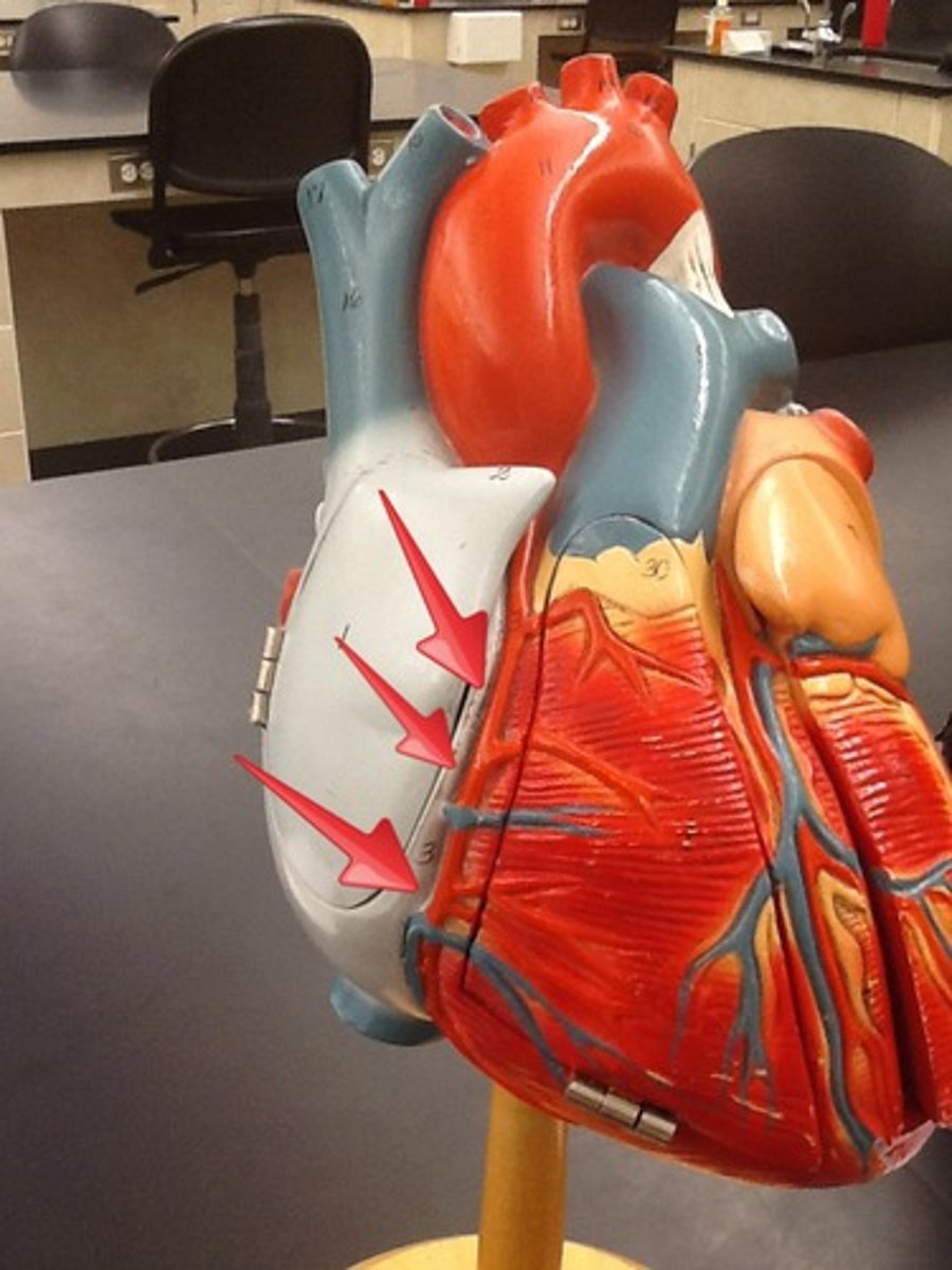
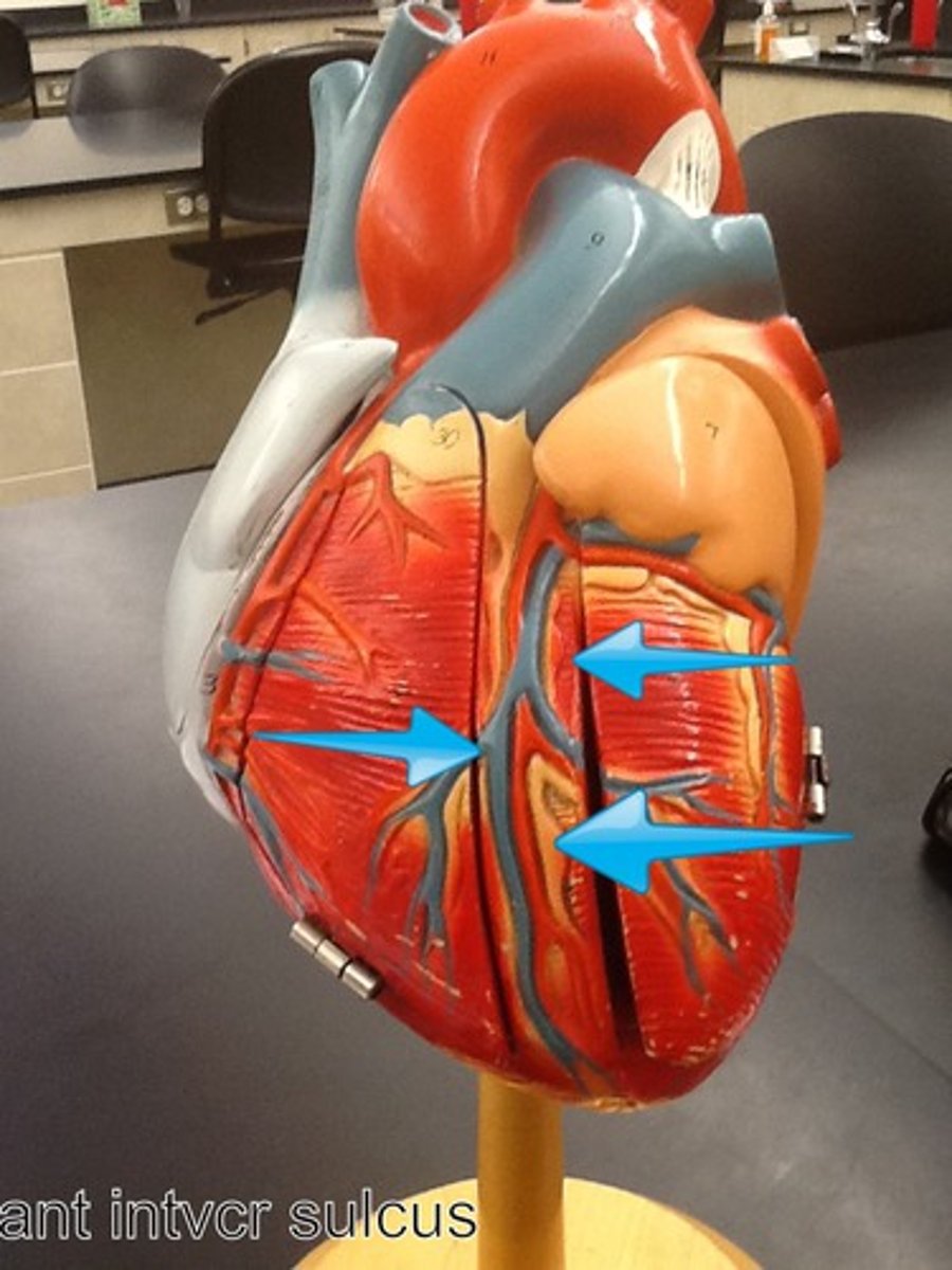
Interventricular Sulcus
Groove separating left and right ventricles.
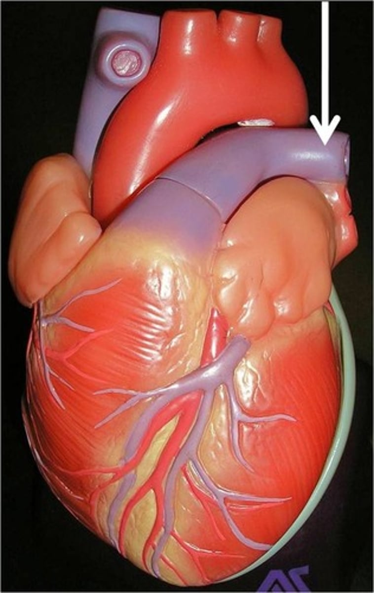
Pulmonary Arteries
Carry deoxygenated blood from heart to lungs.
Pulmonary Veins
Carry oxygenated blood from lungs to heart.
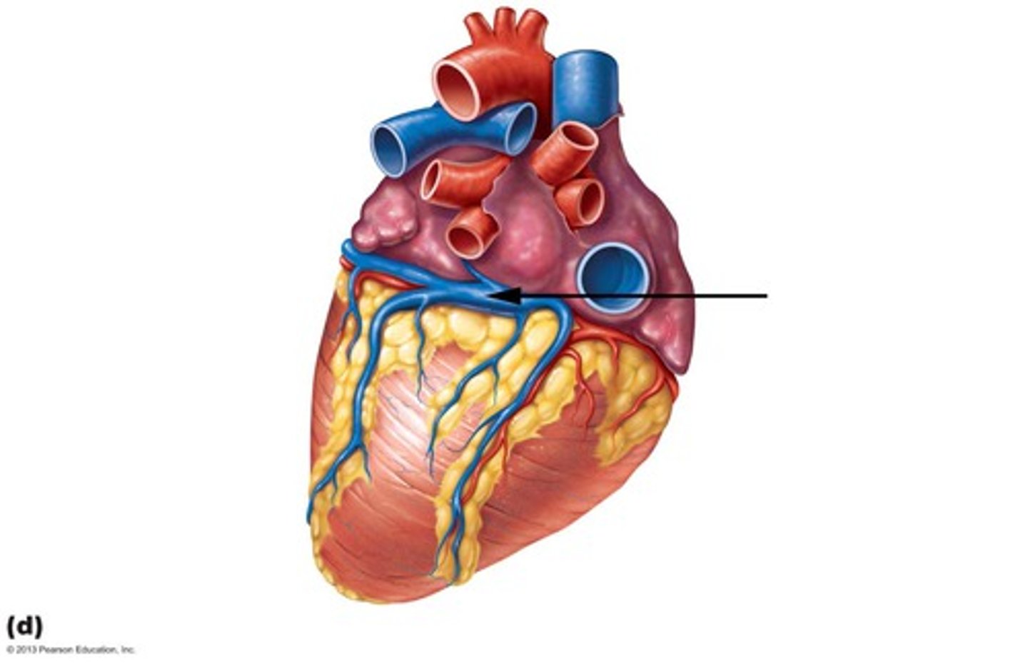
Coronary Sinus
Veins draining into the heart's right atrium.
Myocardial Infarction
Heart attack due to blood flow blockage.
SA Node
Pacemaker initiating heartbeat in the heart.
Atrioventricular Bundle
Conducting fibers splitting into left and right branches.
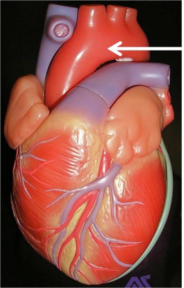
Aortic Arch
Curved section of the aorta leaving the heart.
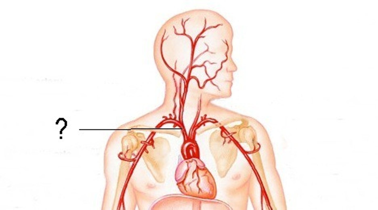
Brachiocephalic Trunk
Artery branching to arms and head.
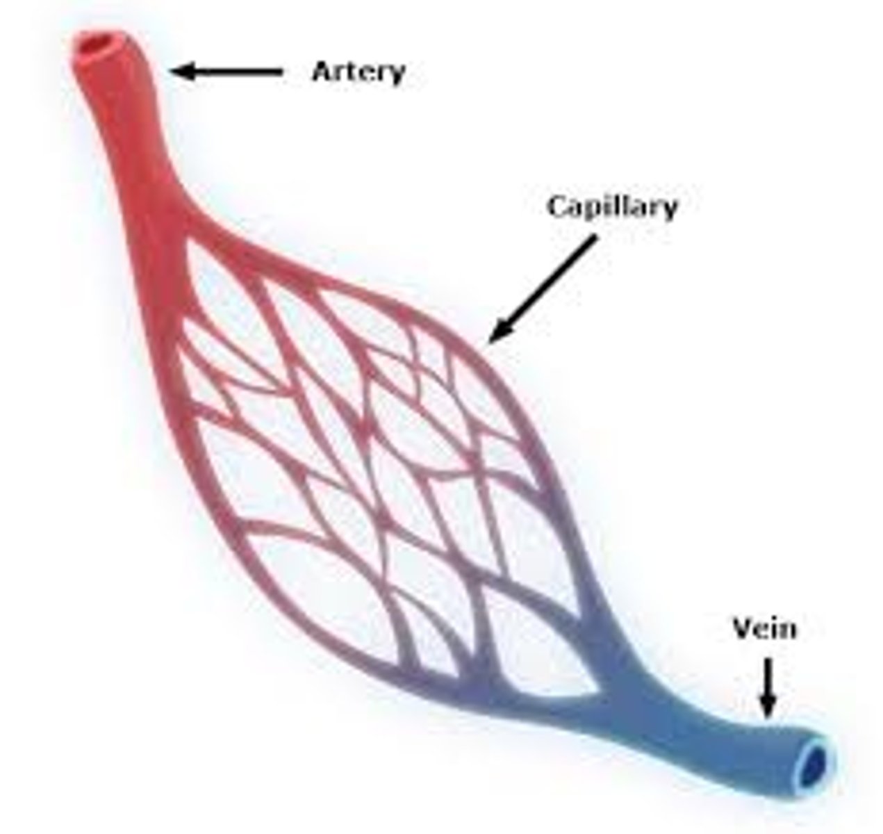
Arteries
Carry blood away from the heart.
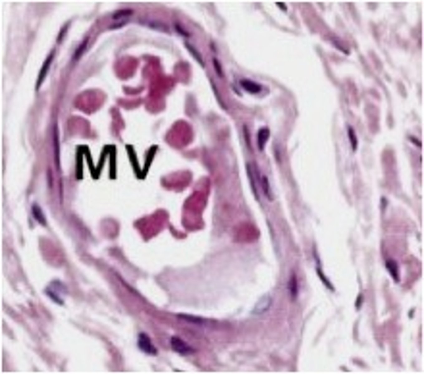
Veins
Return blood to the heart.
Common carotid artery
Branches into external and internal carotid arteries.
Internal carotid artery
More posterior branch of common carotid.
External carotid artery
More anterior branch of common carotid.
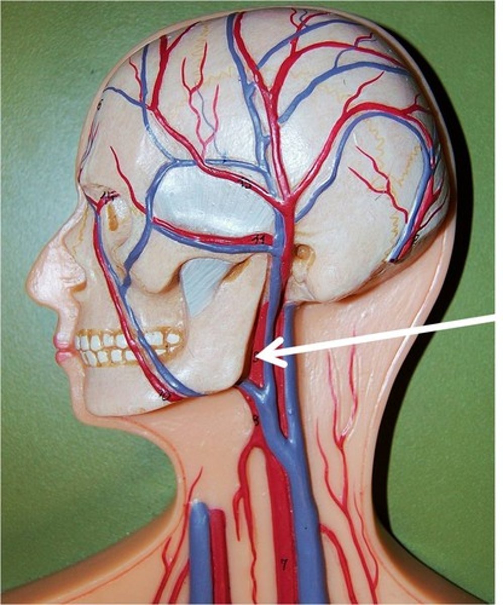
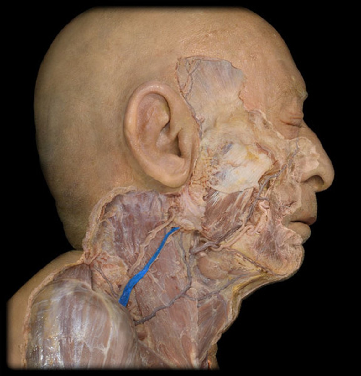
External jugular vein
Lateral and smaller vein.
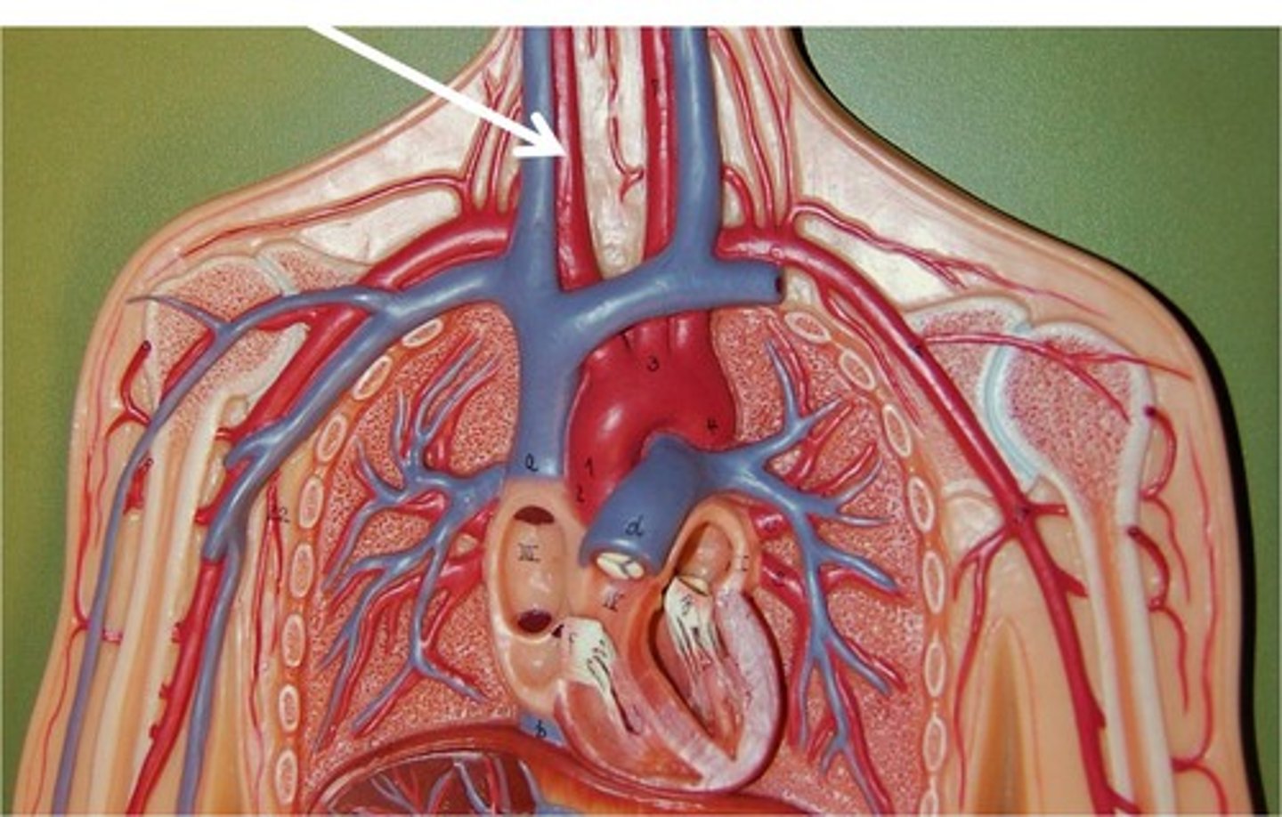
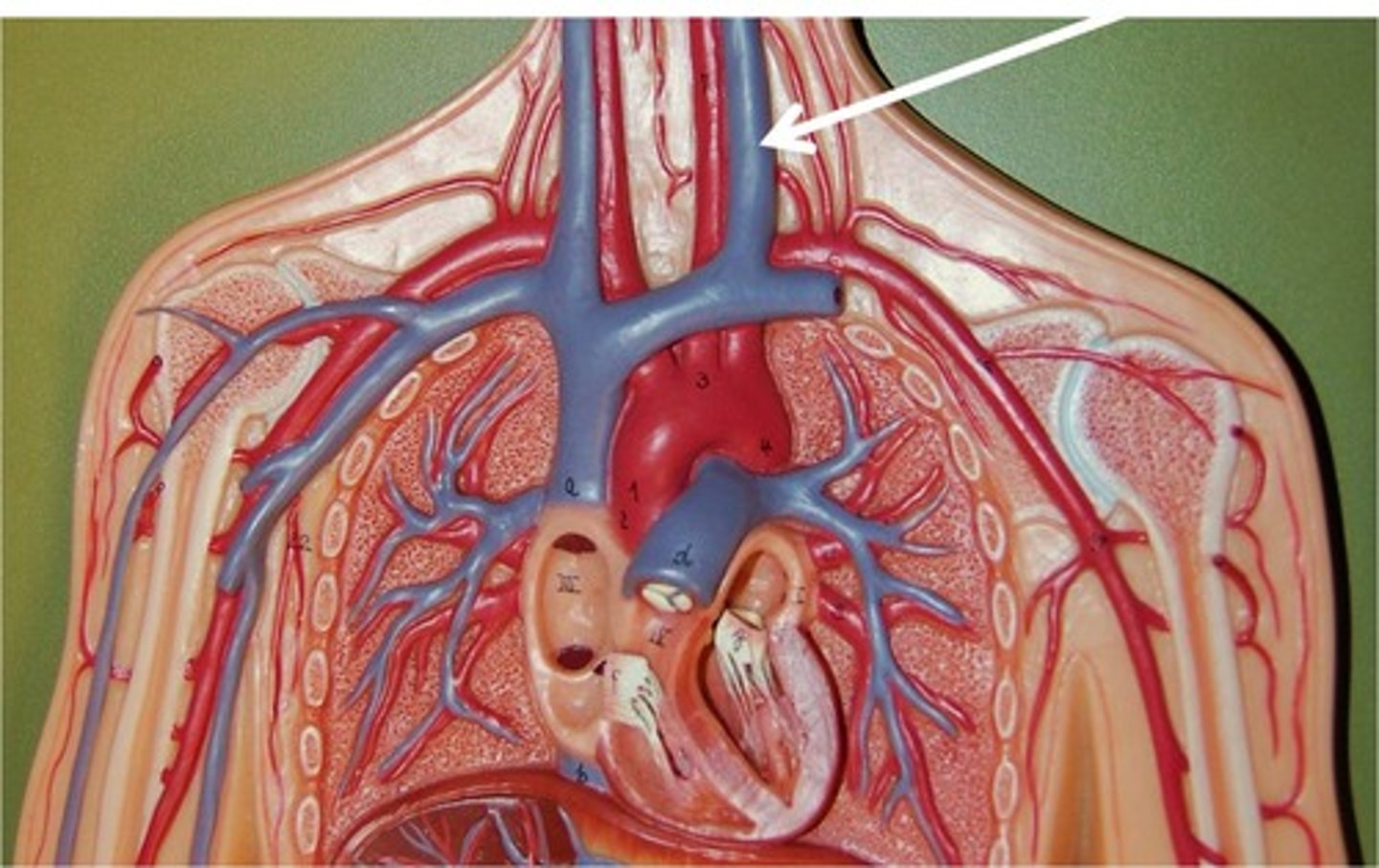
Internal jugular vein
Medial and larger vein.
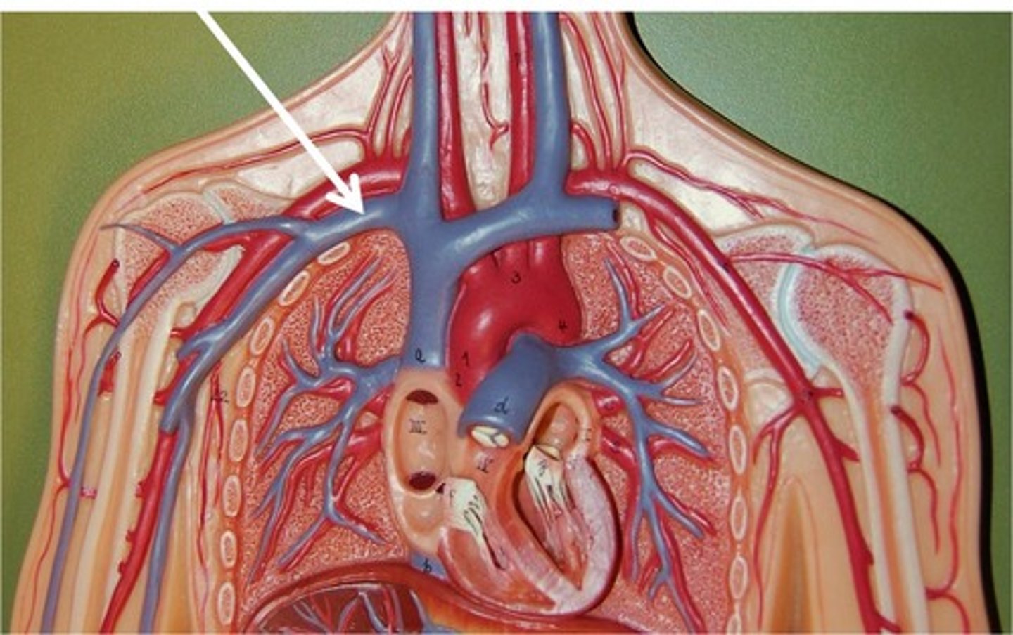
Subclavian vein
Joins with internal jugular vein.
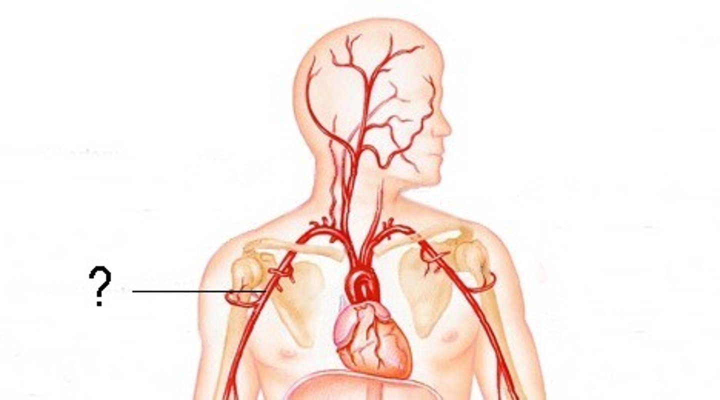
Axillary artery
First portion in armpit region.
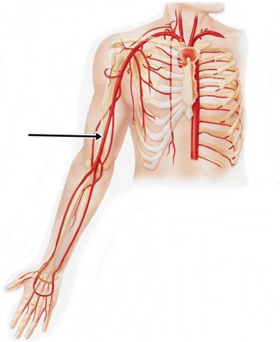
Brachial artery
Continues from axillary artery.
Radial artery
Branches from brachial artery to radius.
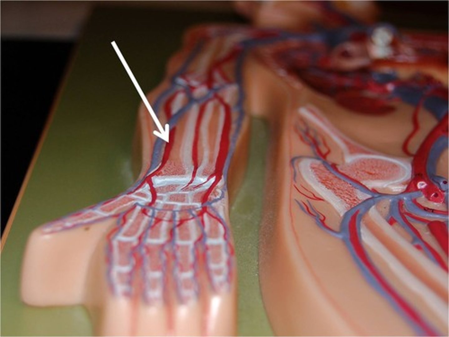
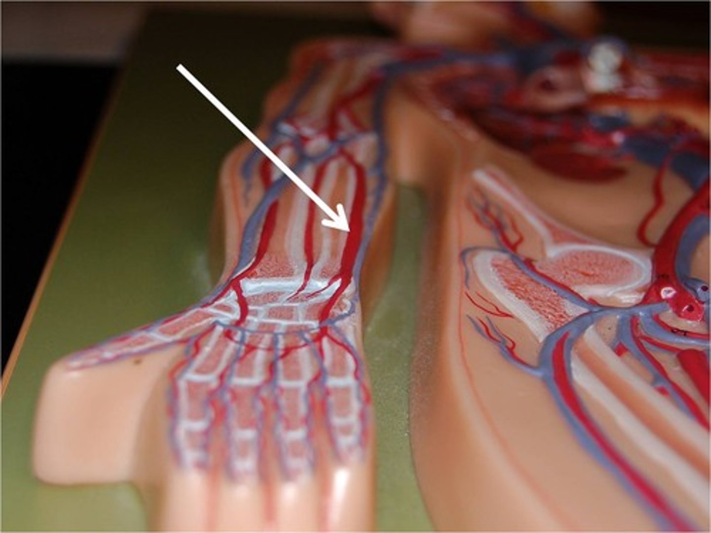
Ulnar artery
Branches from brachial artery to ulna.
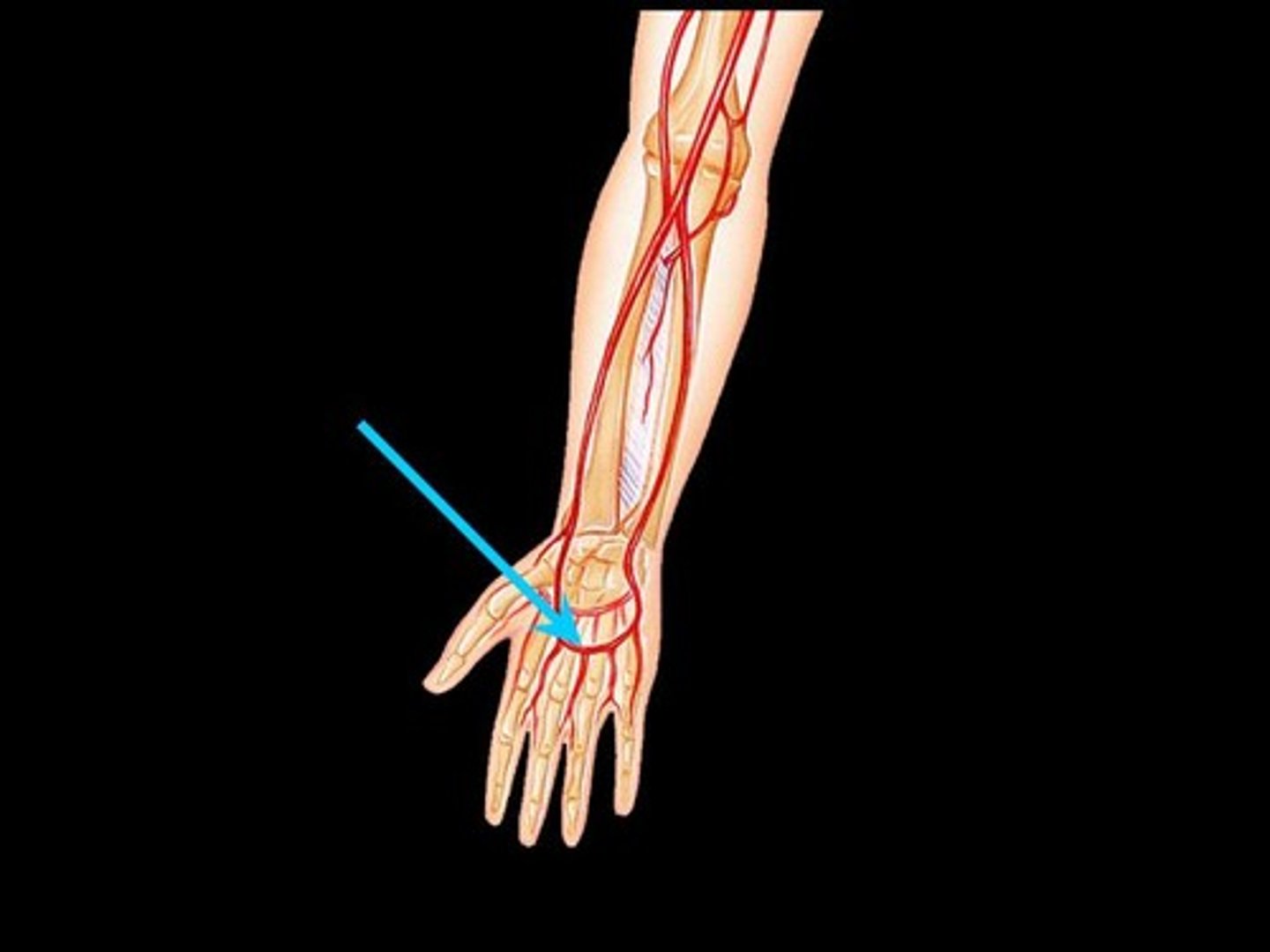
Superficial palmar arch
Formed by radial and ulnar arteries.
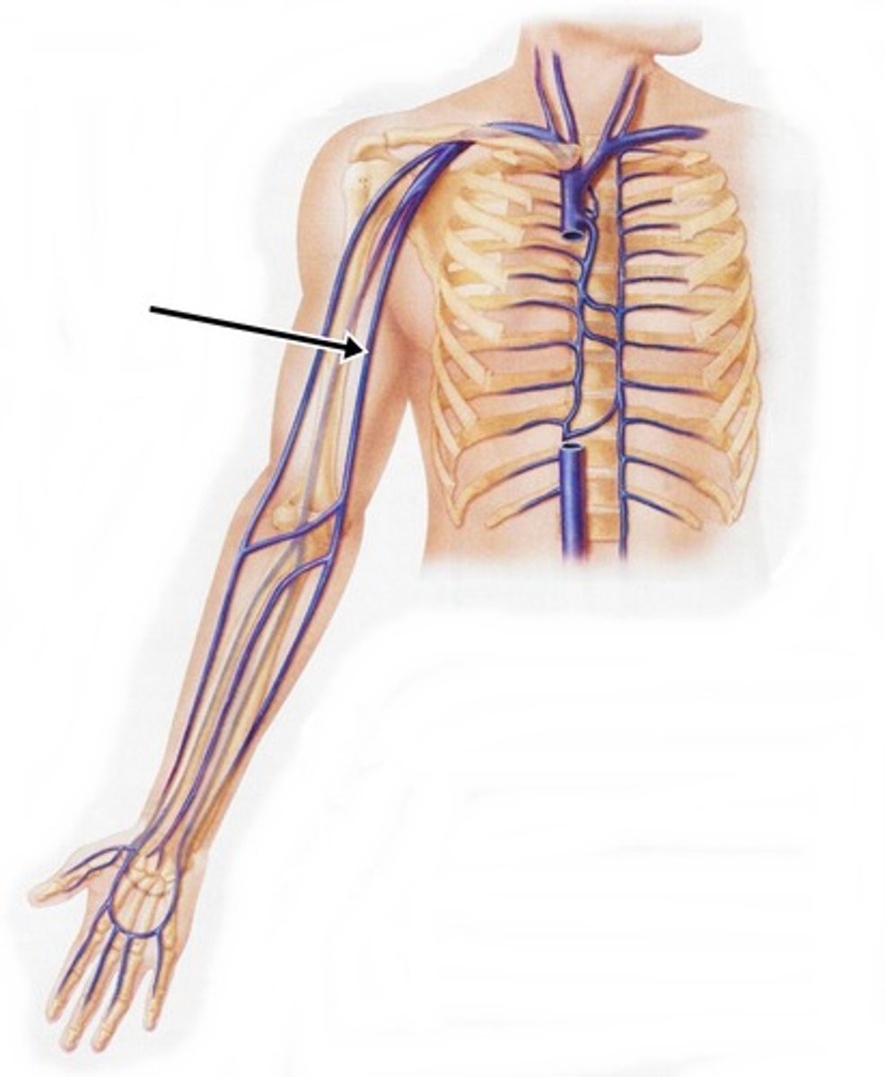
Cephalic vein
Lateral vein of forearm and arm.
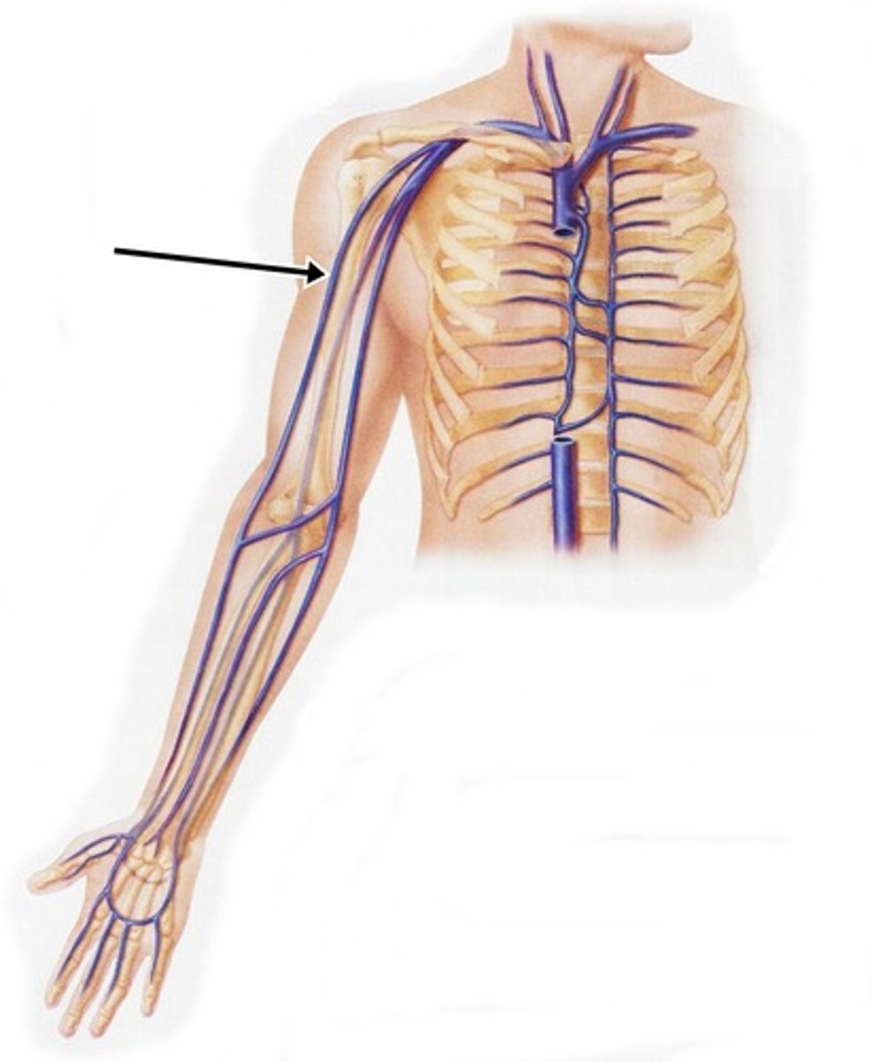
Basilic vein
Medial vein of arm and forearm.
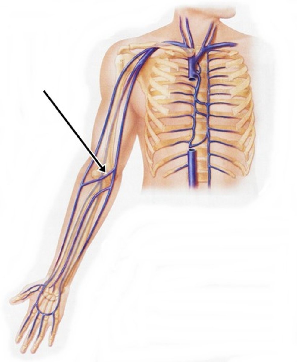
Median cubital vein
Common site for blood draws.
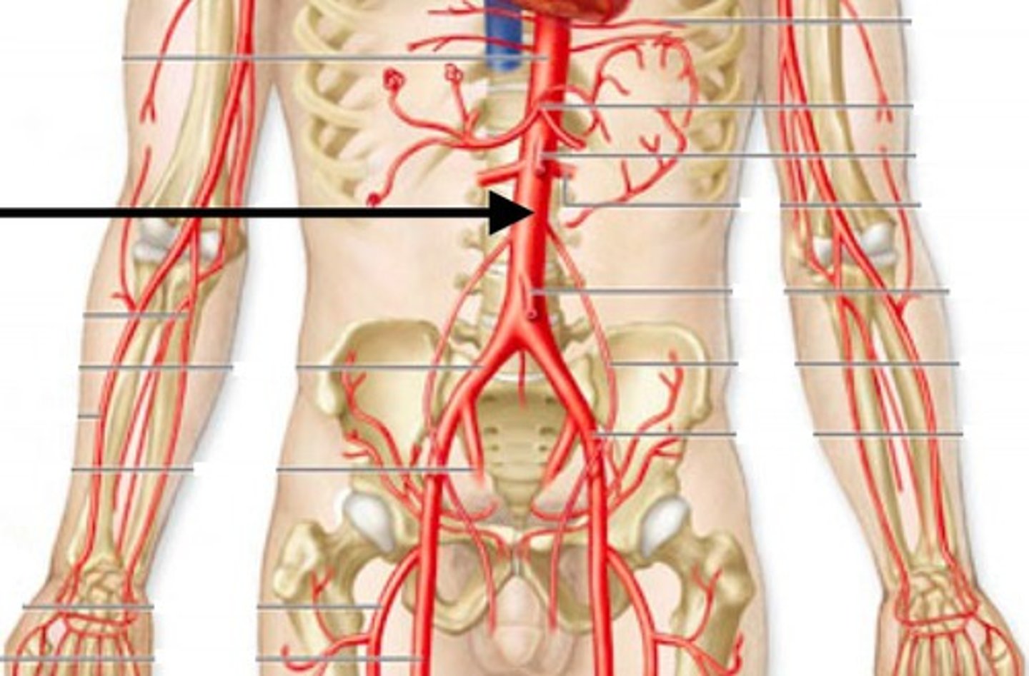
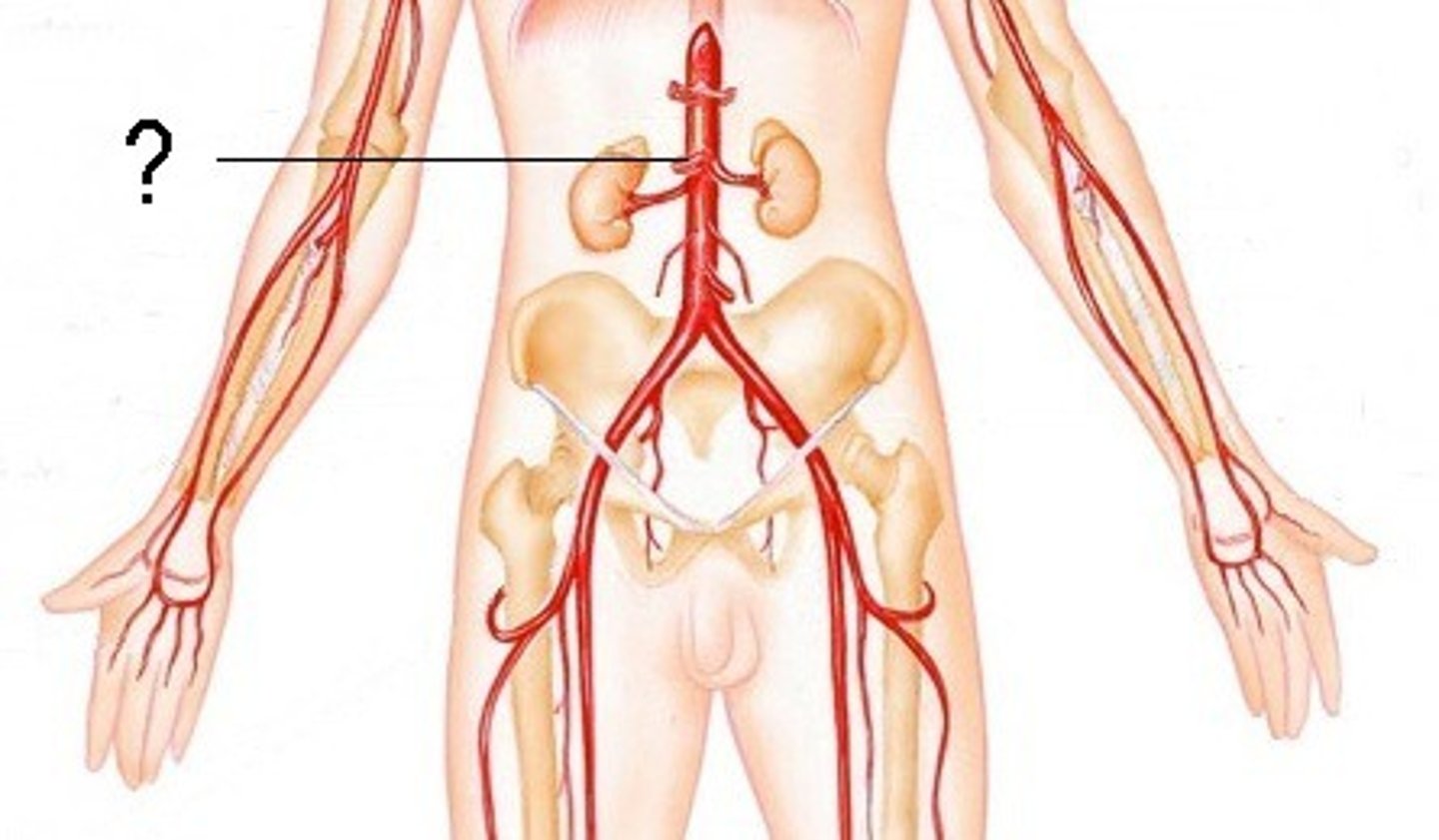
Abdominal aorta
Continuation of thoracic aorta after diaphragm.
Celiac trunk
Branches into major abdominal arteries.
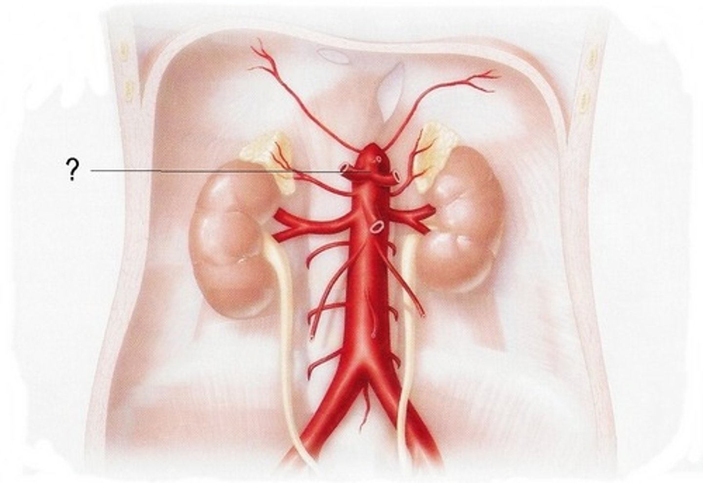
Superior mesenteric artery
Supplies blood to small intestine.
Inferior mesenteric artery
Supplies blood to large intestine.
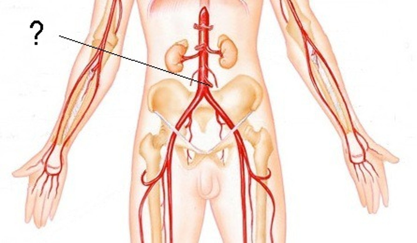
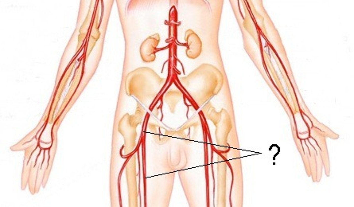
Common iliac arteries
Branches from abdominal aorta.

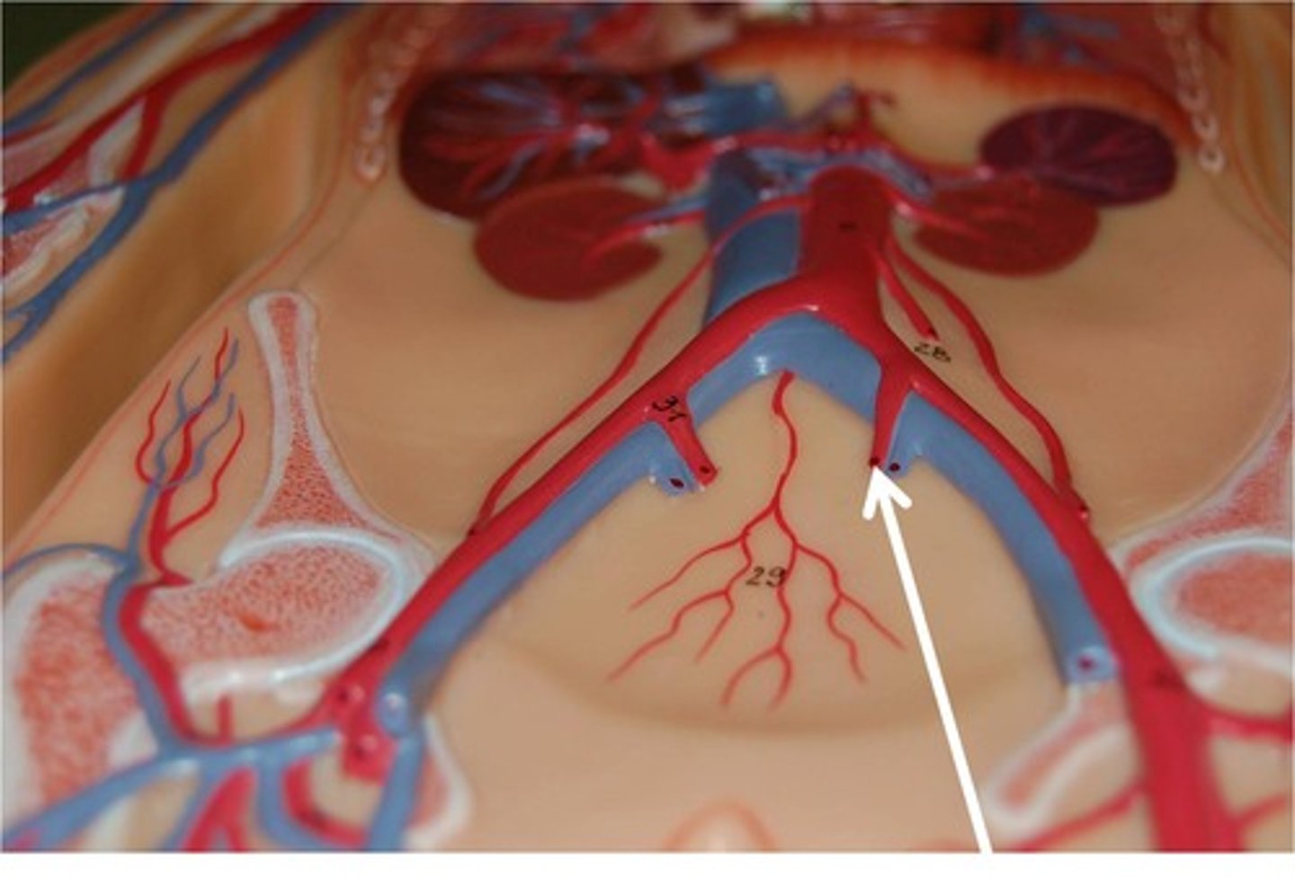
Internal iliac arteries
Supply pelvic organs.
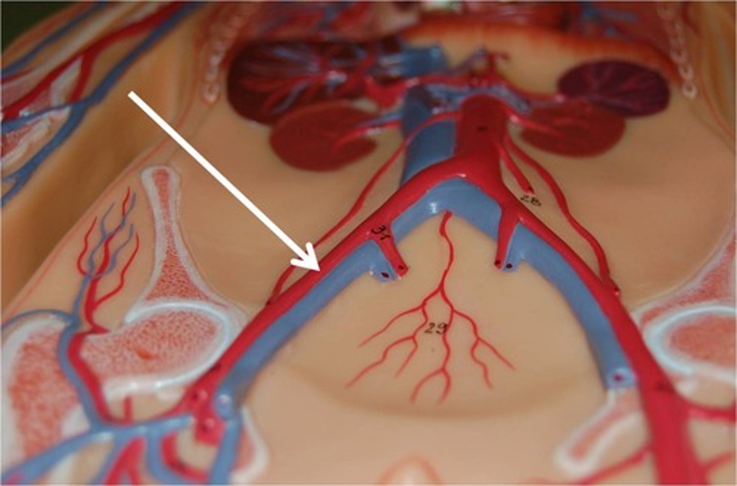
External iliac arteries
Become femoral artery in the leg.
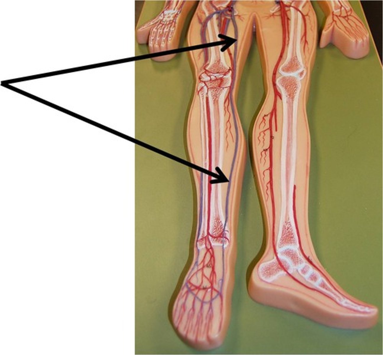
Femoral artery
Continuation of external iliac artery.
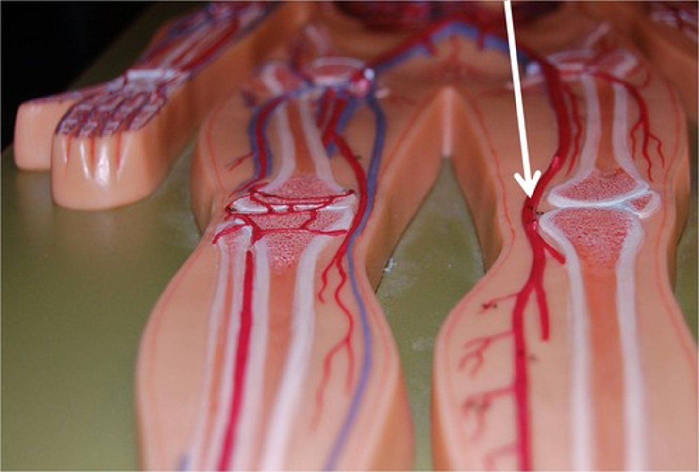
Popliteal artery
Branches from femoral artery behind knee.
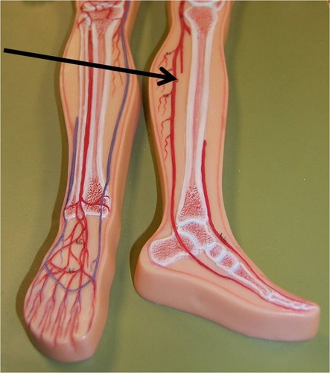
Anterior tibial artery
Branches from popliteal artery, more lateral.
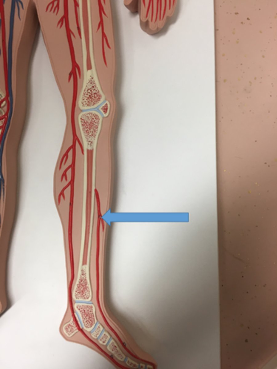
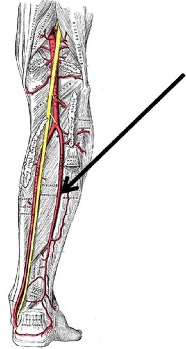
Posterior tibial artery
Branches from popliteal artery, more medial.
Fibular artery
Branches from posterior tibial artery.
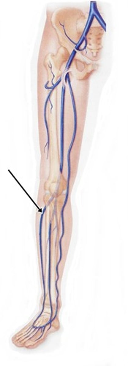
Great saphenous vein
Longest vein in the body.
Small saphenous vein
Visible only posteriorly.
Lymph
Fluid containing excess substances from blood.
Lymph nodes
Checkpoints filtering lymph fluid.
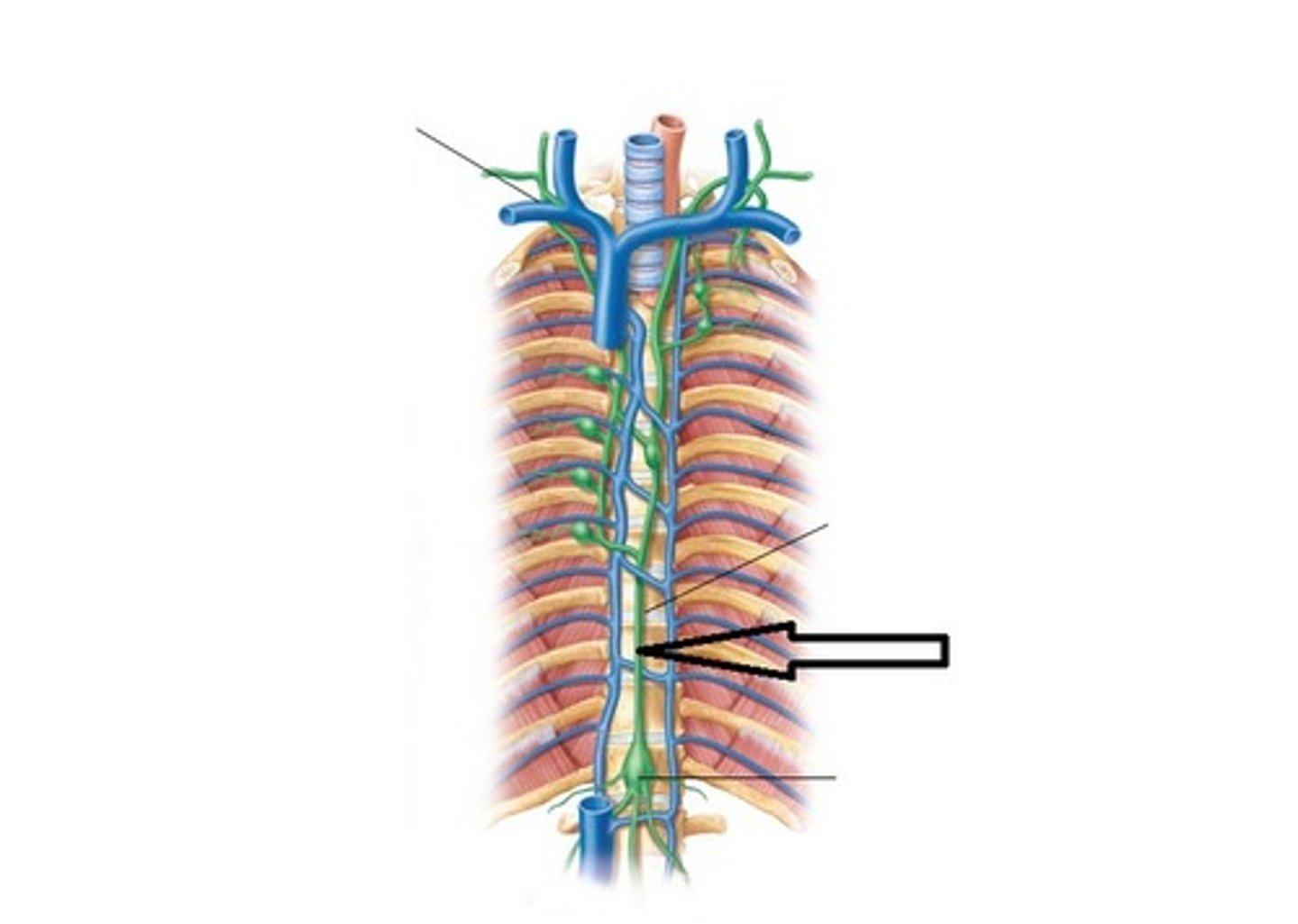
Thoracic duct
Returns lymph to venous system.
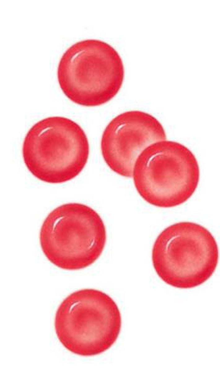
Erythrocytes
Red blood cells transporting oxygen.
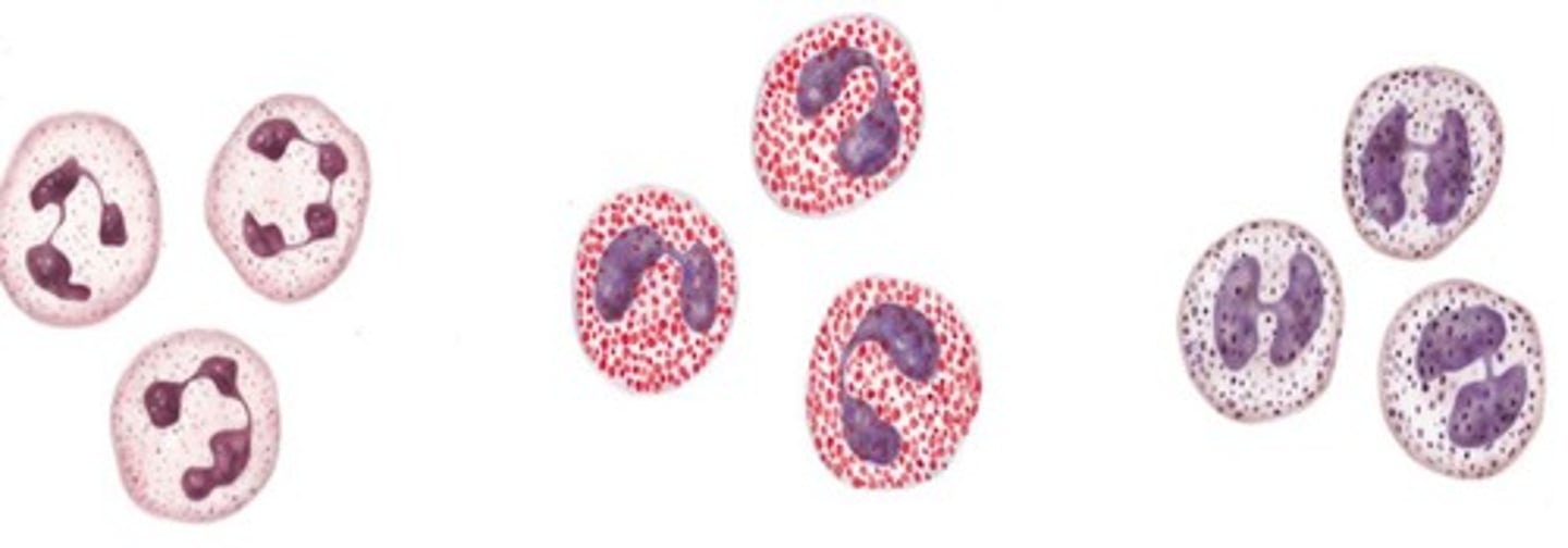
Leukocytes
White blood cells involved in immune response.
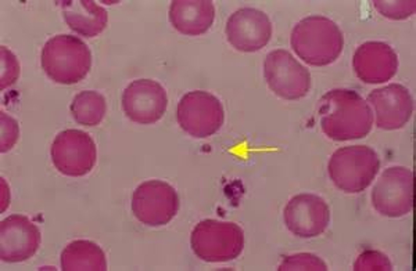
Platelets
Cell fragments aiding in blood clotting.
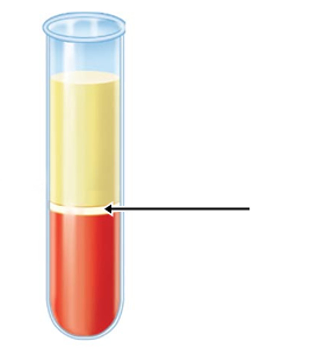
Buffy coat
Layer containing white blood cells and platelets.
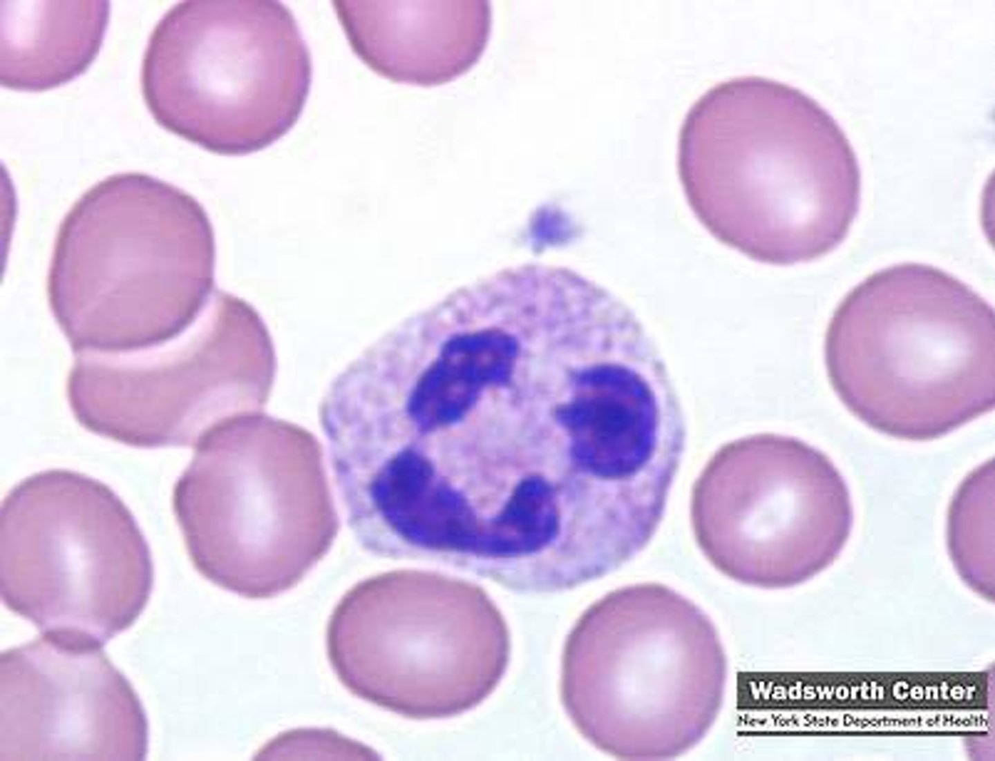
Neutrophil
Most numerous white blood cell, fights bacteria.
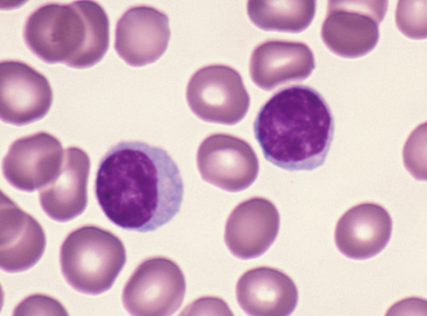
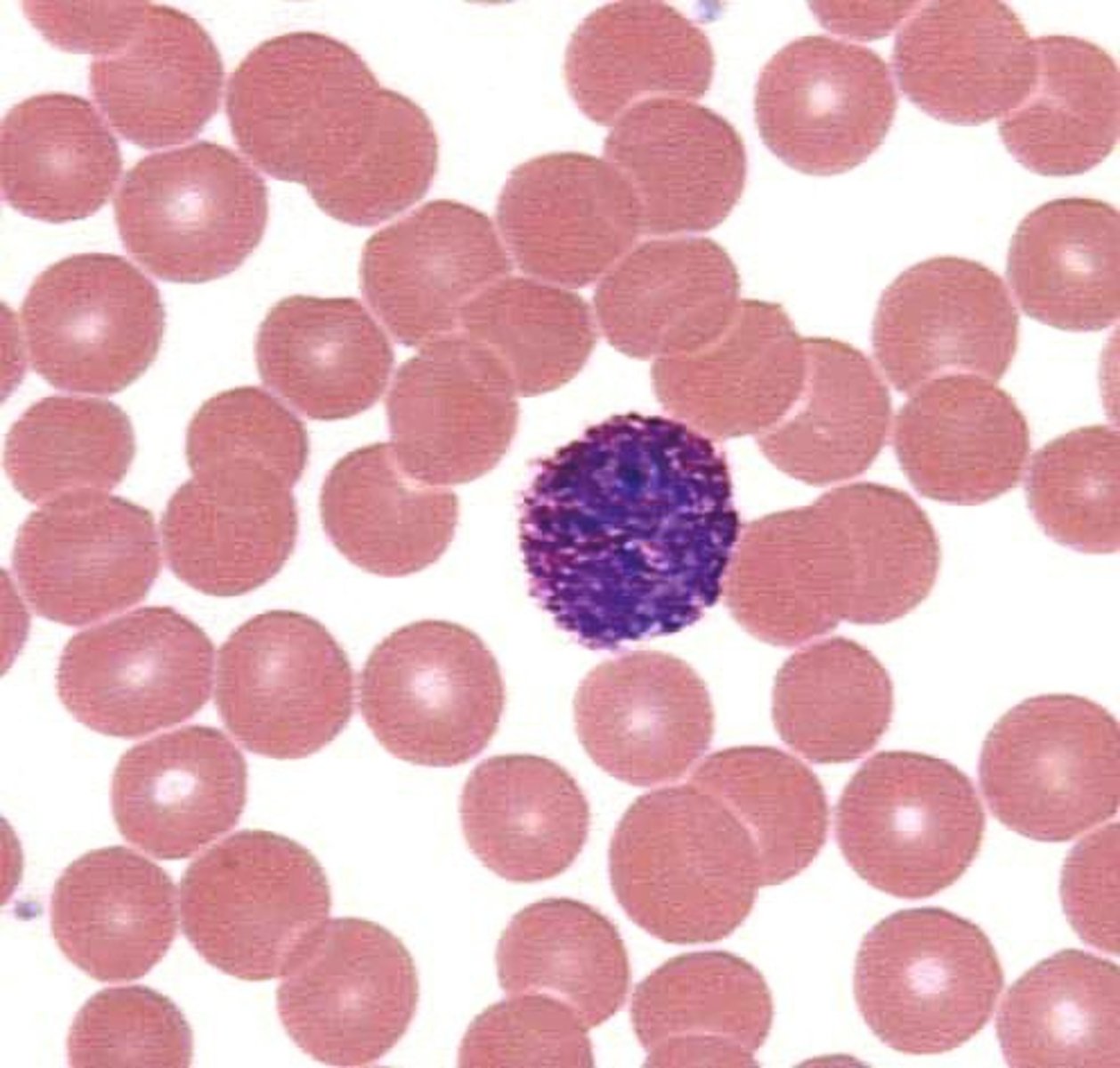
Lymphocyte
Involved in adaptive immunity, produces antibodies.
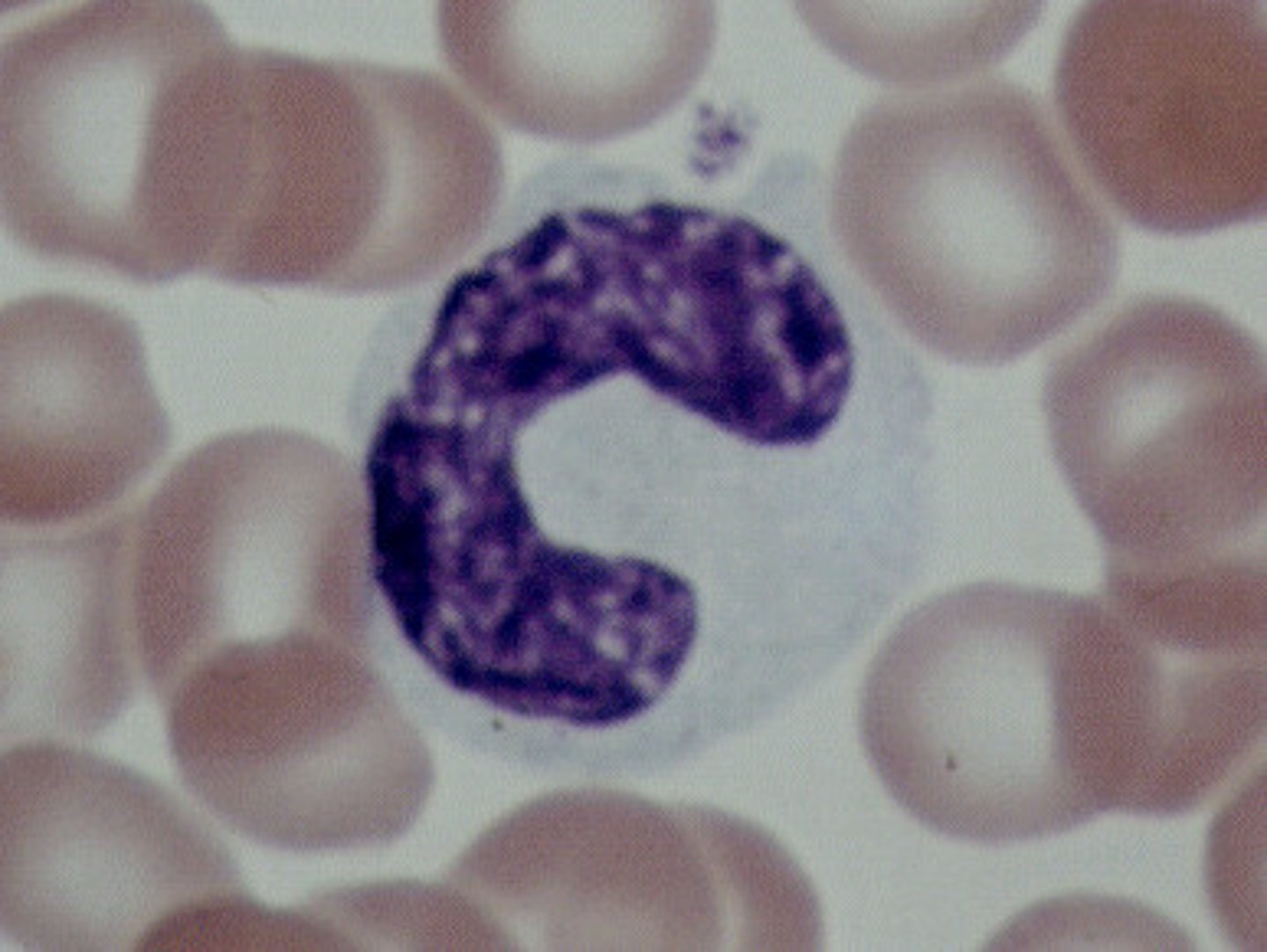
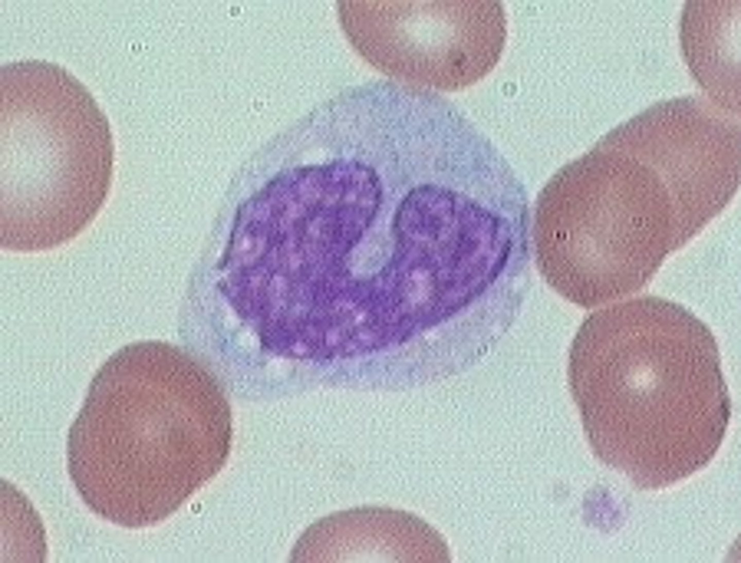
Monocyte
Largest leukocyte, becomes macrophage in tissues.
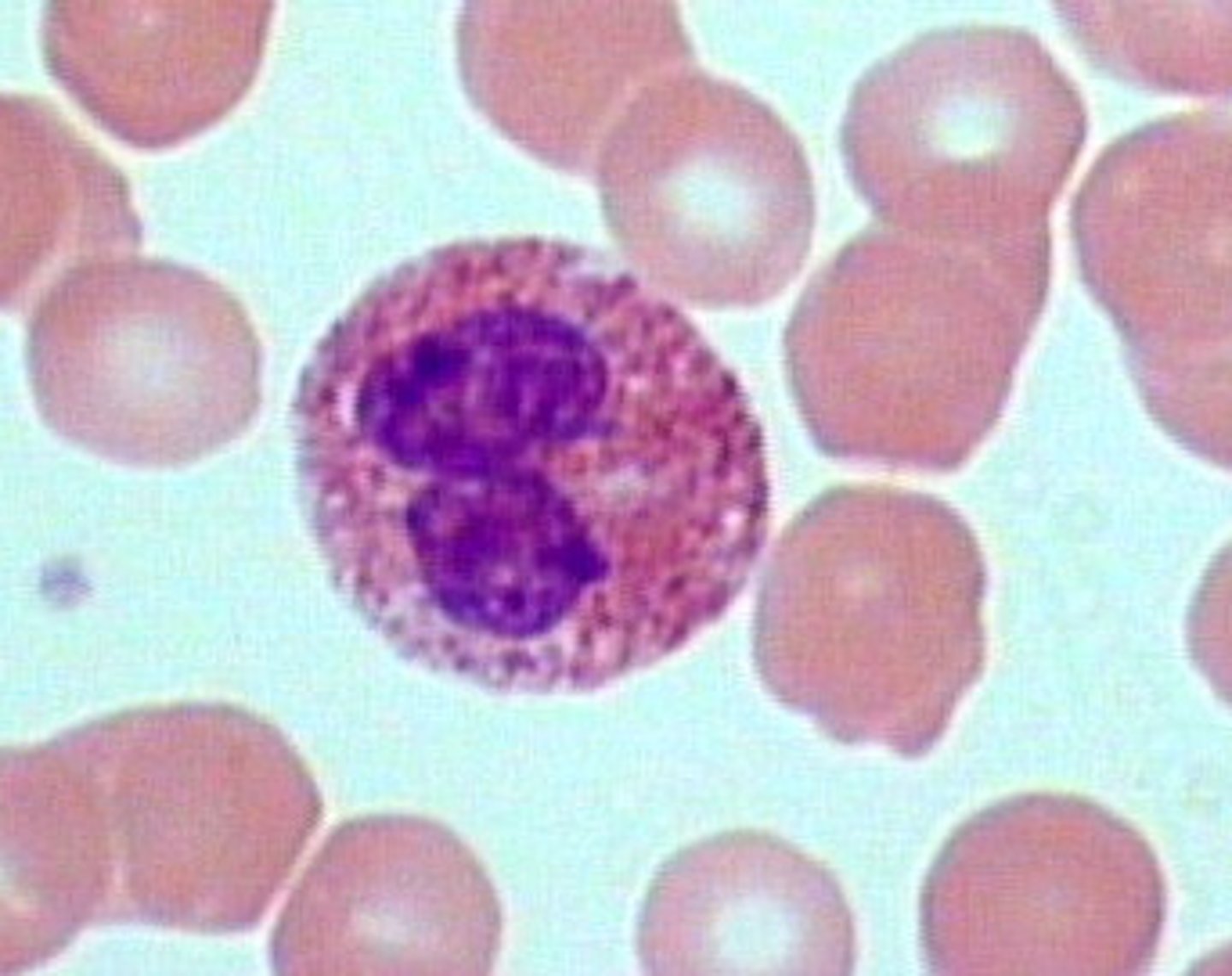
Eosinophil
Phagocytizes allergens and parasitic worms.
Basophil
Releases histamine during allergic reactions.
Capillaries
Small vessels for gas exchange.
Venules
Collect blood from capillaries. smaller veins
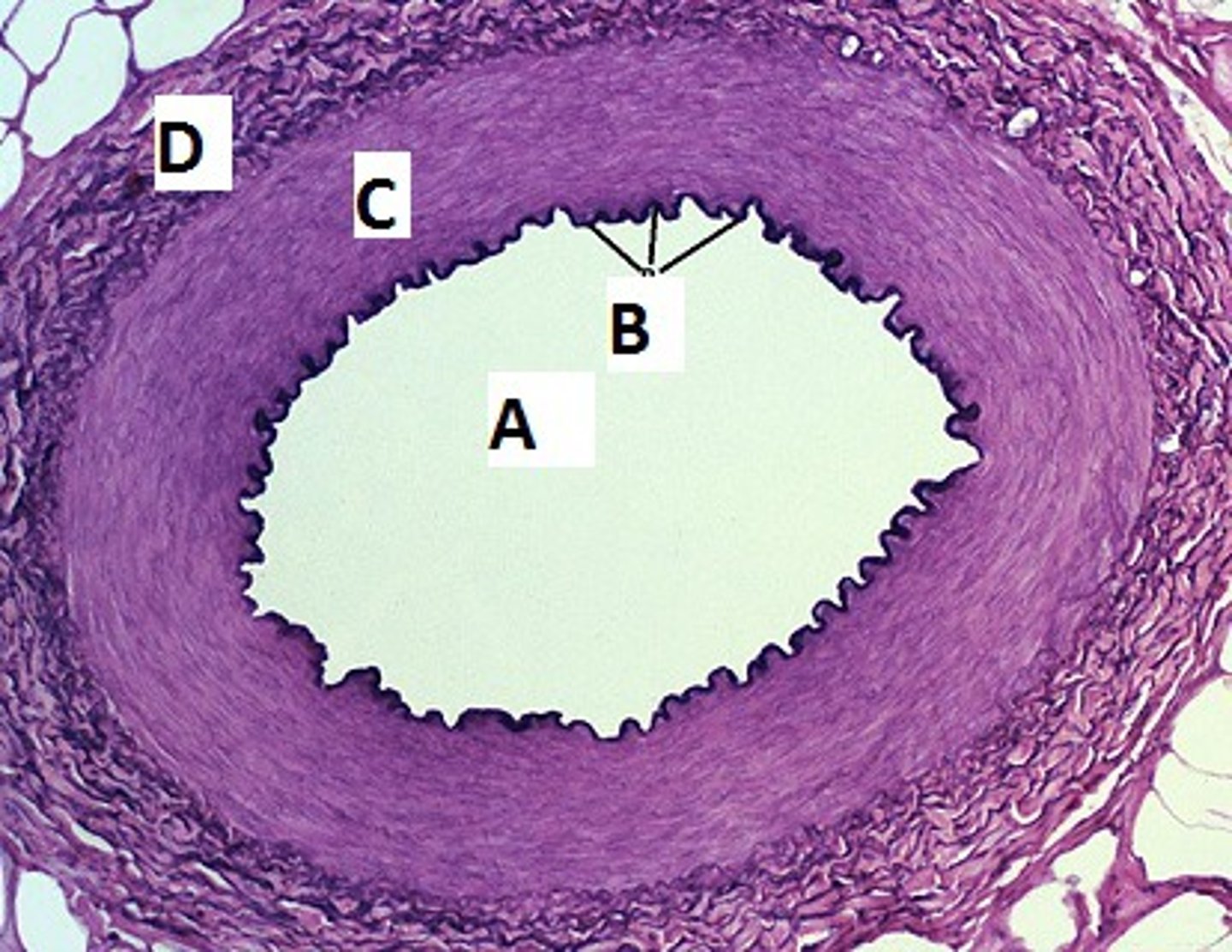
Tunica externa
Outermost layer of blood vessels.
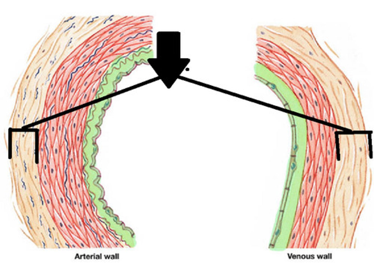
Tunica media
Middle layer, mainly smooth muscle.
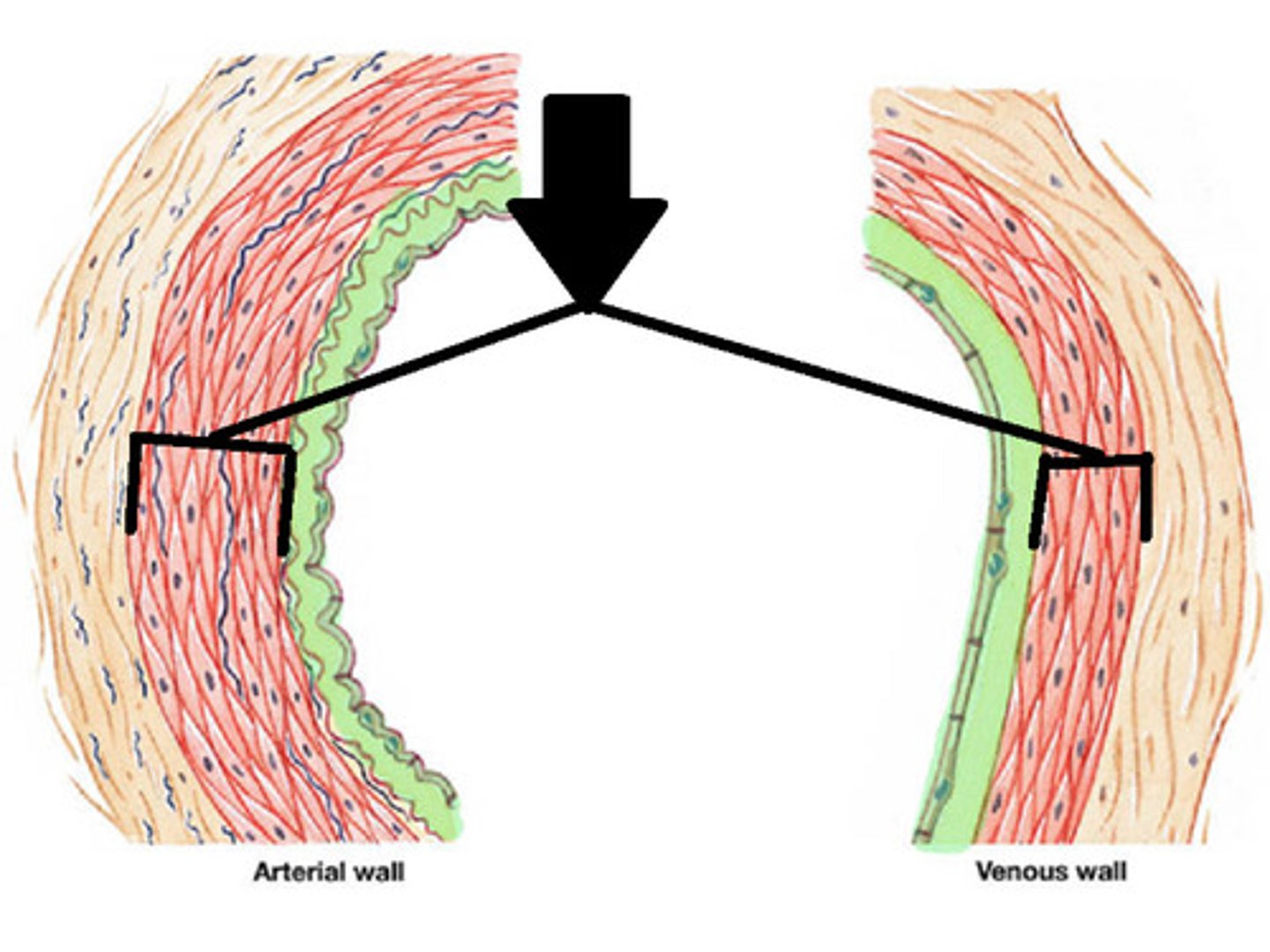
Tunica intima
Innermost layer, forms valves.
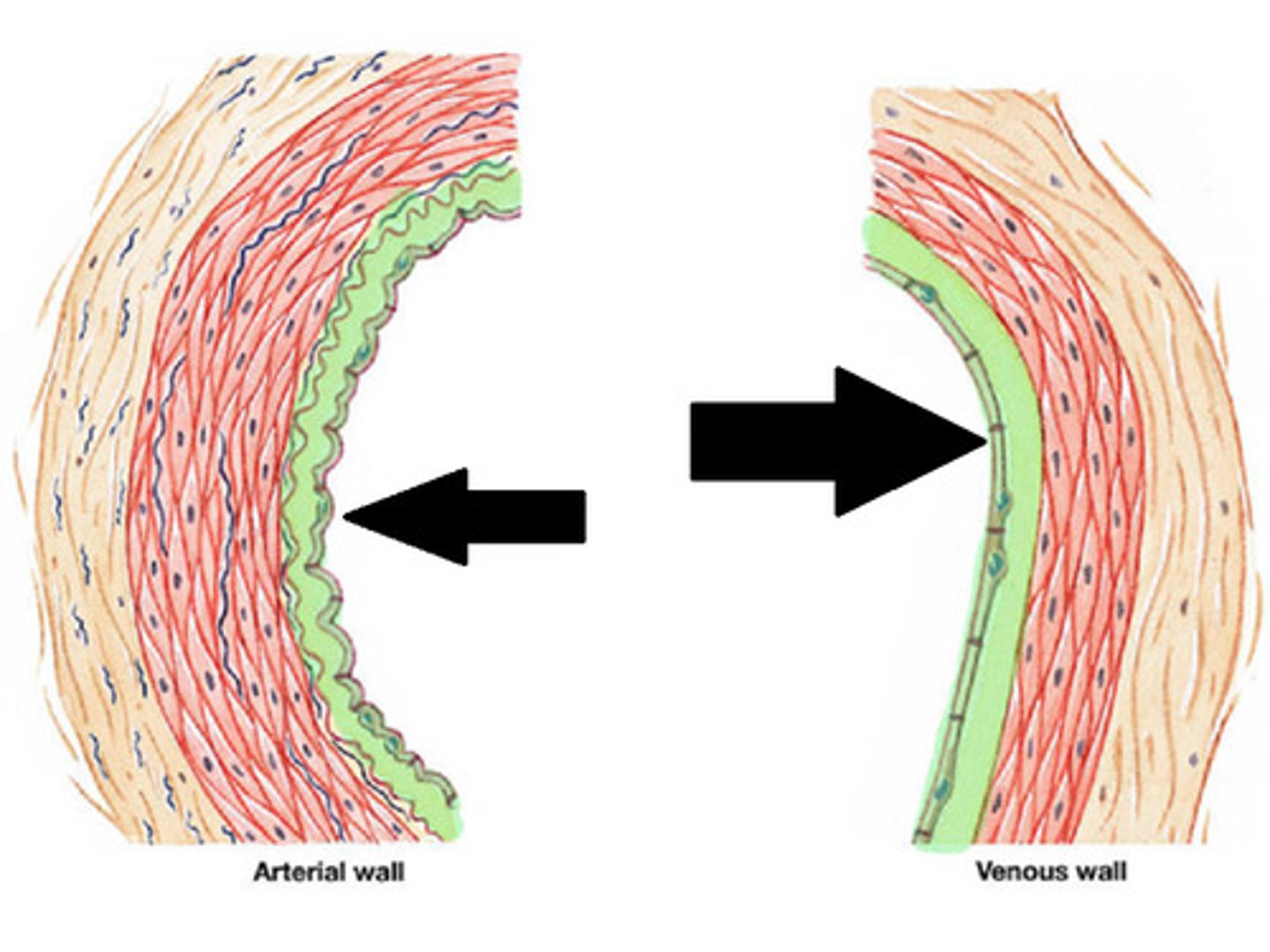
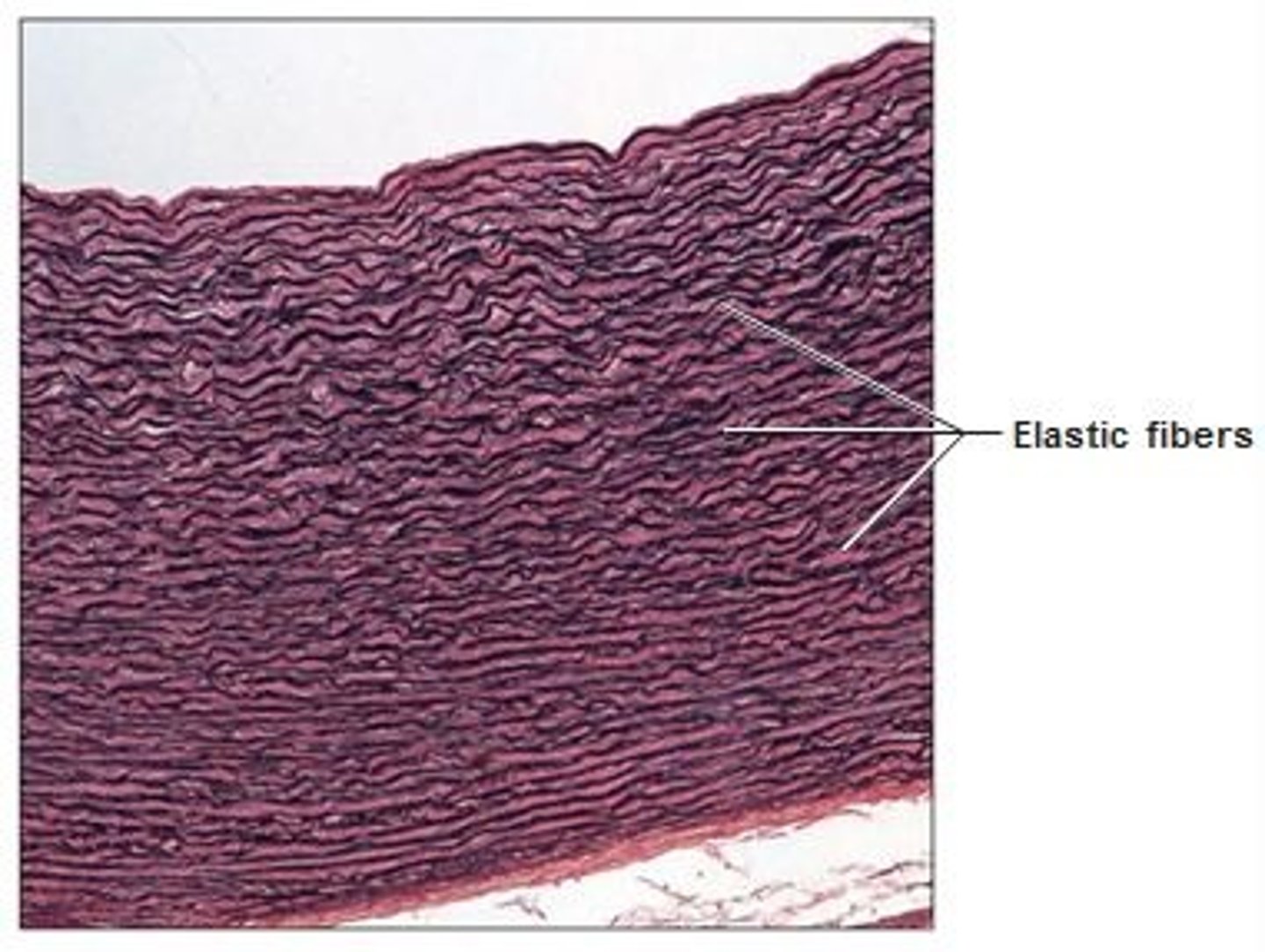
Elastic arteries
Contain large amounts of elastic fibers.
Muscular arteries
More smooth muscle, less elastic fibers.
Arterioles
Smallest arteries leading to capillaries.
Innate immunity
Non-specific defense mechanism present at birth.
Adaptive immunity
Specific defense developed after antigen exposure.
T lymphocytes
Coordinate immune response and attack infected cells.
B lymphocytes
Produce antibodies against antigens.
Dendritic cells
Present antigens to lymphocytes.
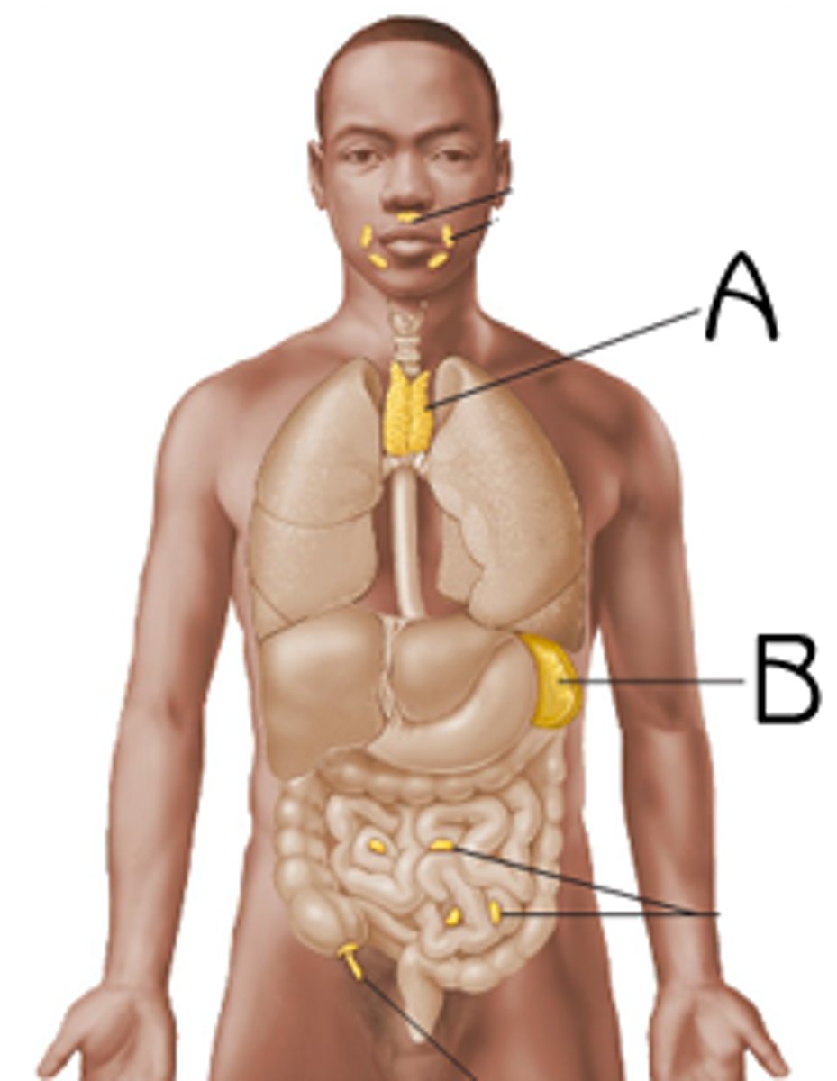
MALT
Mucosa-associated lymphoid tissue in various tracts.
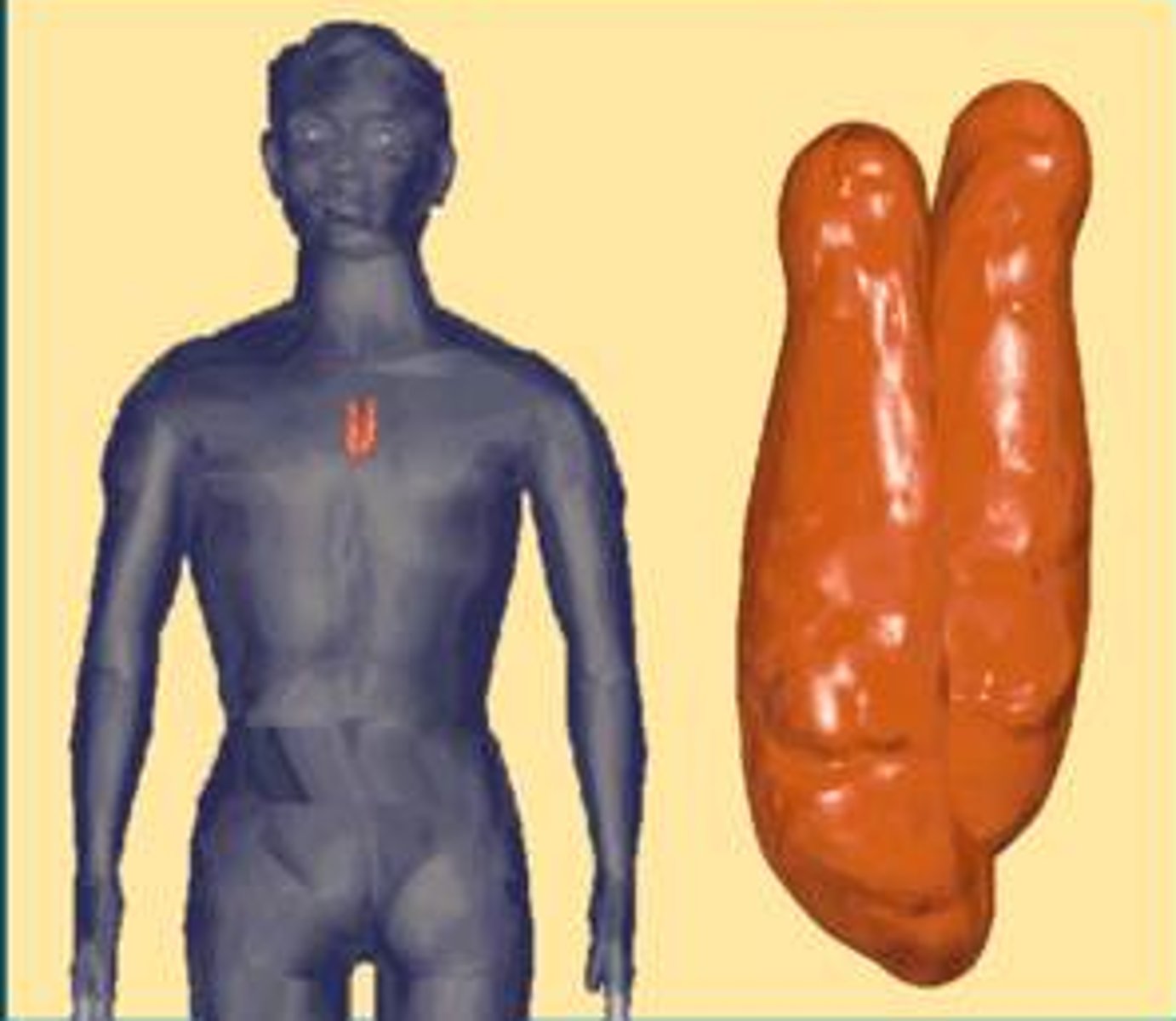
Thymus
Site of T cell maturation.
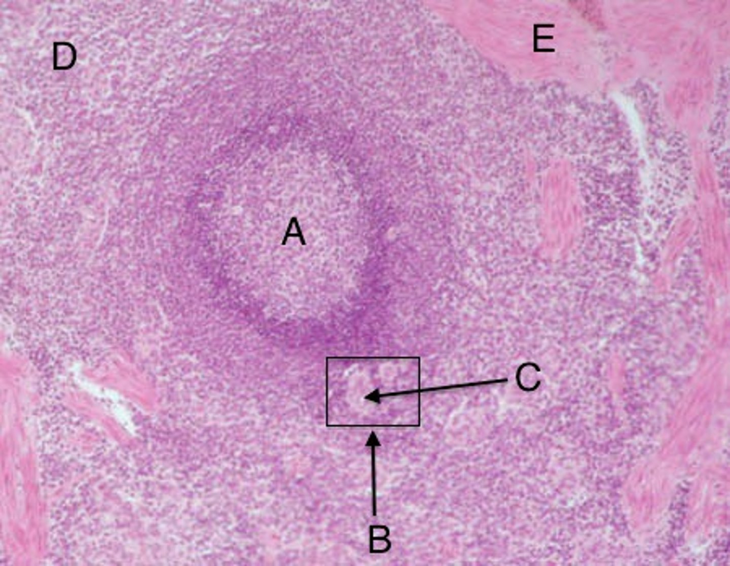
Spleen
Largest lymphoid organ in the body.
White pulp
Contains lymphocytes for immune response.
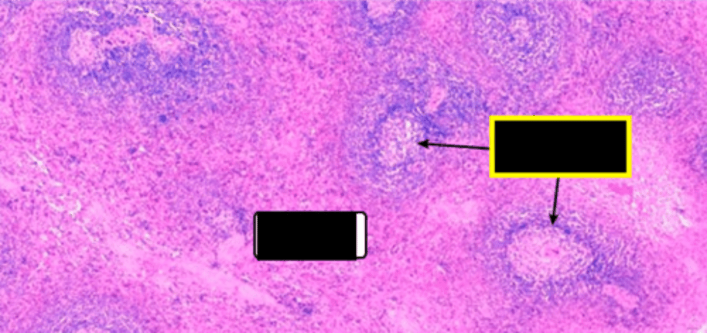
Red pulp
Rich in blood; removes old cells.
Macrophages
Cells that break down old erythrocytes.
Respiration
Process of gas exchange in the body.
Inhalation
Breathing in oxygen from the environment.
Exhalation
Breathing out carbon dioxide from the body.
Conducting portion
Air passage without gas exchange.
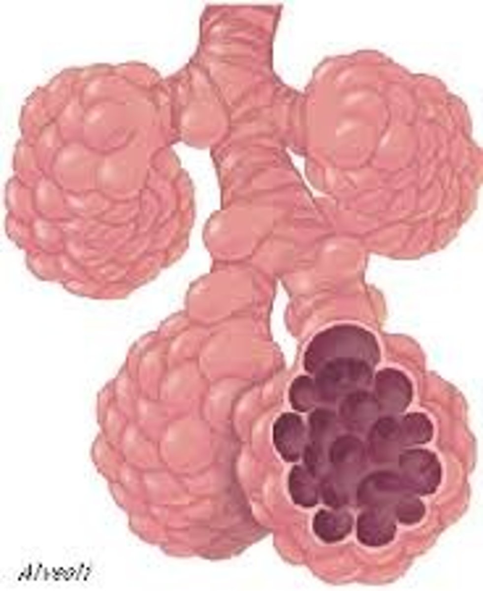
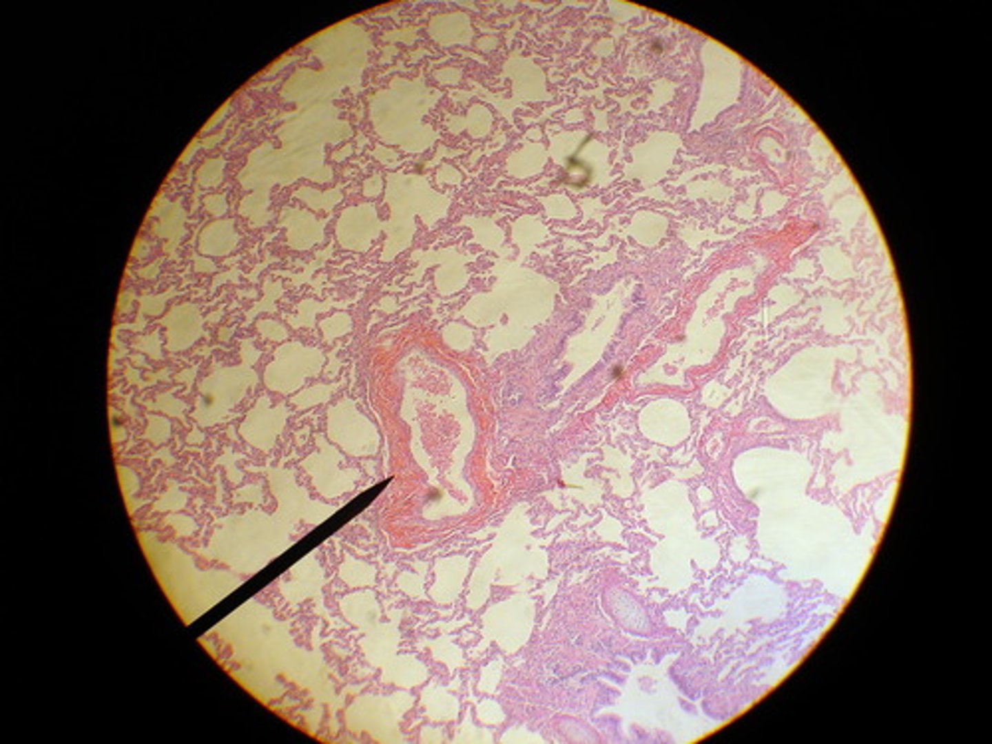
Respiratory portion
Site of gas exchange in lungs.
Alveoli
Tiny air sacs for gas exchange.