Chap. 9-Anatomy of Hearing (CHAPTER REVIEW)
1/79
Earn XP
Description and Tags
Chapter Nine of Seikel, J. A., Drumright, D. G., Hudock, D. J. (2021). Anatomy and physiology for speech, language, and hearing. Sixth Edition. Plural Plus
Name | Mastery | Learn | Test | Matching | Spaced | Call with Kai | Chat |
|---|
No analytics yet
Send a link to your students to track their progress
80 Terms
“Energy Transducer”
(of the ear) converts acoustic energy into electrochemical energy
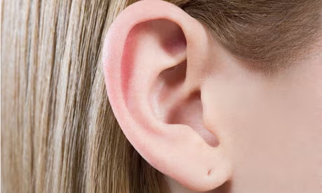
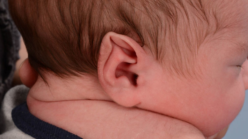
Pinna
(aka auricle) of the outer ear, collector of sound to be processed deeper. Structure provided by cartilaginous framework. Latin for FEATHER.
Helix
Forms curled margin of pinna, marking its most distal borders. Greek for COIL.
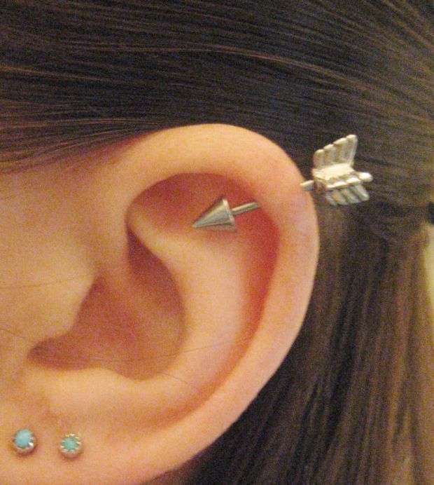
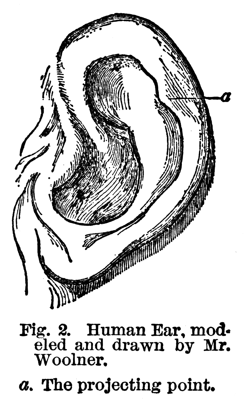
Auricular (Darwin’s) tubercle
Superior-posterior bulge on the helix.
Antihelix
Immediately anterior to the helix, fold of tissue marking entrance to the concha.
Scaphoid Fossa
Between helix and antihelix.
Crura Anthelicis
Produced by the antihelix bifurcating superiorly.
Cymba Conchae
Anterior extension of helix, marks anterior entrance to concha.
Cavum conchae
Deep portion of the concha
Concha (external auditory meatus)
Entrance to the ear canal
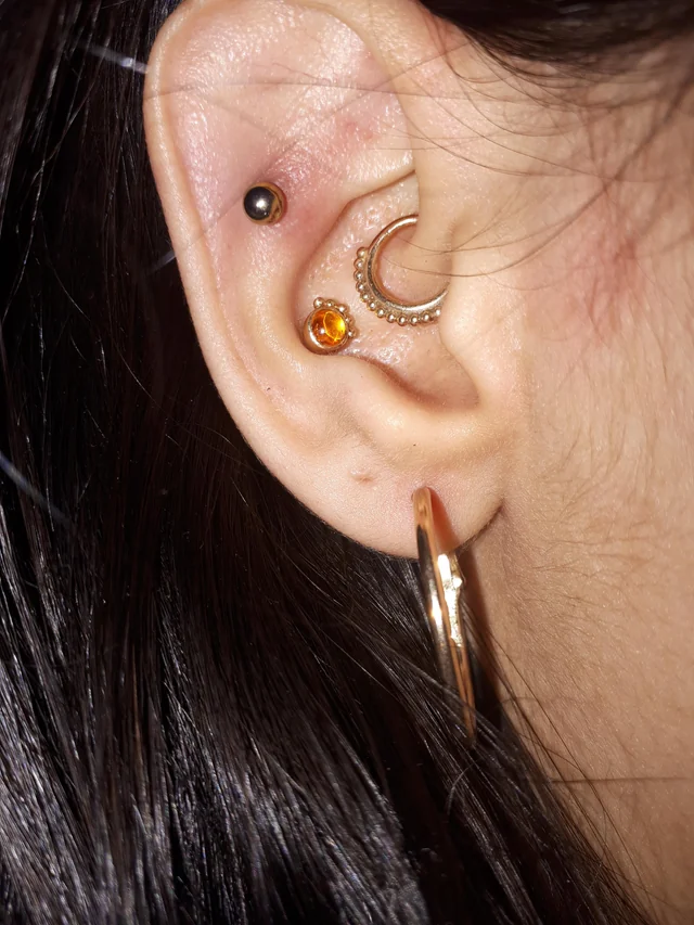
Tragus
Flap of epithelium-covered cartilage, may have covered the meatus entrance in early version of auditory mechanism
Tuberculum supratagicum
Superior to the tragus
Antitragus
Posterior and inferior to the tragus
Intertragic incisure
Region between tragus and antitragus
Lobule
Below the antitragus
Auricular cartilage
Unitary structure, covered with a layer of epithelial tissue invested with fine hairs, useful for keeping insects and dirt out of the ear canal.
External auditory meatus
7 mm diameter, 2.5 cm long from depth of concha.
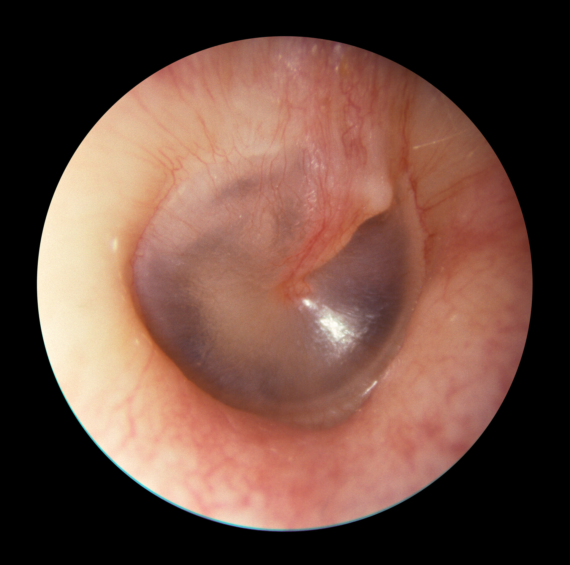
Tympanic membrane (eardrum)
thin, oval, semi-transparent membrane that separates the outer ear from the middle ear.
Cerumen
Ear wax, along with hairs in the EAM, they trap insects and dirt, protecting the tympanic membrane. Secreted by glands into the ear canal.
Umbo
Most distal point of attachment of inner TM to the malleus in middle ear.
Cone of light
Inferior and anterior to the TM, Reflects the light of the audiologist’s otoscope.
Otitis externa
Inflammation of external ear skin. May result from bacterial infection after trauma, abrasion, or viral infection (herpes zoster virus) Very painful infection that can lead to facial paralysis or hearing loss when facial or vestibulocochlear nerves are involved.
Edema
Swelling caused by inflamed tissue
Auricular malformations
Arise from issues in embryological development
Deformations
Arise from effects of physical forces on the prenatal structures.
Chondrogenesis
Failure of cartilage to develop
Meatal Atresia
cartilage doesn’t develop, born without the external auditory meatus
Microtia
Small auricle
Polyotia
When parts of auricle are duplicated, sometimes leading person to develop a second tragus- may occur in hemifacial microsomia.

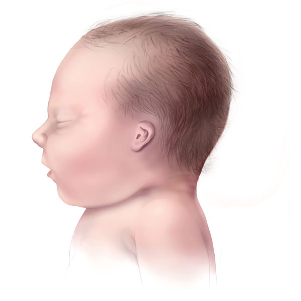
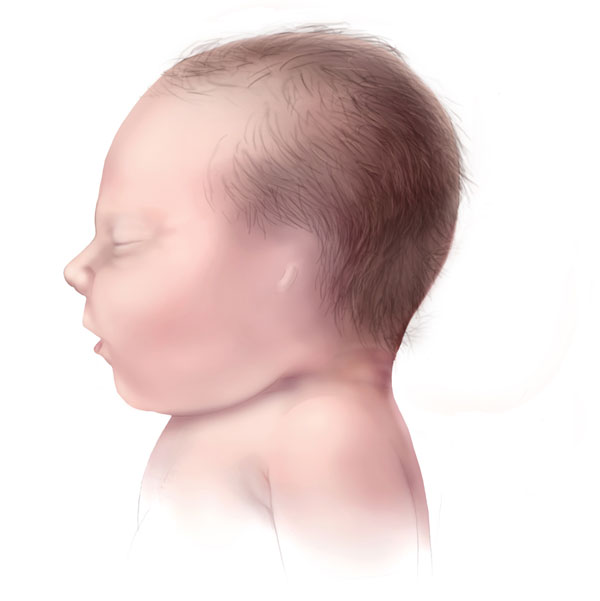
Preauricular Tags
Prominences that form prenatally anterior to the pinna

Cryptotia
Congenital absences or maldevelopment of upper portion of the ear
Anotia
Complete absence of pinna
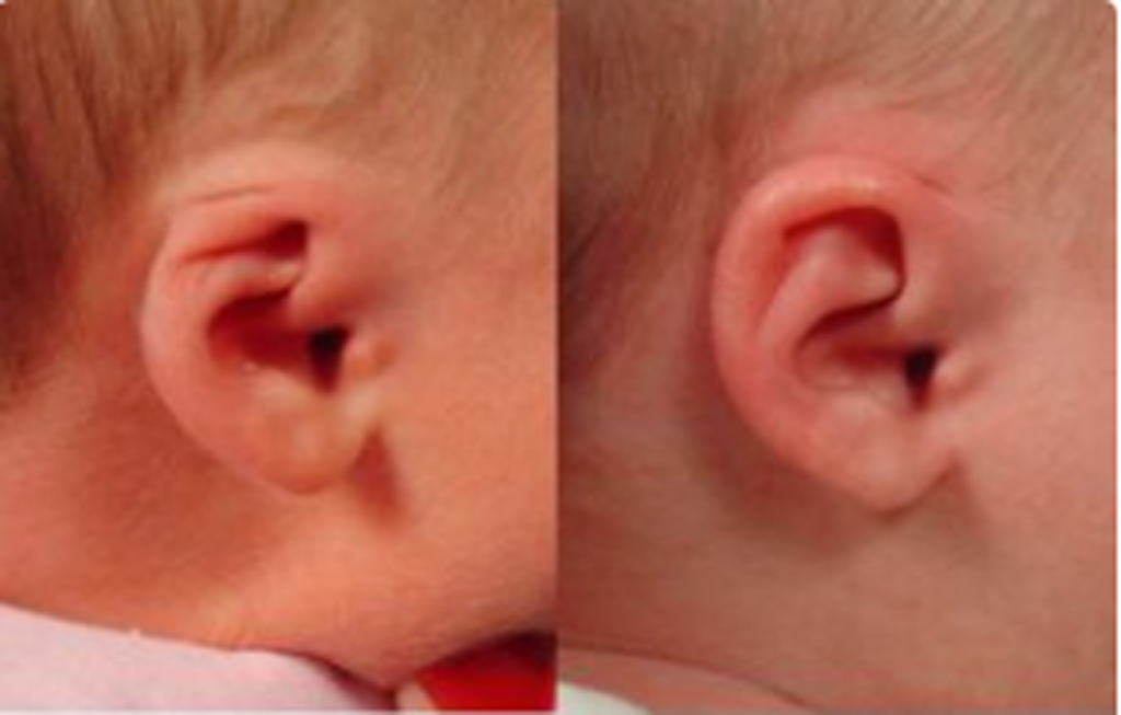
Stahl’s ear
pointy, elfin-shaped ears.
Pars flaccida
“flaccid part” of the TM in the superior quadrant.
Anterior and posterior malleolar folds
the recess on either side of the pars flaccida
Tympanic sulcus
Groove in the temporal bone, incomplete in superior aspect to accommodate the anterior and posterior malleolar folds.
Cuticular Layer
Outer layer of the TM tissue, continuation of epithelial lining of EAM and pinna
Fibrous layer
intermediate layer of the TM tissue, composed of TWO layers
Superficial layer (of TM fibrous layer)
Composed of fibers that radiate out from handle of malleus to the periphery
Deep Layer (of TM fibrous layer)
Made up of circular fibers that are found mostly in the periphery of the membrane
Mucous layer
Inner layer of the TM, continuous with mucosa of middle ear
Ossicles
bones of the ear (malleus, incus, and stapes)
Ossicular chain
made of three articulated bones, provide means for transmission of acoustic energy striking on the TM to inner ear
Malleus
Largest of ossicles, provides point of attachment with TM
Manubrium
Handle of the malleus, long process, separated from the head by a thin neck
Anterior and lateral processes
Provide attachment for ligaments
Caput
Head, head of malleus protrudes into epitympanic recess of middle ear.
Incus
“The anvil” provides intermediate communicating link of ossicular chain.
Incus body
articulates with the head of the malleus through the malleolar facet, allowing long process of incus to be parallel with manubrium of malleus
Lenticular process
process of the incus where the stapes articulates
Stapes
“stirrup”, caput articulates with lenticular process of incus, and neck bifurcates to become the crura.
Bifurcate
to cause to DIVIDE into TWO branches or parts
Serous (secretory) otitis media
any condition where fluid accumulates in middle ear cavity
Auditory (pharyngotympanic) tube
allows oxygen into the middle ear space (aeration)
Tensor tympani
muscle, inserts into upper manubrium malli and pulls the malleus anteromedially.
Medial wall of middle ear cavity landmarks
oval window, round window, promontory of cochlea, and prominence of the facial nerve
Anterior wall
Houses entrance to auditory tube
Posterior wall
houses the prominence of the stapedial pyramid.
Stapedius muscle
inserts into posterior neck of stapes and pulls stapes posteriorly
Ligaments of ossicular chain
superior, anterior, and lateral of the malleus ligaments (SAL), and posterior and superior ligaments of incus (PS)
Inner ear
Houses sensors for balance (vestibular) and hearing (cochlea)
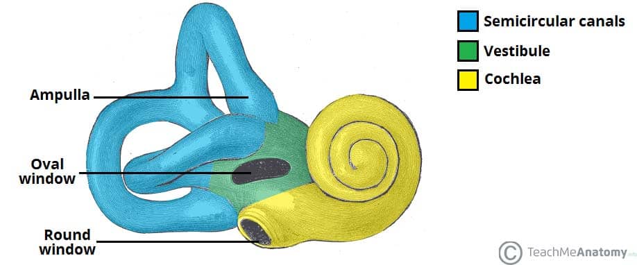
Vestibule
entrance to cochlea
Osseous labyrinth
(the snail) made up of entryway to the labyrinth, vestibule, semicircular canals, and osseous cochlear canal
Two incomplete chambers of the labyrinth
scala vestibuli and scala tympani
Osseous spiral lamina
incomplete bony shelf protruding from modiolus. divides the scala vestibuli and scala tympani
Round window
provides communication between scala tympani and middle ear space
Mediolus
latin for hub
Oval window
permits communication between the scala vestibuli and middle ear space
Cochlear aqueduct
connects the upper duct and the subarachnoid space
Crista
receptor organ for movement within vestibular mechanism
Membranous labyrinth
fluid filled sac, rests within the cavity of the osseous labyrinth, filled with endolymph. Houses the vestibular organ.
Ampulla
expanded region of the semicircular canals, contains crista ampullaris. In the vestibule lies the utricle and saccule.
Scala media
Made up of the membranous labyrinth of cochlea residing between the scala vestibuli and scala tympani
Reissner’s membrane
forms upper boundary of the scala media, and the basilar membrane forms the floor.
Organ of Corti
has four rows of hair cells resting on a bed of Deiter’s cells for support
Outer hair cells
separated from the inner hair cell row by the tunnel of Corti
Tunnel of corti
separates outer hair cells from inner hair row
Stereocilia
graces the upper surface of each hair cell, connected by tip links