Cancerous and Precancerous Histopathology Laboratory
1/96
There's no tags or description
Looks like no tags are added yet.
Name | Mastery | Learn | Test | Matching | Spaced | Call with Kai |
|---|
No analytics yet
Send a link to your students to track their progress
97 Terms
Colon
Desmoplasia (Colon)
Colon
Infiltrative edge (Colon)
Colon
Tumor Buds (Colon)
Colon
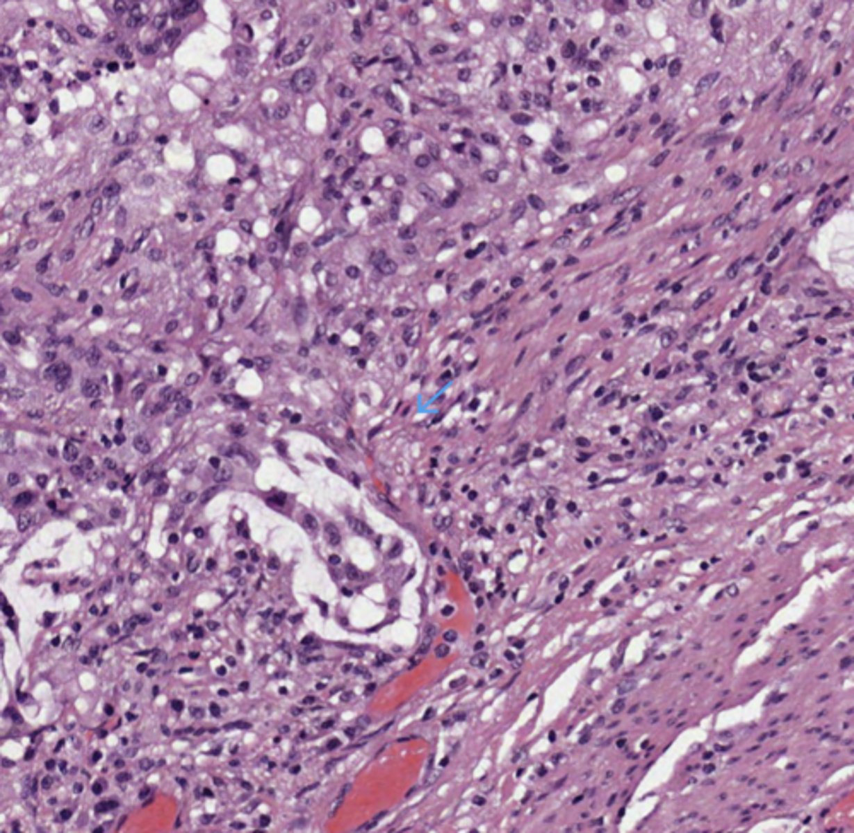
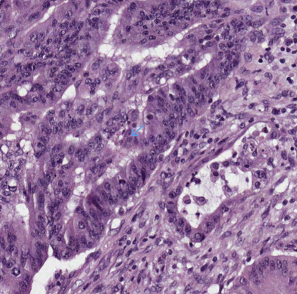
Colorectal adenocarcinoma (Colon)
Colon
Mucinous differentiation (Colon)
Colon
TILs (tumor-infiltrating lymphocytes, Colon)
Colon
Tumor Budding (Colon)
Colon
Acute perivisceral inflammation (Colon)
Colon
"Dirty necrosis" (Colon)
Colon
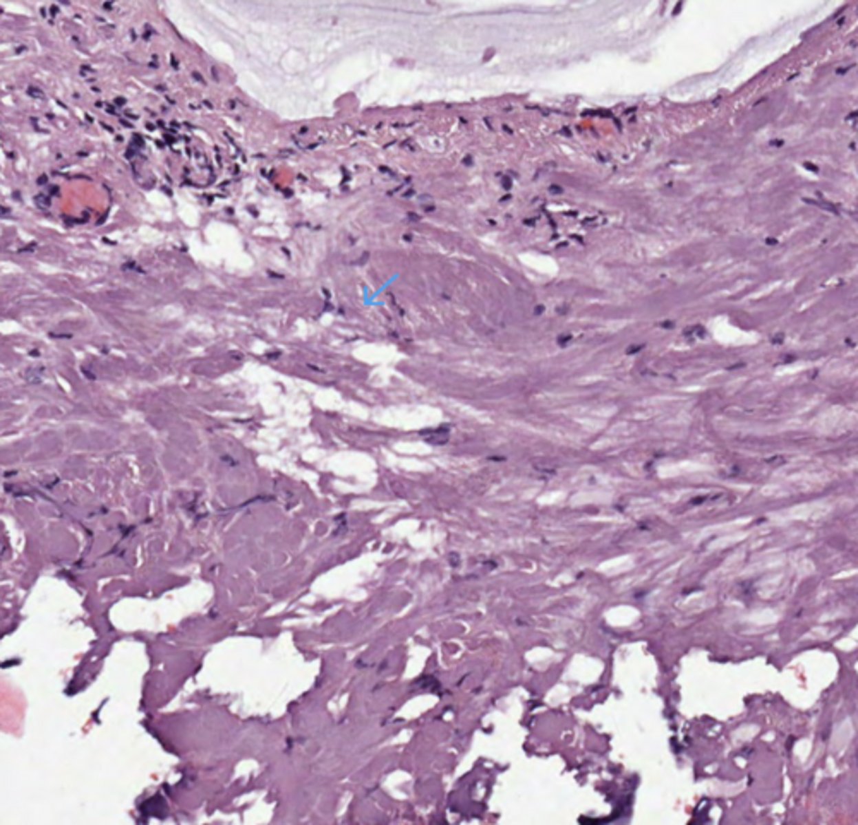
Desmoplasia (Colon)
Colon
Mucinous differentiation (Colon)
Colon
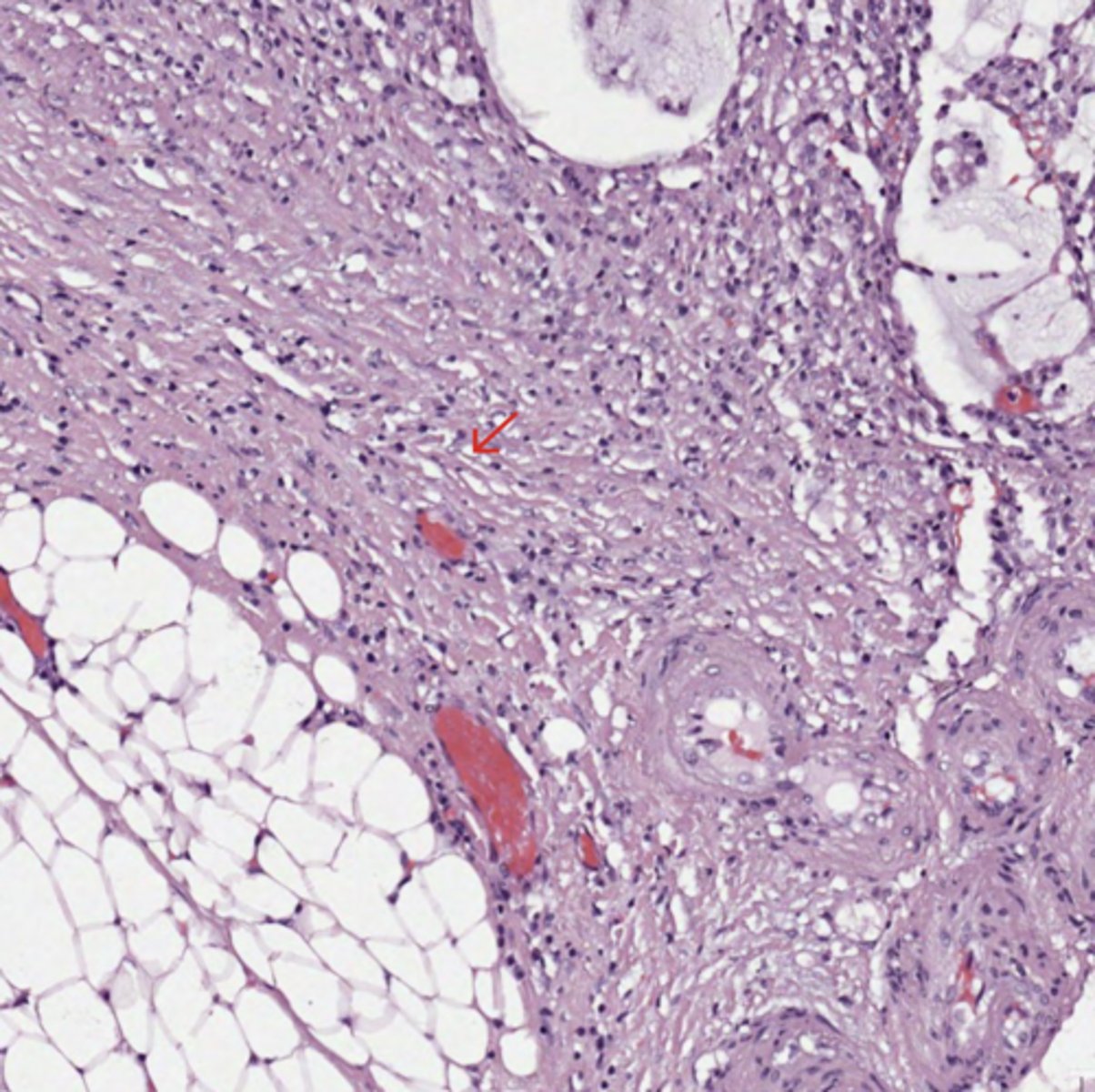
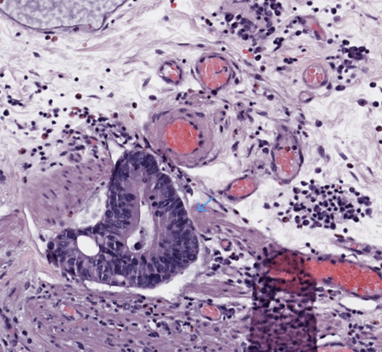
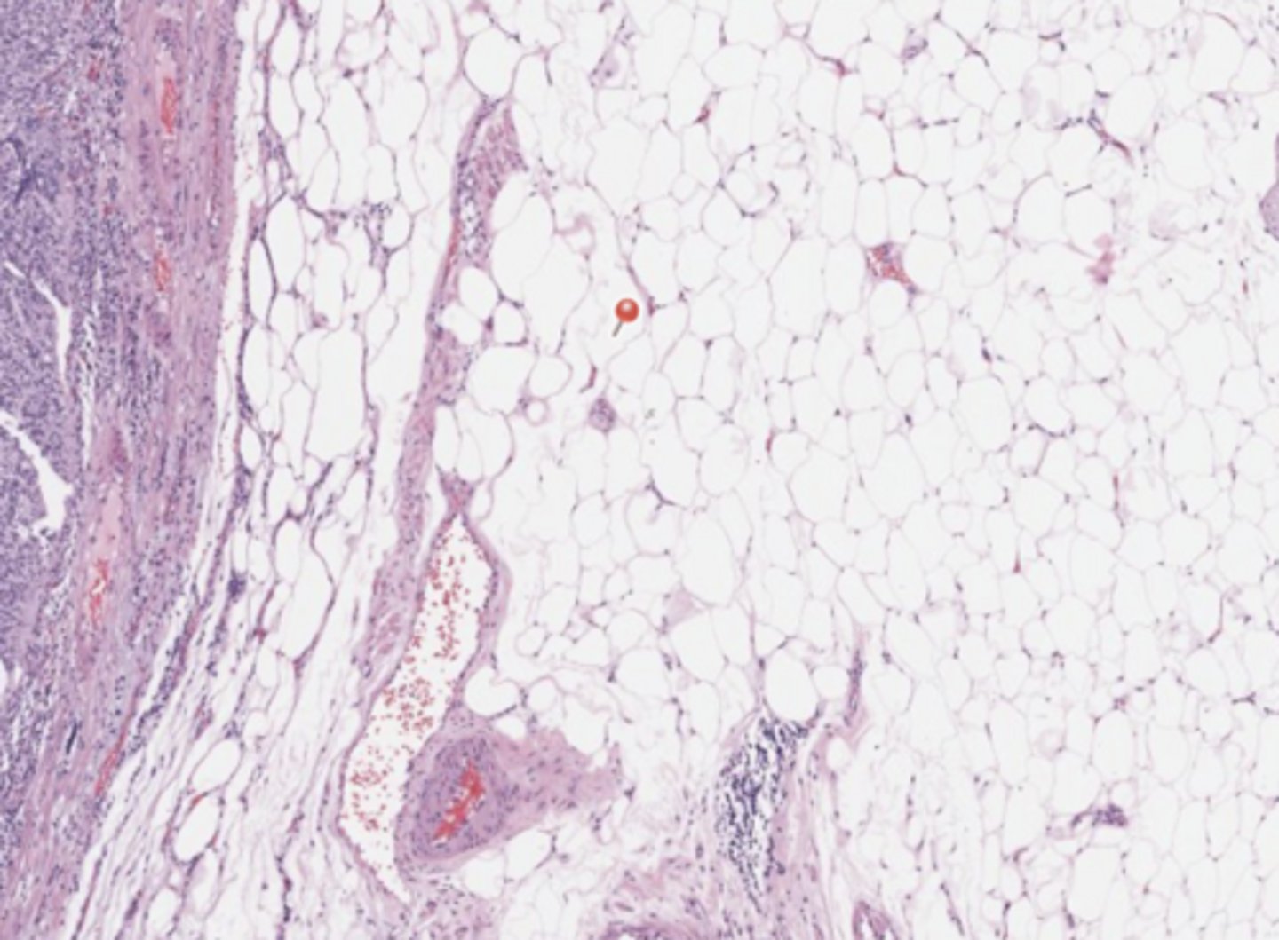
The tumor infiltrates through the muscularis propria into the pericolorectal/pericolic adipose tissue. (Colon)
* My note - the arrow actually points at desmoplasia, the infiltration is seen around it on the left and right fields
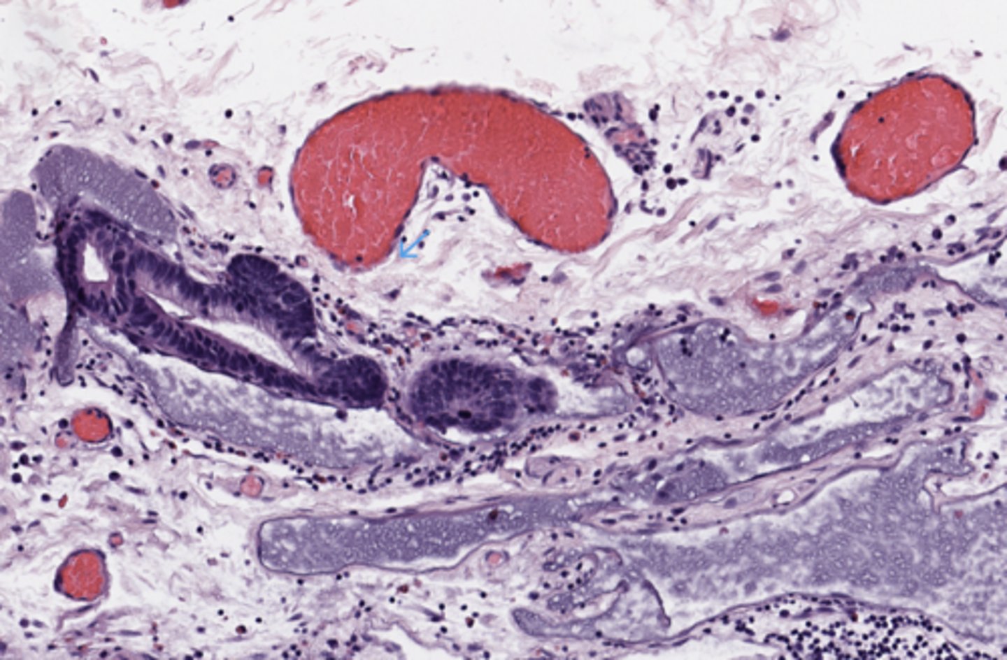
Stomach
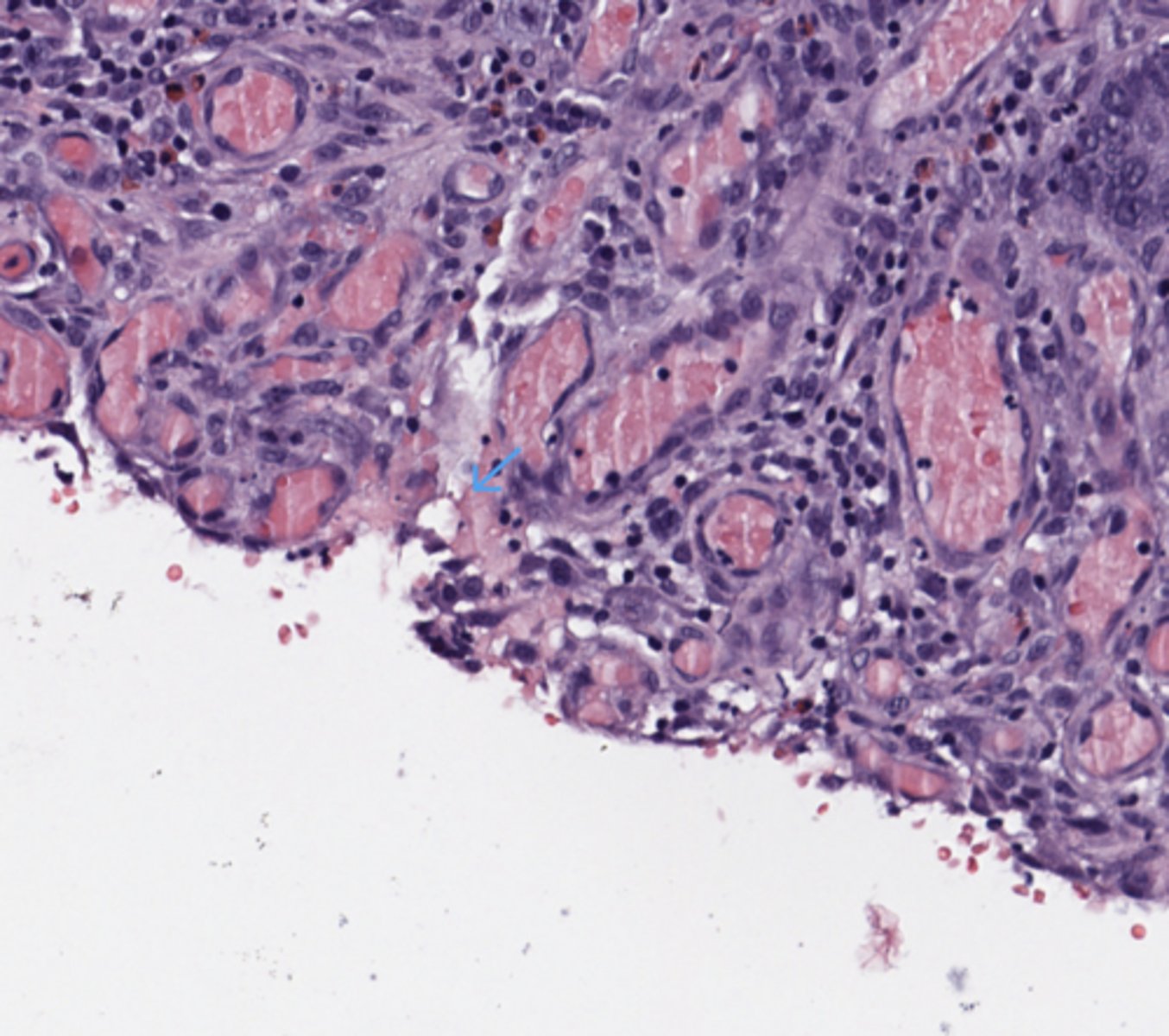
Lymphovascular invasion (Stomach)
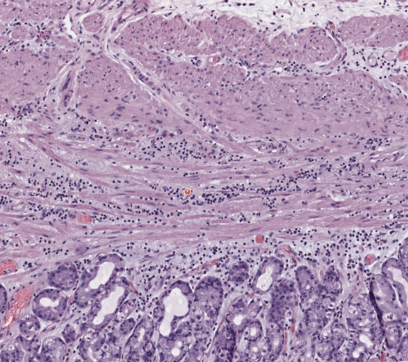
Stomach
Muscolaris Mucoase (Stomach)
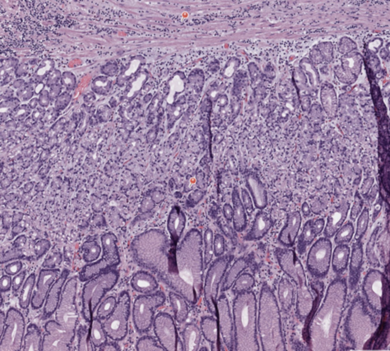
Stomach
Gastric Mucosa (Stomach)
Stomach
Submucosae (Stomach)
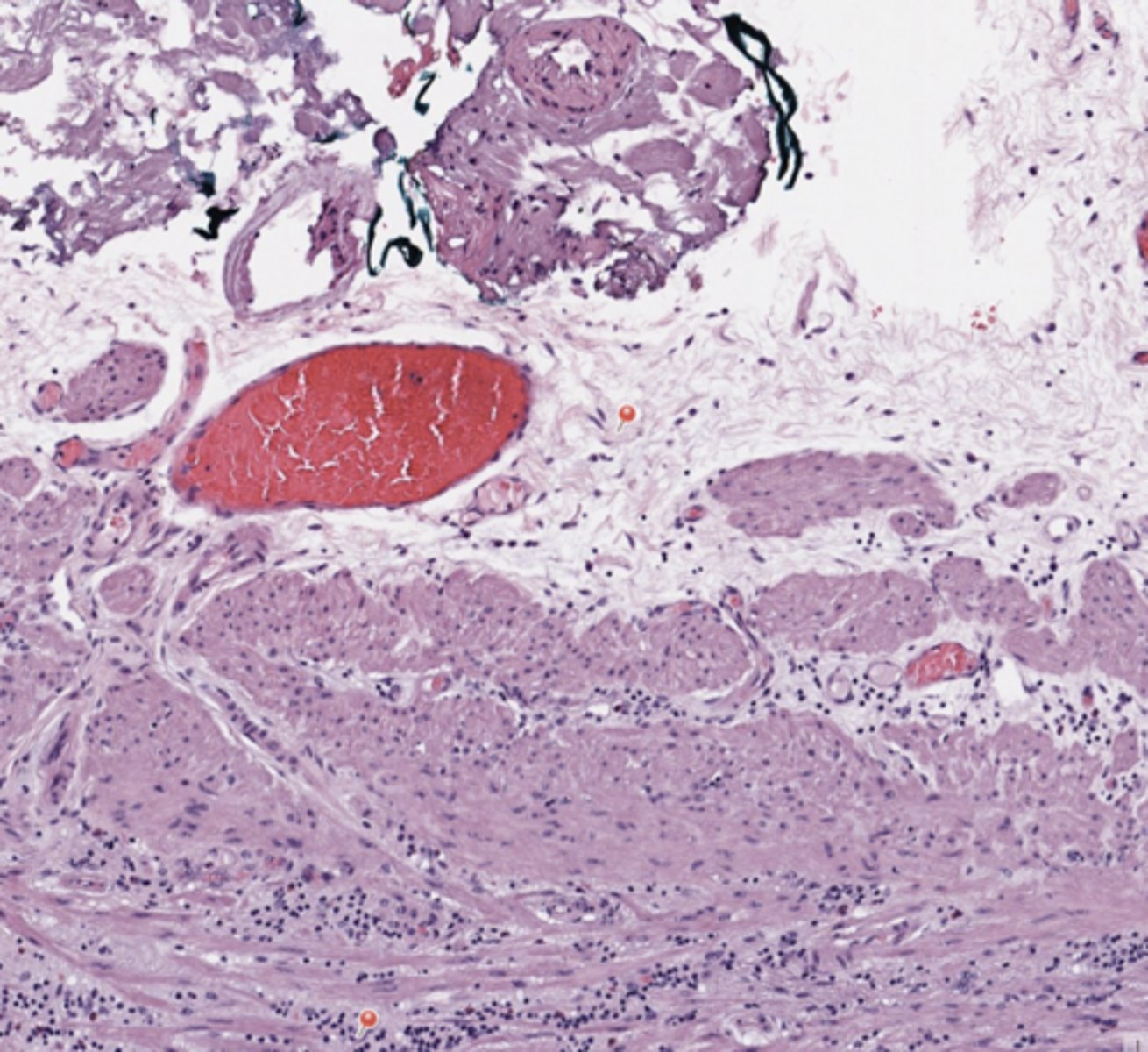
Stomach
Lymphovascular invasion (Stomach)
Stomach
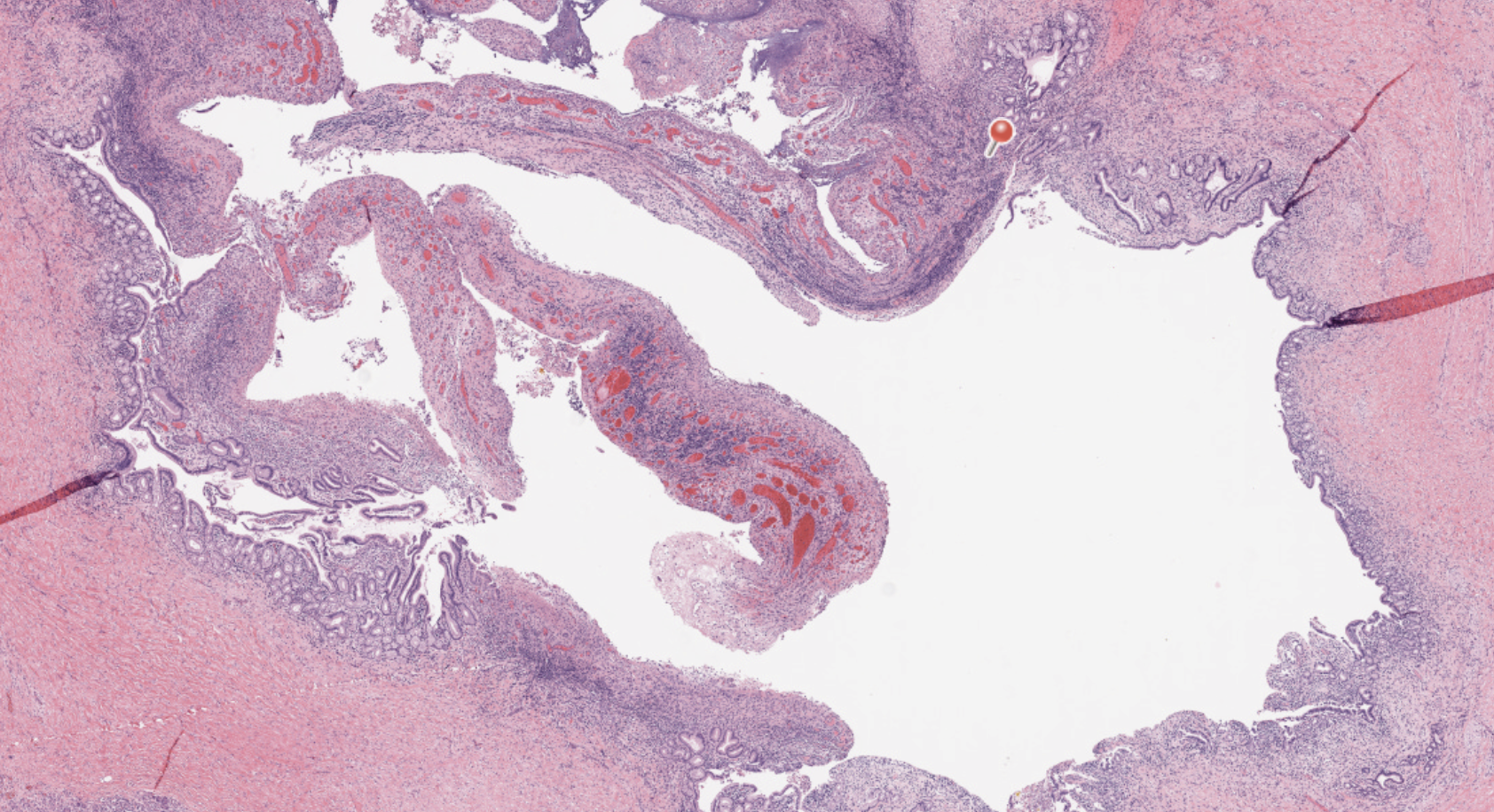
Gastric ulcer (Stomach)
Stomach
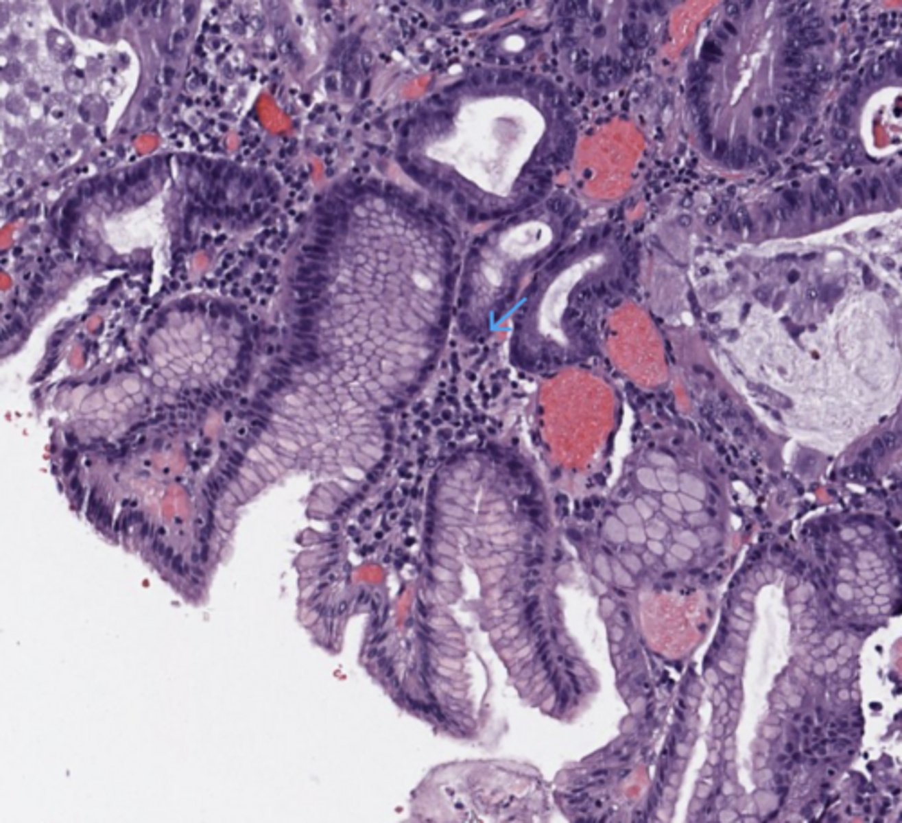
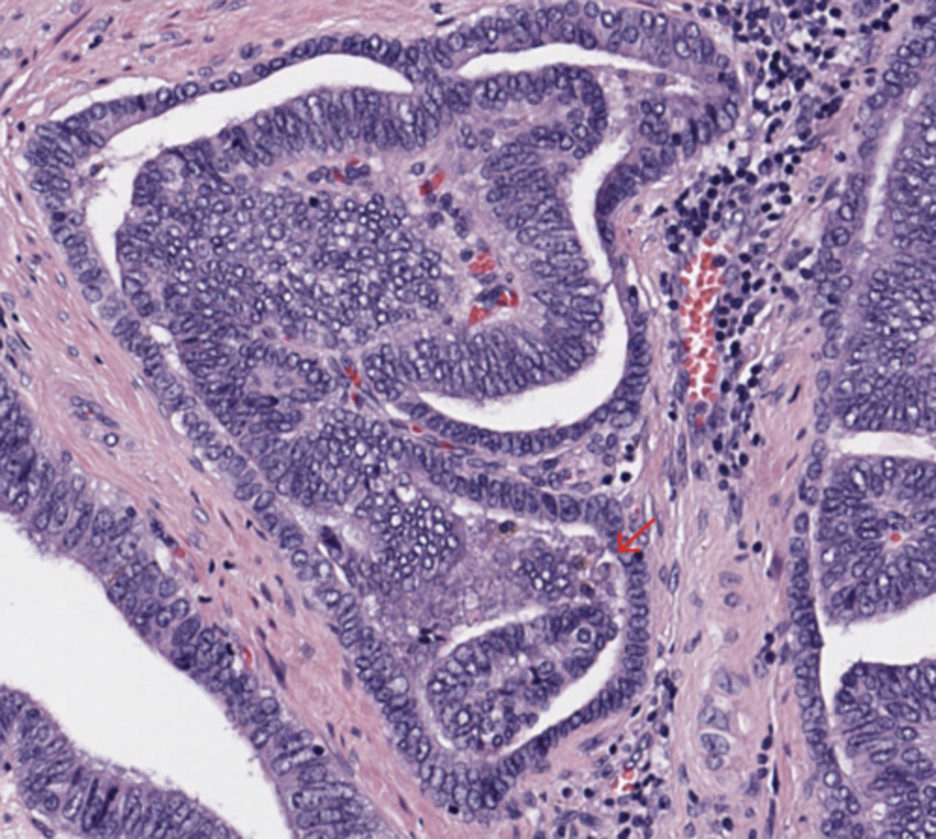
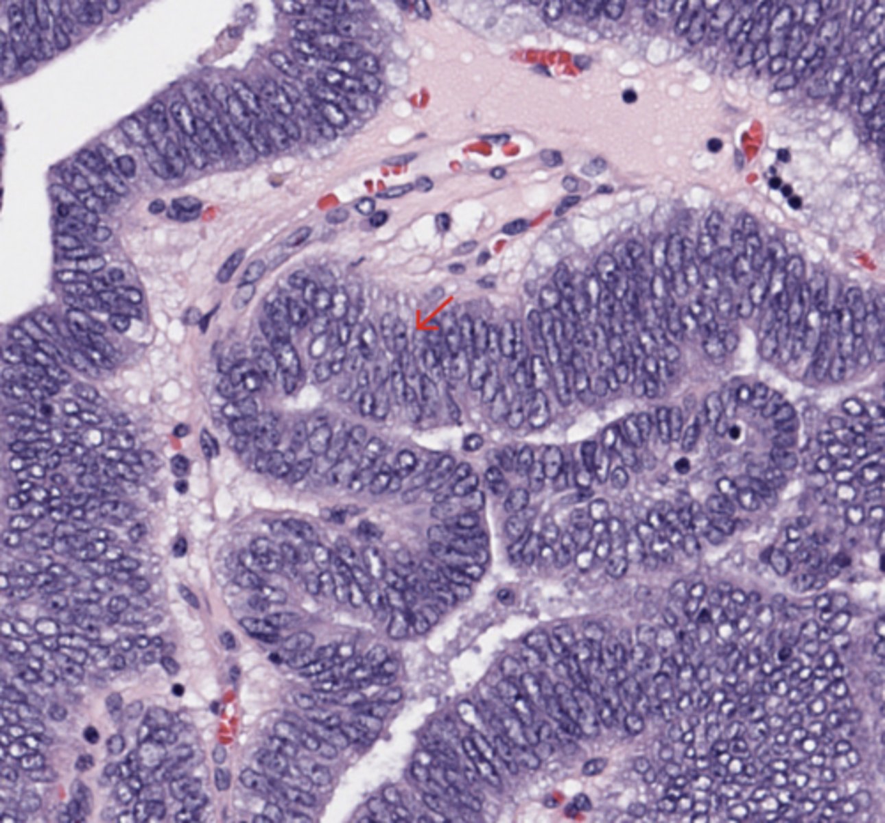
Gastric dysplasia (Stomach)
Stomach
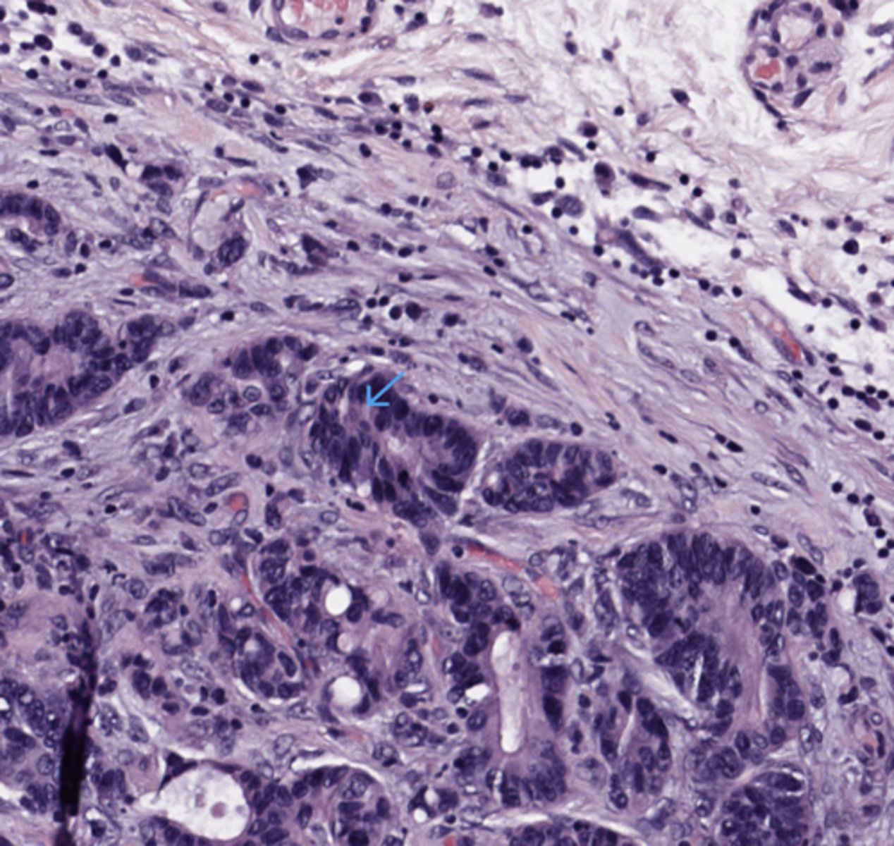
Infiltrating adenocarcinoma (Stomach)
Breast
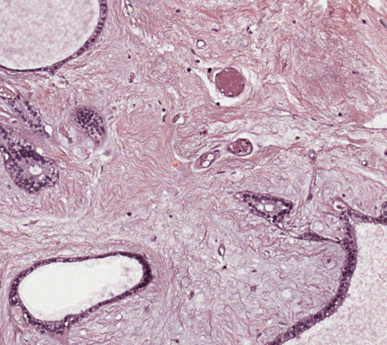
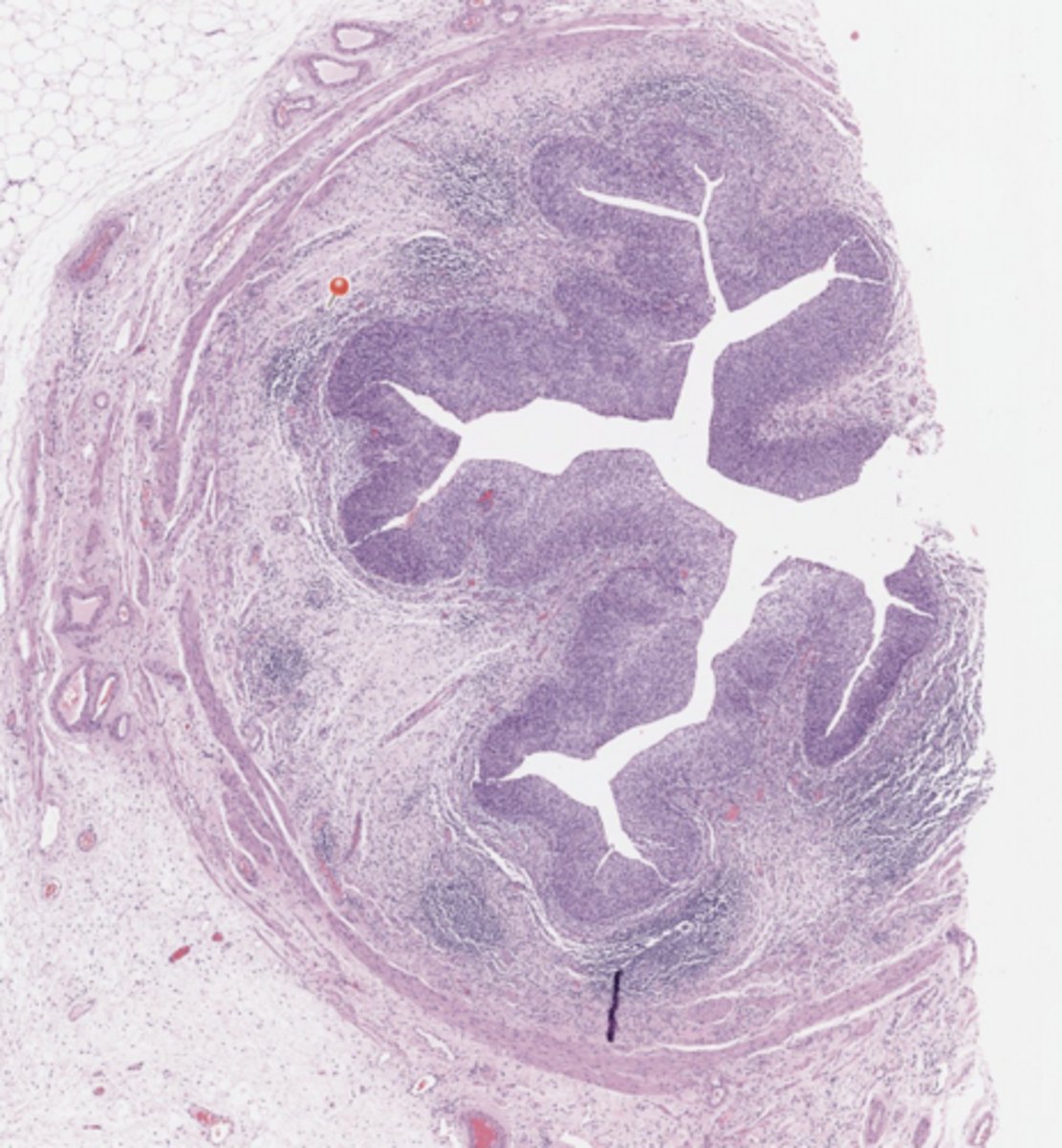
Fibroadenoma (Breast)
Breast
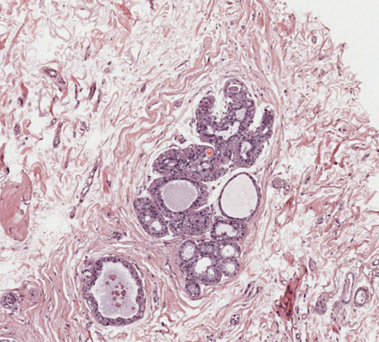
Normal lipoinvoluted breast gland parenchyma (Breast)
Breast
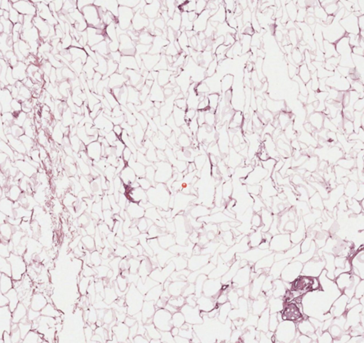
Adipose tissue (Breast)
Breast
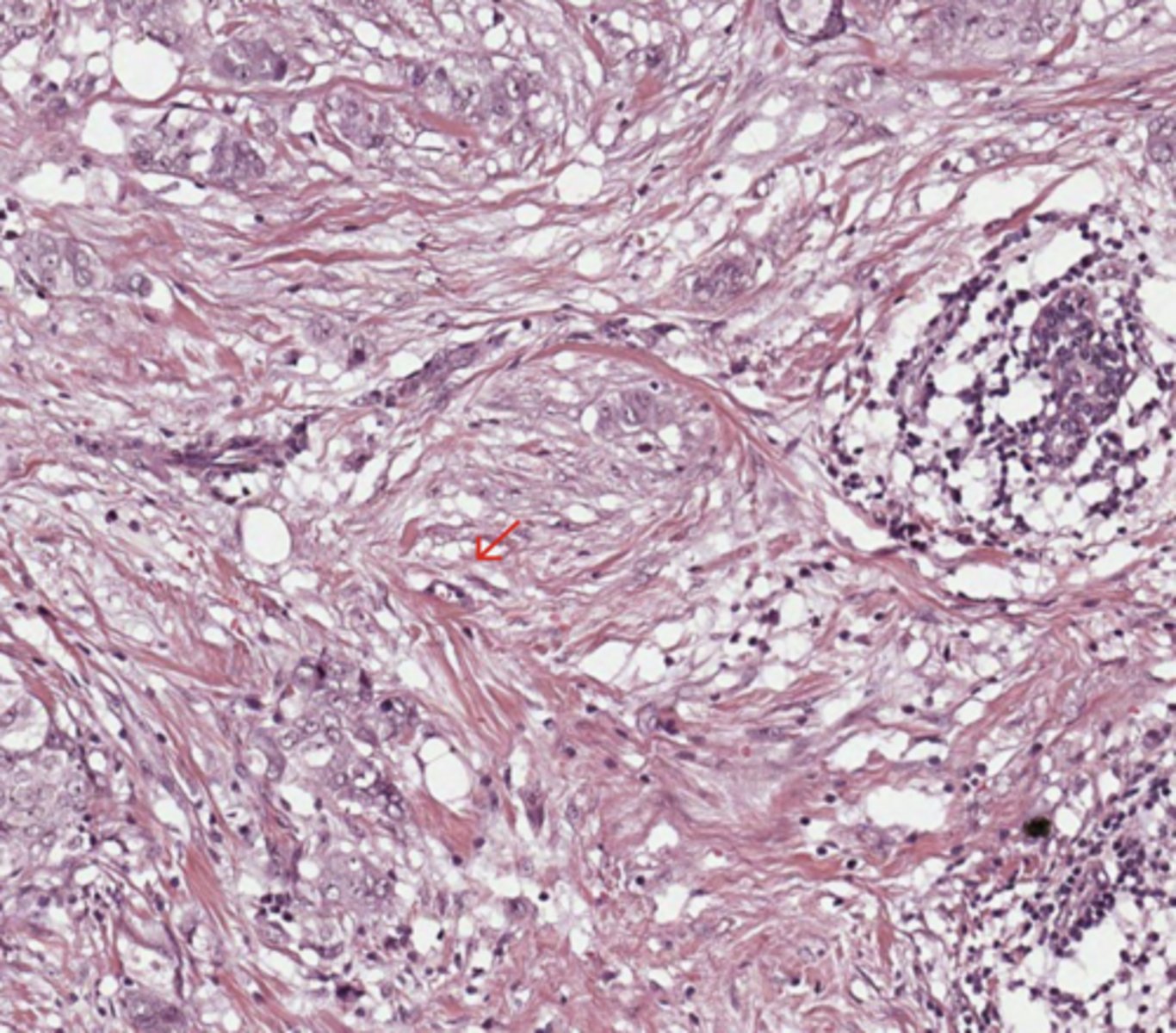
invasive breast carcinoma (IBC) of no special type (NST)
(Breast)
Breast
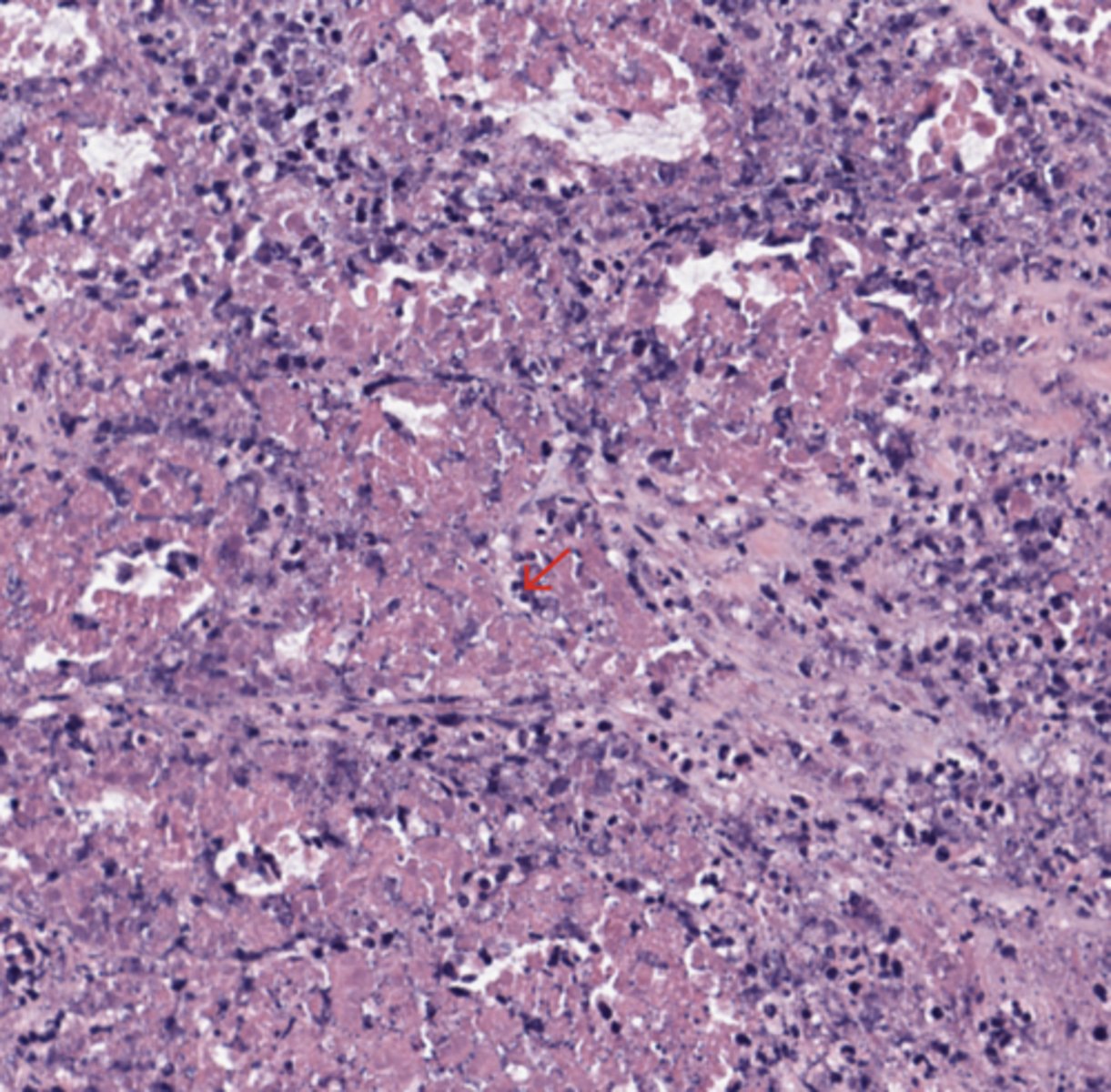
Necrosis + Calcification - Comedonic Carcinoma (Breast)
breast
TILs (Breast)
Breast
In situ carcinoma with necrosis in breast carcinoma (Breast)
Breast
Striated muscle (Breast)
Breast
Mamma ducts (Breast)
Breast
Mitosis (Breast)
Kidney
Kidney cancer - Vessels
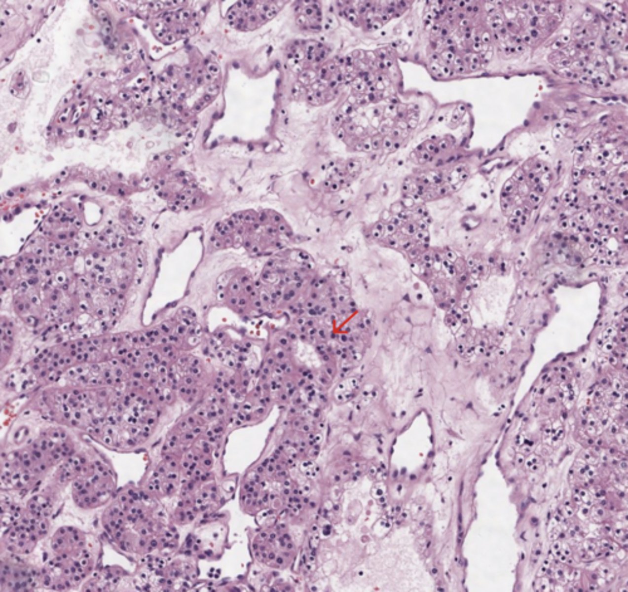
Network of arborizing(branching) small, thin walled vessels (important diagnostic feature for cases with granular eosinophilic cytoplasm)
(Kidney)
*My note - this is "chicken wire" pattern, we see many vessels bc tumors are highly vascularized.
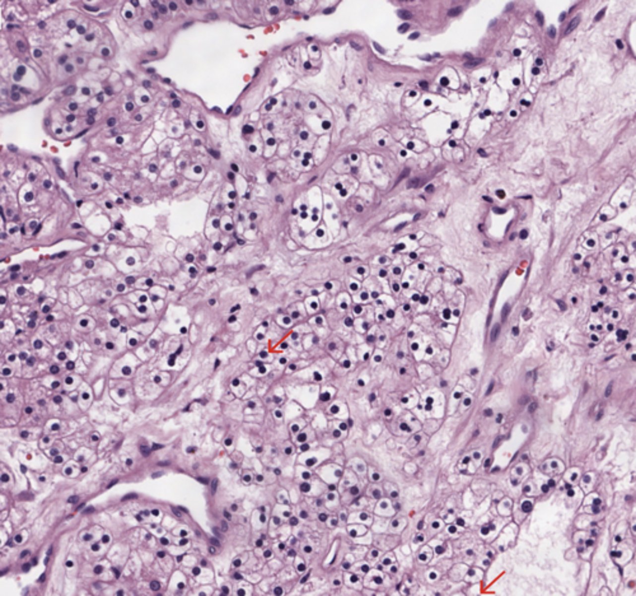
Kidney
Kidney cancer - Cells
Nests of large cells with cleared out and vacuolated cytoplasm and nuclear atypia.
(Kidney)
*My note - this is Clear Cell Renal Cell Carcinoma (ccRCC), the most common type of kidney cancer.
Clear cell carcinoma is called “clear cell” because the tumor cells contain lipid-rich cytoplasm, which becomes clear during histological processing.
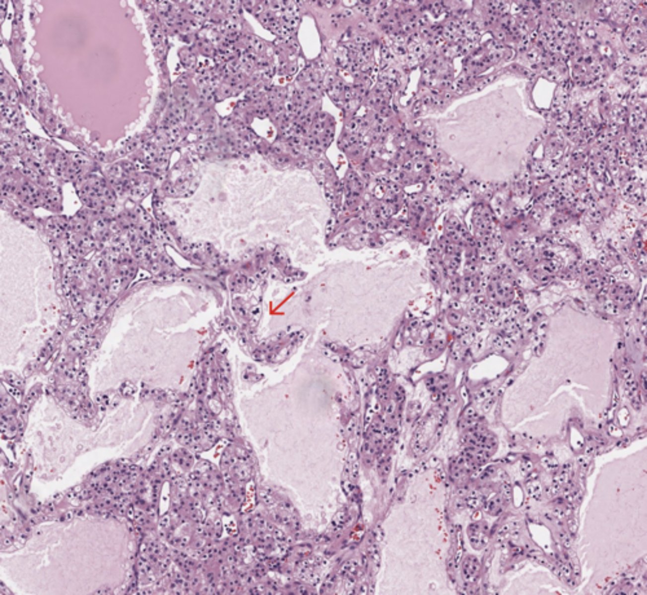
Kidney
Kidney cancer - Architectural patterns
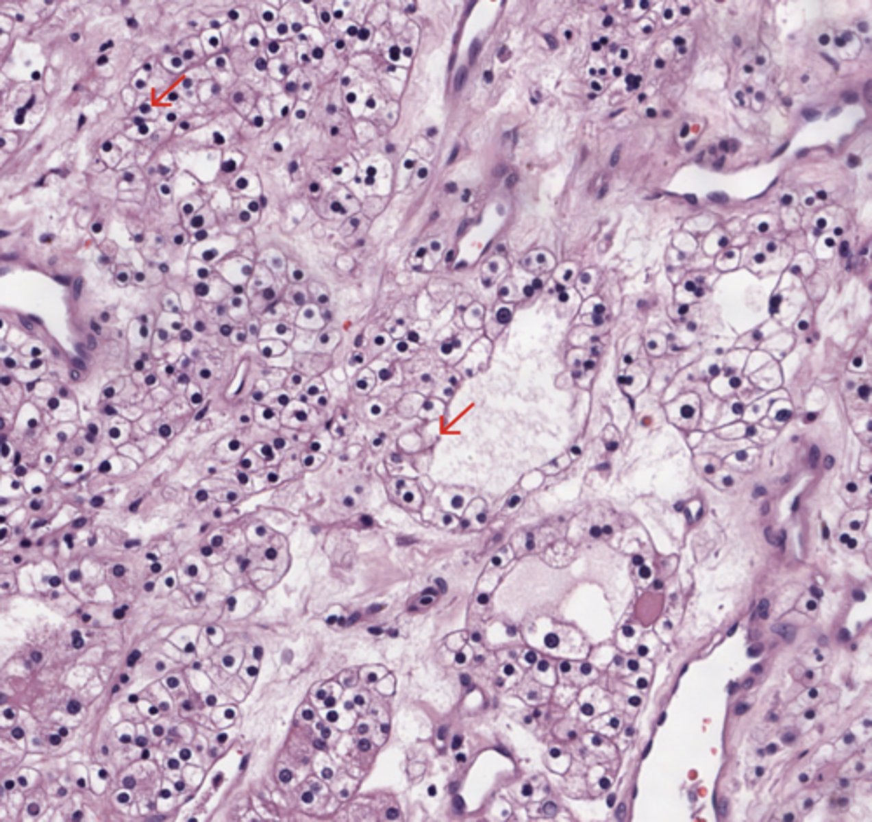
Architectural patterns: solid, alveolar (nested), acinar (tubular), microcystic (containing extravasated red blood cells or eosinophilic fluid) and occasionally macrocystic
(Kidney)
* My note - Architectural Diversity: The cells assemble into organized geometric configurations. These present as alveolar nests, gland-like acini (tubules), solid sheets, or fluid-filled microcysts.
Kidney
Kidney cancer - Membrane
Typically compact nests and sheets of cells with clear cytoplasm and distinct membrane
(Kidney)
* My note - Distinct Membrane: The thick, dark pink boundaries create a rigid perimeter around each cell. This mimics a plant cell or a crisp "honeycomb" network.
Pancreas
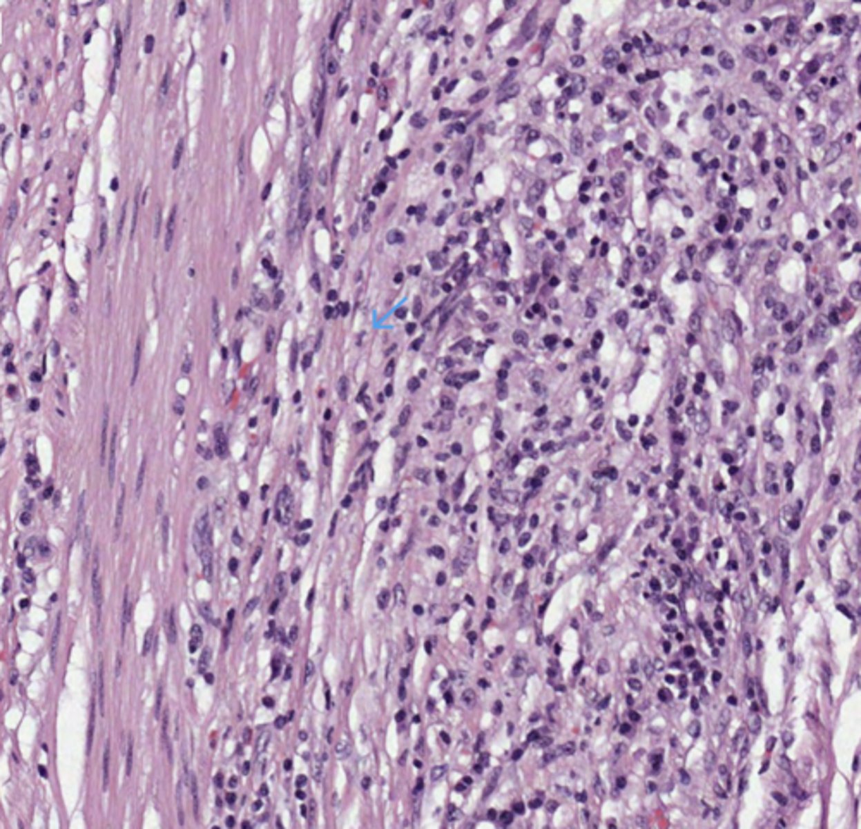
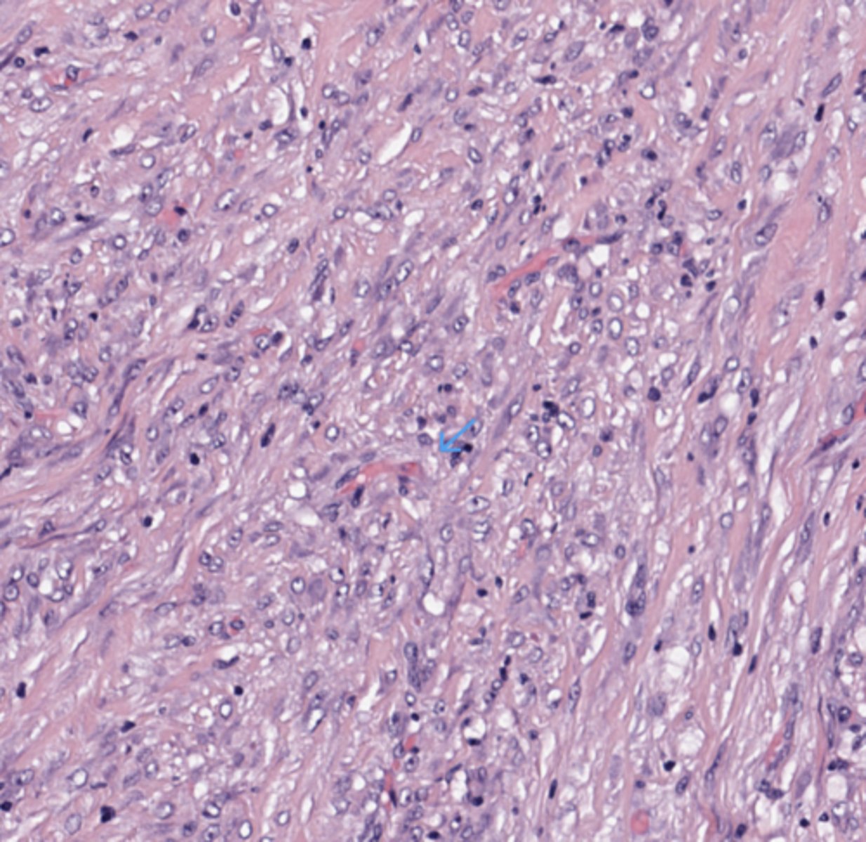
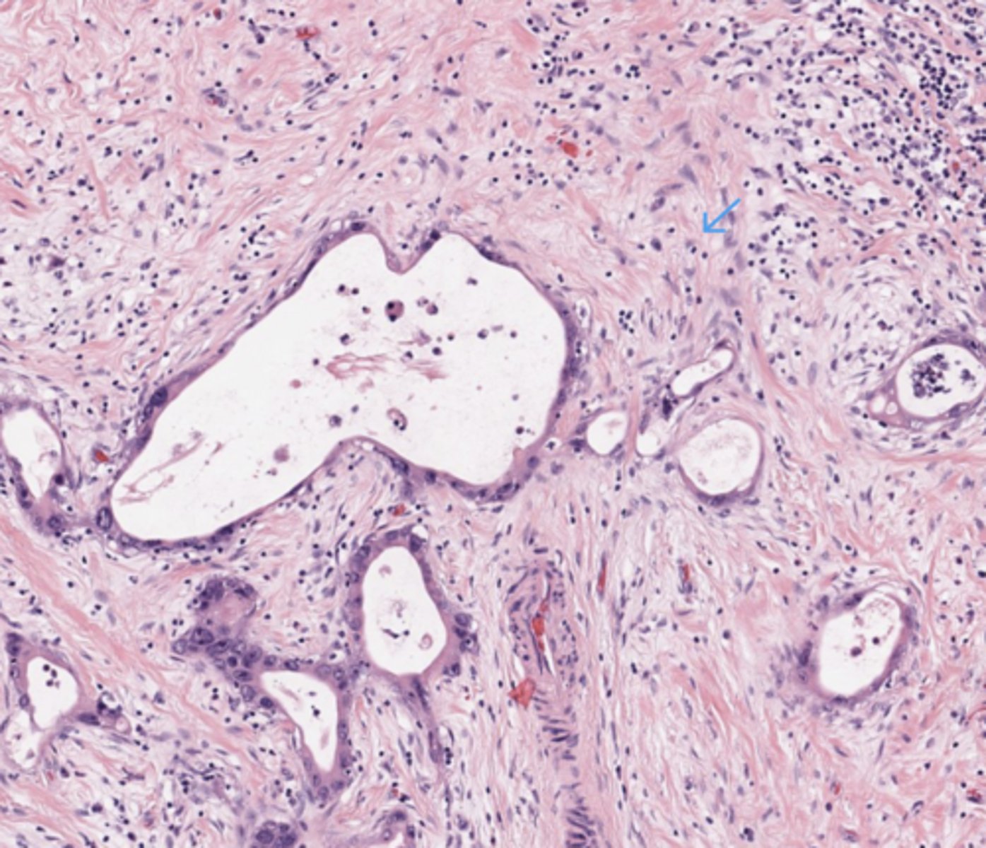
Invasive PDAC (Pancreatic Ductal Adenocarcinoma)
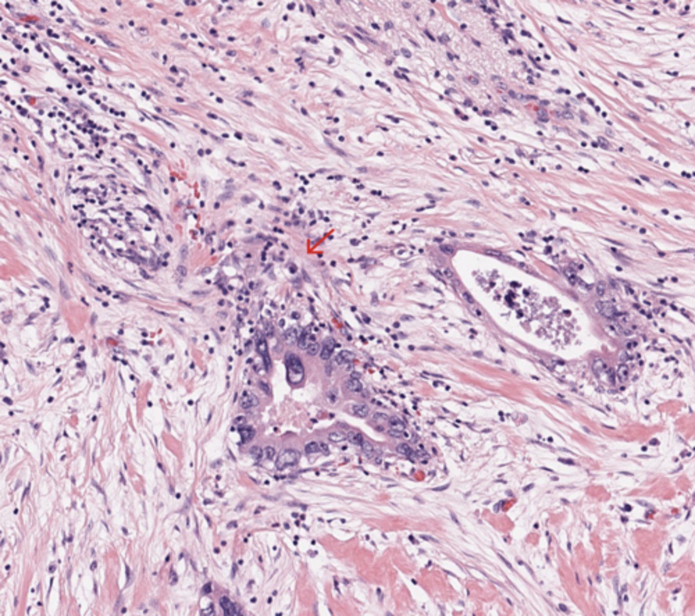
Moderately differentiated adenocarcinoma. Foci of smaller and more irregular glands and some individual pleomorphic cells are often found at the tumour margins. Common to both well-differentiated and moderately differentiated carcinomas is a desmoplastic stroma that encompasses the neoplastic glands, sometimes in a ductocentric pattern.
(Pancreas)
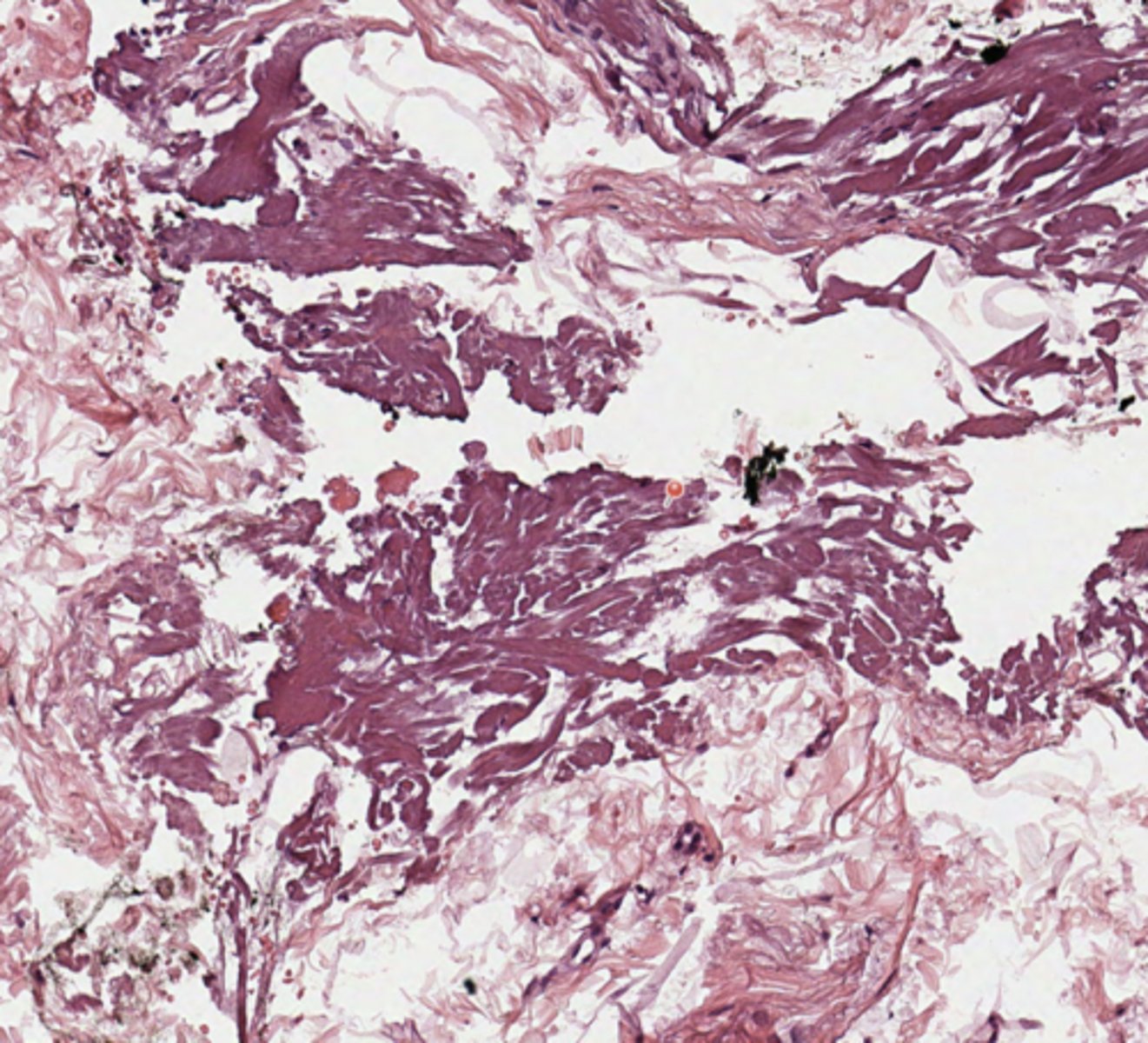
Pancreas
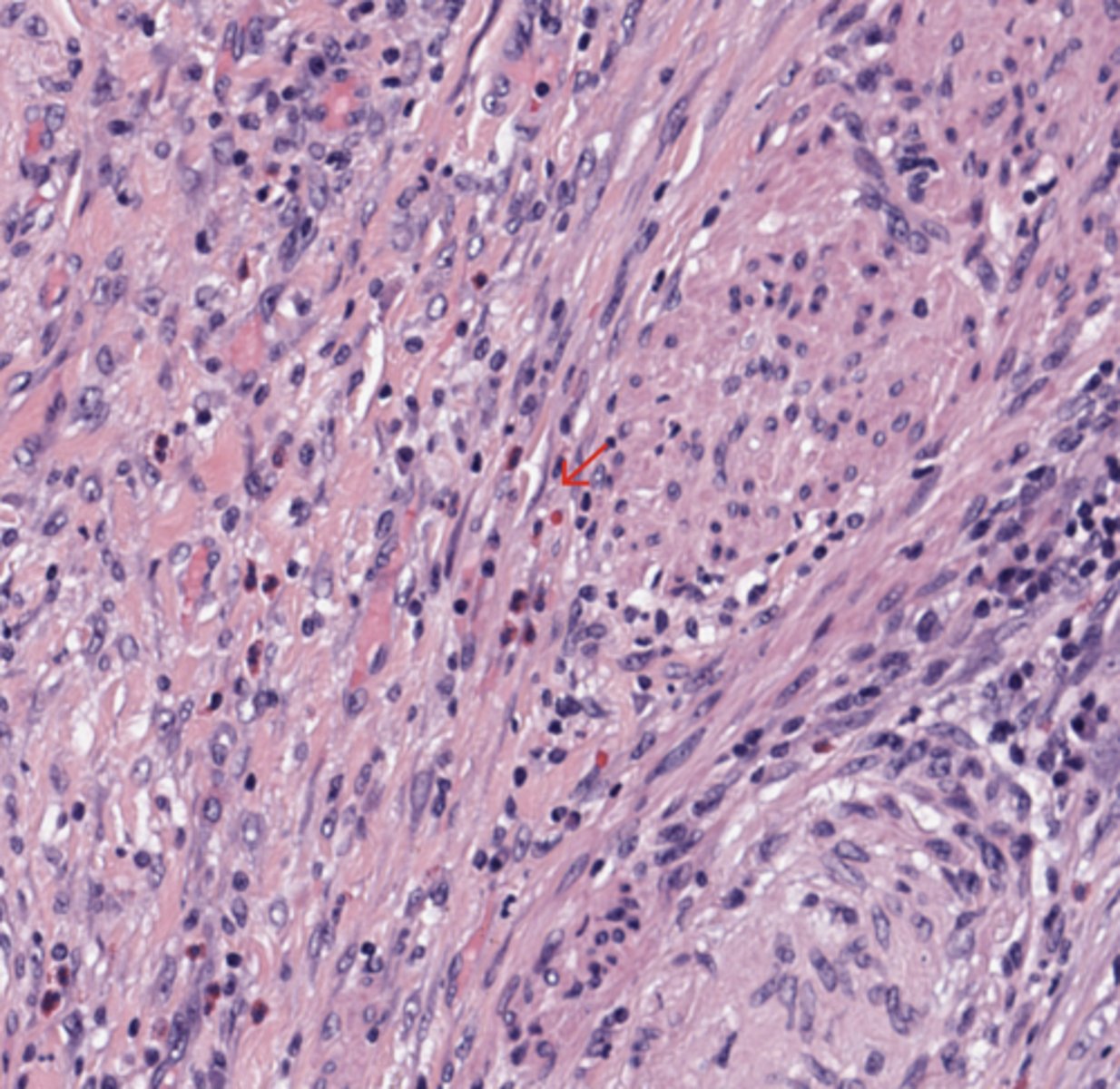
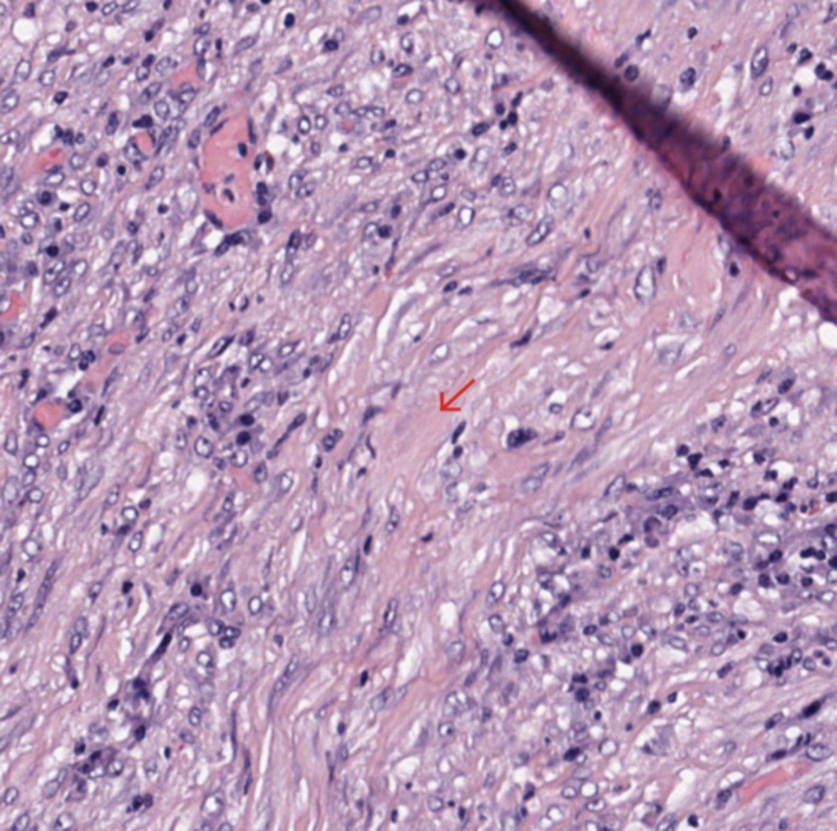
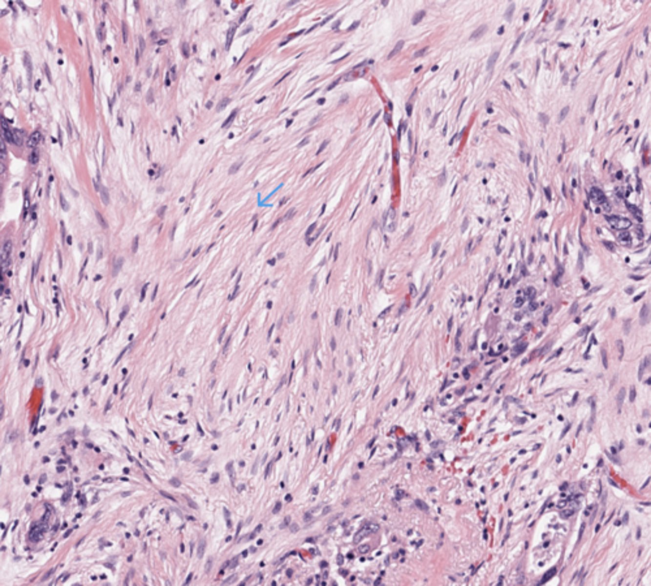
Desmoplastic stroma
The stroma is one of the most important histological features.
In pancreatic ductal adenocarcinoma, the tumor glands are often embedded in abundant dense fibrous tissue.
This desmoplastic stroma contains fibroblasts, inflammatory cells, extracellular matrix, and blood vessels.
It is not just a passive background. It actively interacts with tumor cells and contributes to tumor progression, invasion, and resistance to therapy.
For students, the simple idea is: pancreatic ductal adenocarcinoma is often a tumor made of irregular malignant glands in a dense fibrotic stroma.
(Pancreas)
Pancreas
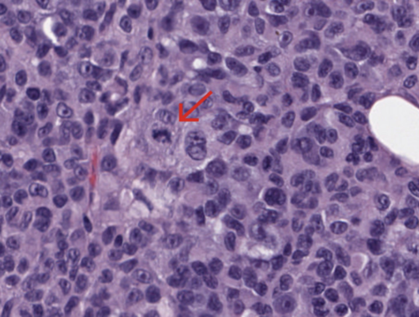
Cytological features
The neoplastic glands are lined by atypical epithelial cells.
The cells may show:
- enlarged nuclei
- irregular nuclear membranes,
- hyperchromasia
- prominent nucleoli
- loss of polarity (of nucleus)
- mitotic figures
- foamy cytoplasm.
The degree of atypia can vary. Some tumors are well differentiated and form recognizable glands. Others are poorly differentiated and may form solid nests or single infiltrating cells.
(Pancreas)
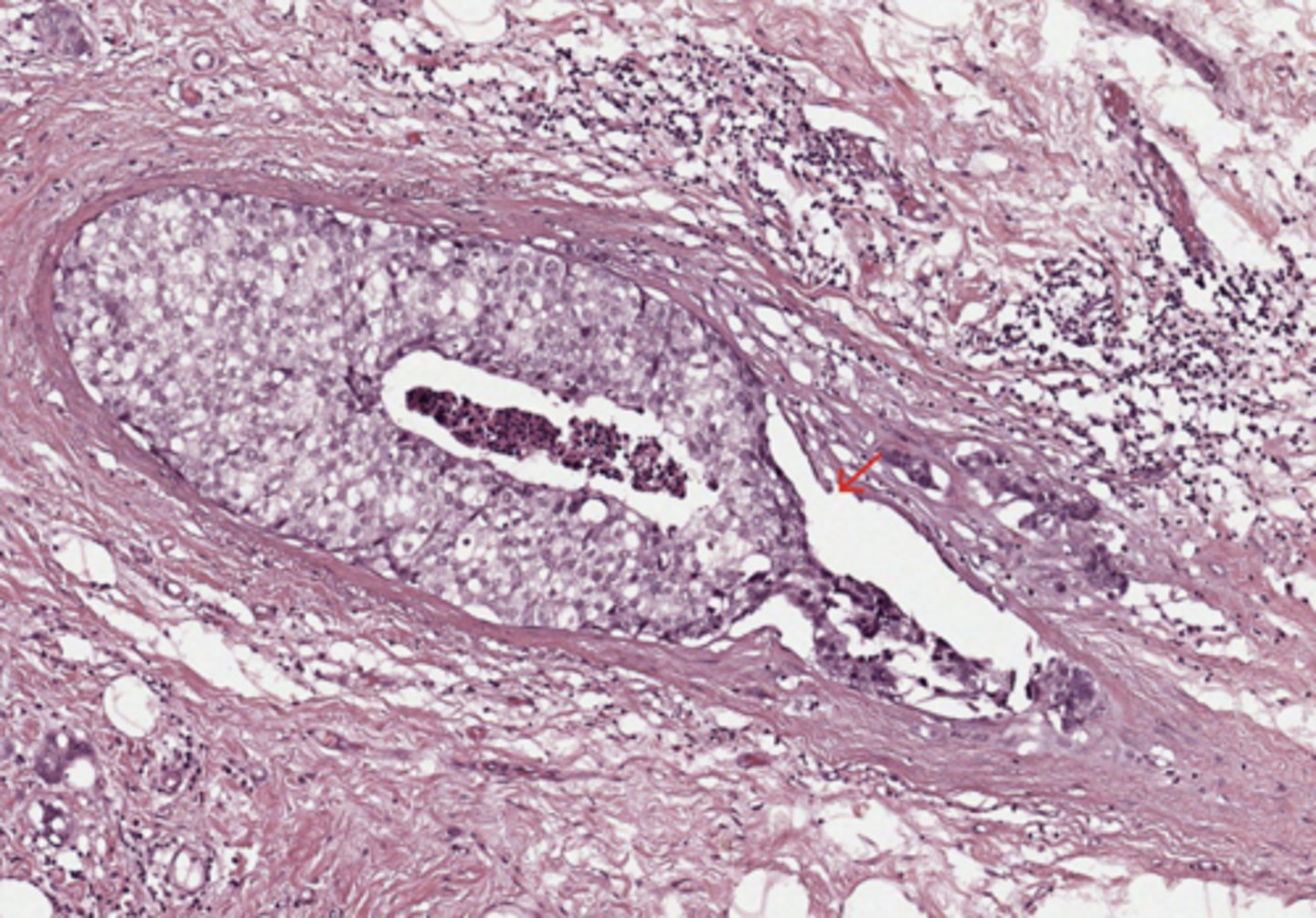
Pancreas
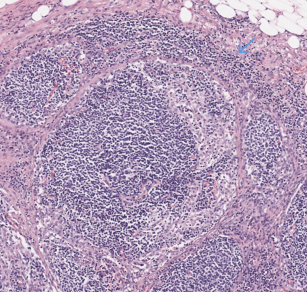
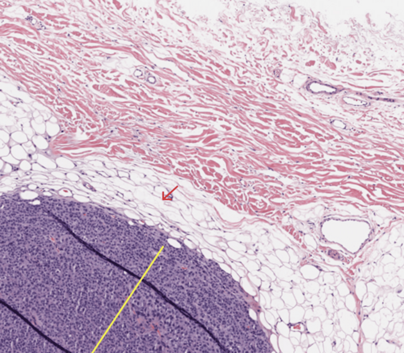
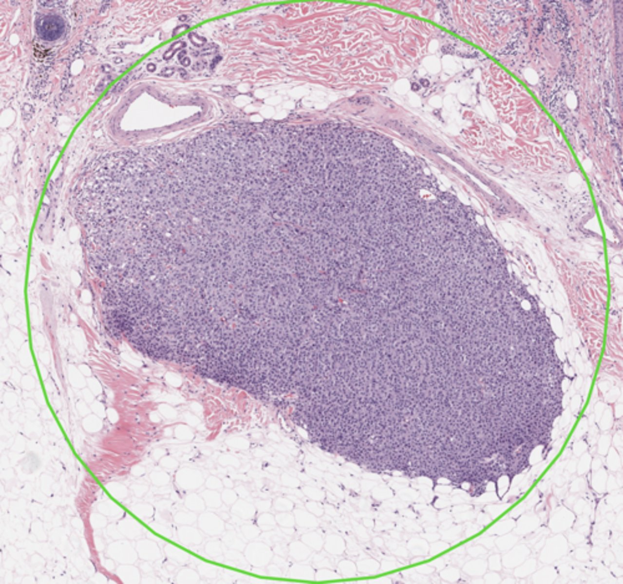
Lymph node
(Pancreas)
Pancreas
Lymphnode metastasis
(Pancreas)
*My note - large, irregular, open white spaces lined by atypical, dark purple epithelial cells.
Pancreas
Duodenum
The duodenum—the first section of the small intestine—features the four standard gastrointestinal layers: mucosa, submucosa, muscularis externa, and adventitia. Its defining histological characteristic is the presence of Brunner's glands in the submucosa, which secrete alkaline mucus to neutralize acidic stomach chyme.
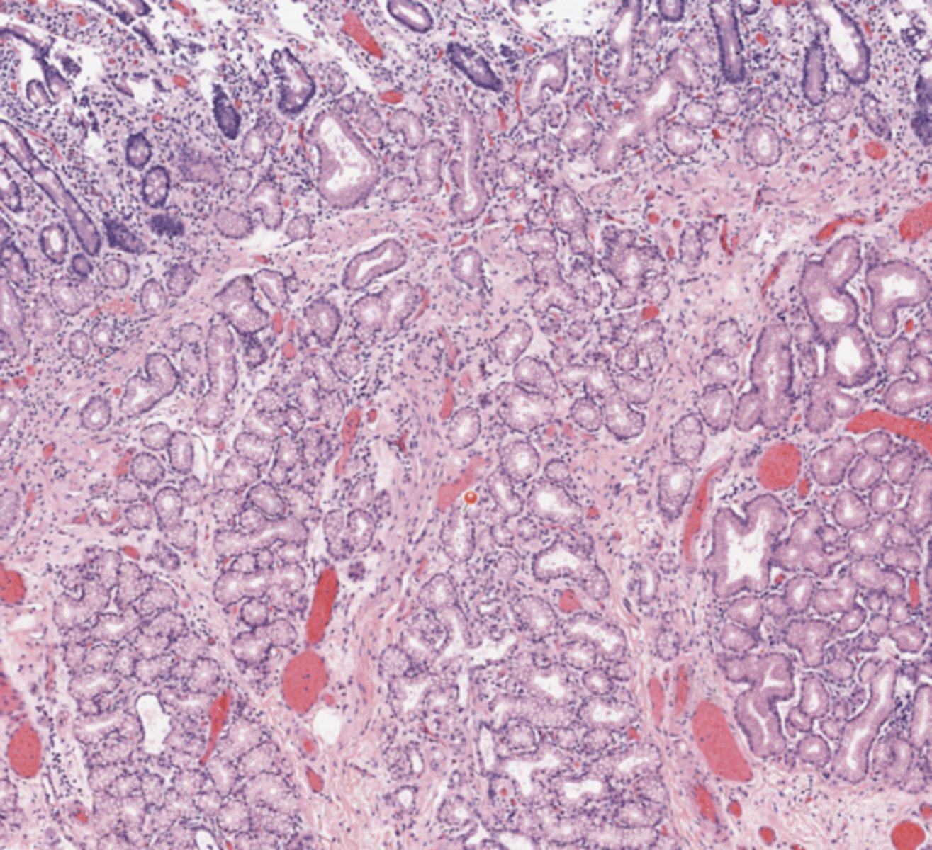
Pancreas
Pancreatic Parenchyma
(Pancreas)
*My note - The pancreatic parenchyma is a compound tubuloacinar gland that consists of two distinct functional units: the exocrine component, which makes up about 98-99% of the tissue, and the endocrine component, which forms scattered clusters throughout. It is enclosed by a thin connective tissue capsule that sends septa into the organ, dividing it into functional lobules.
Pancreas
Intrapancreatic biliary duct
(Pancreas)
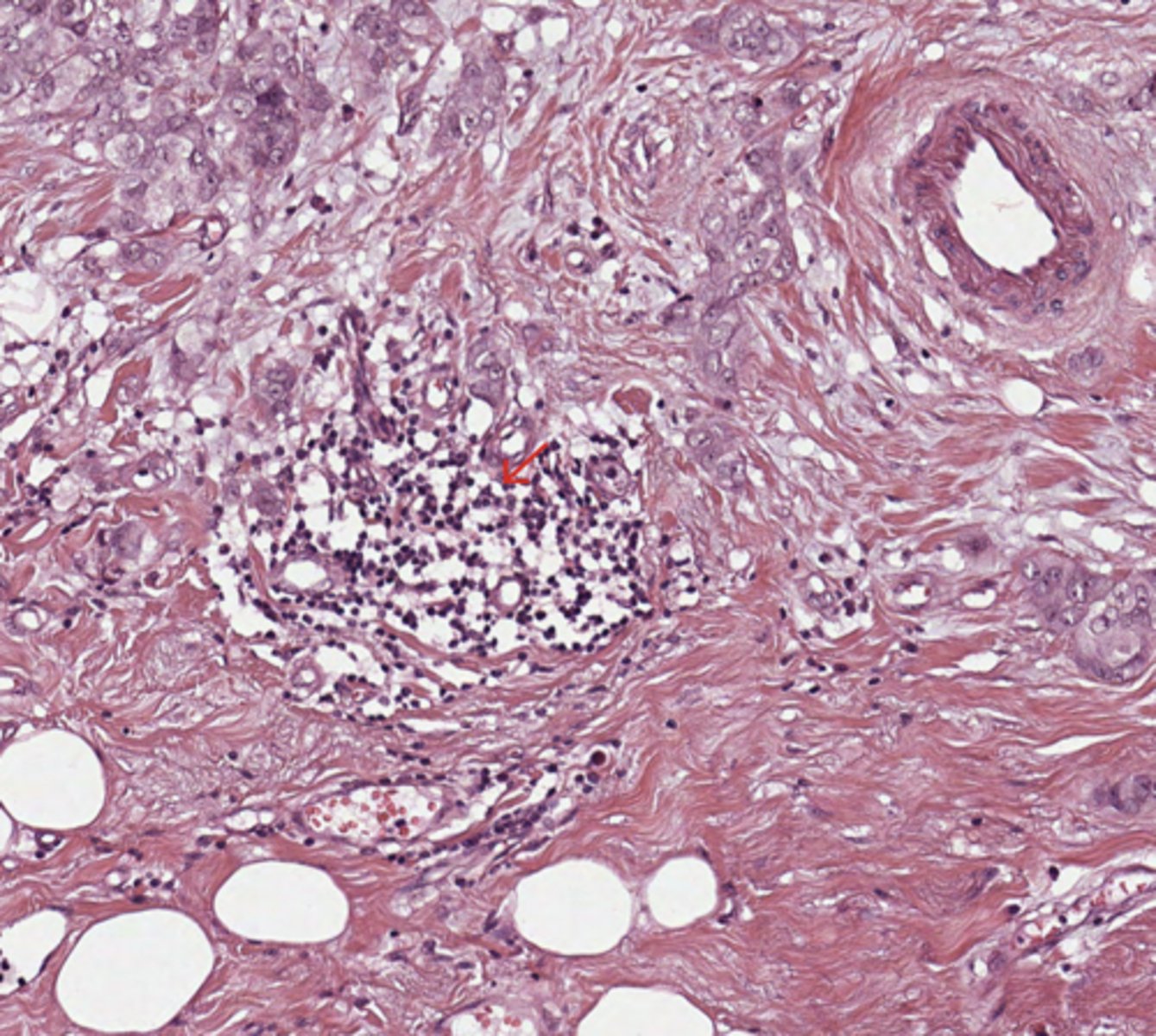
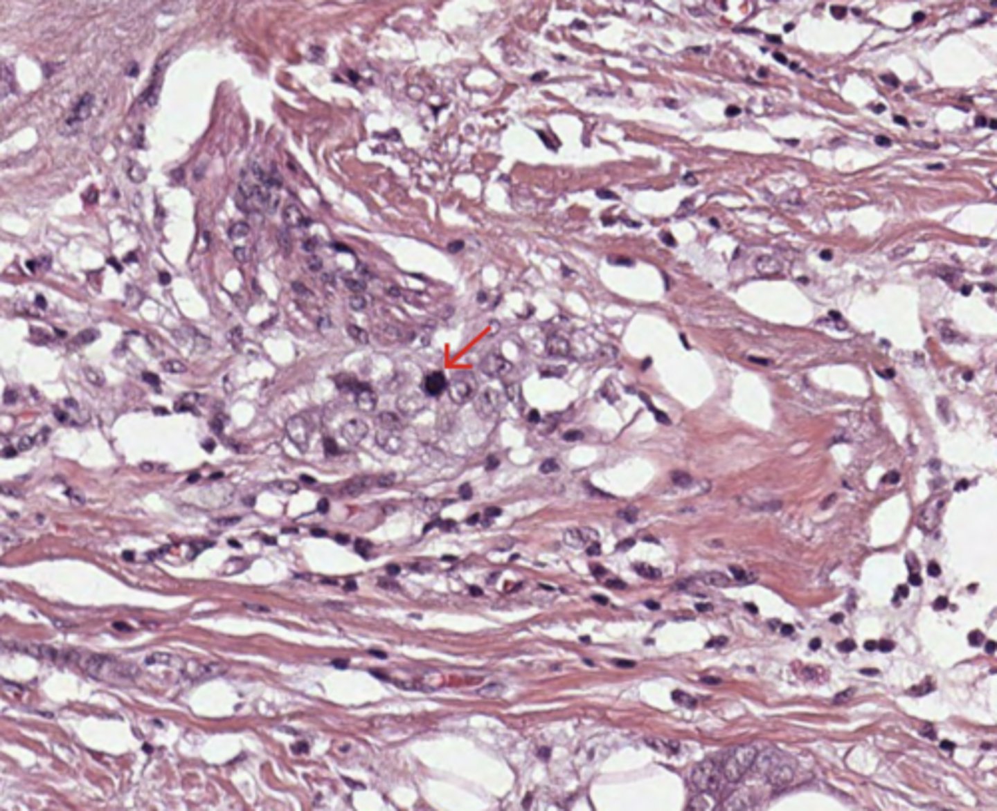
Pancreas
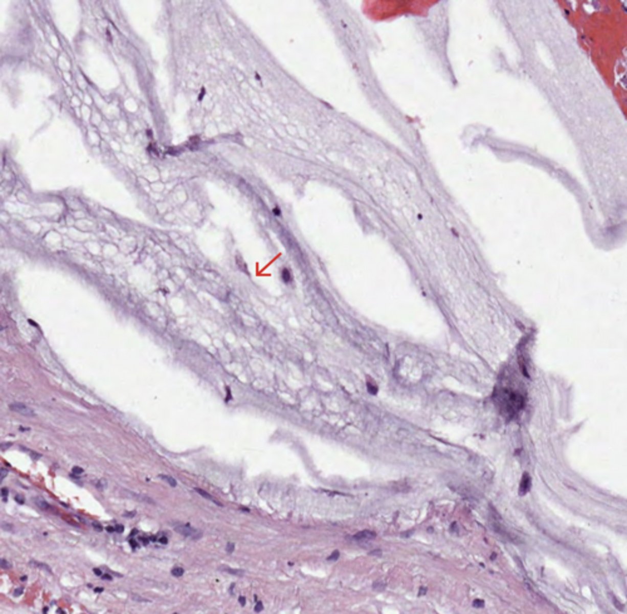
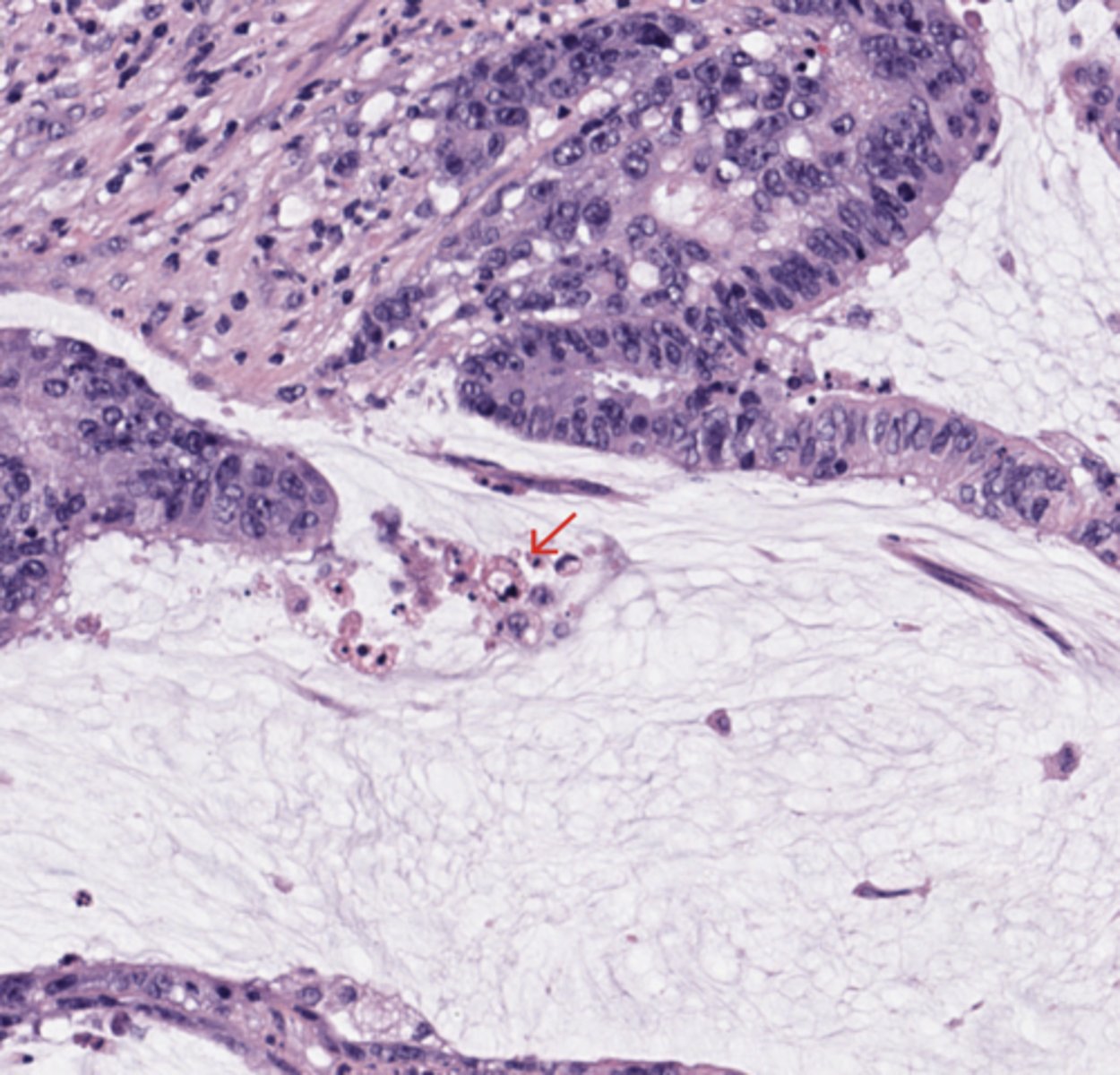
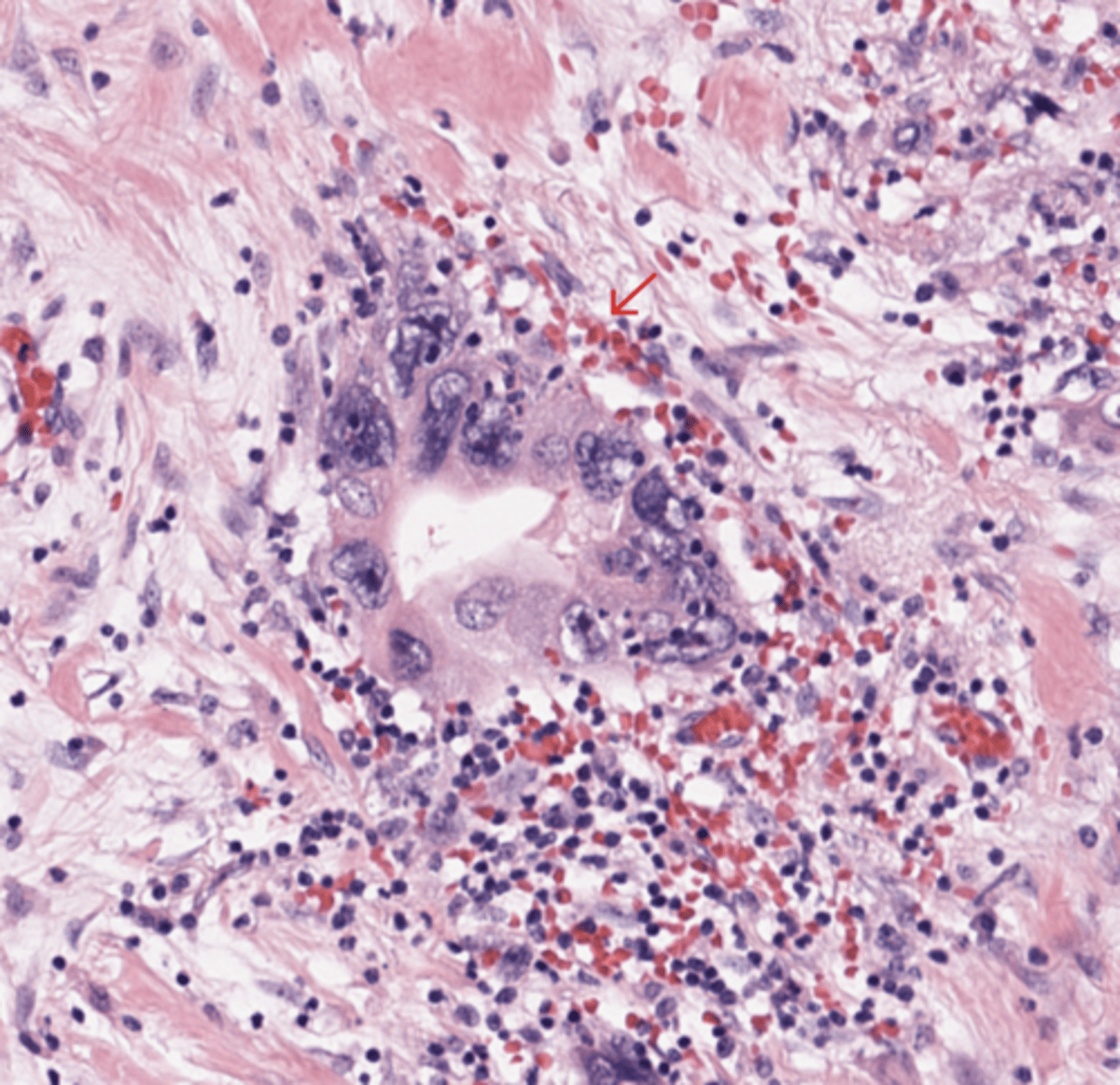
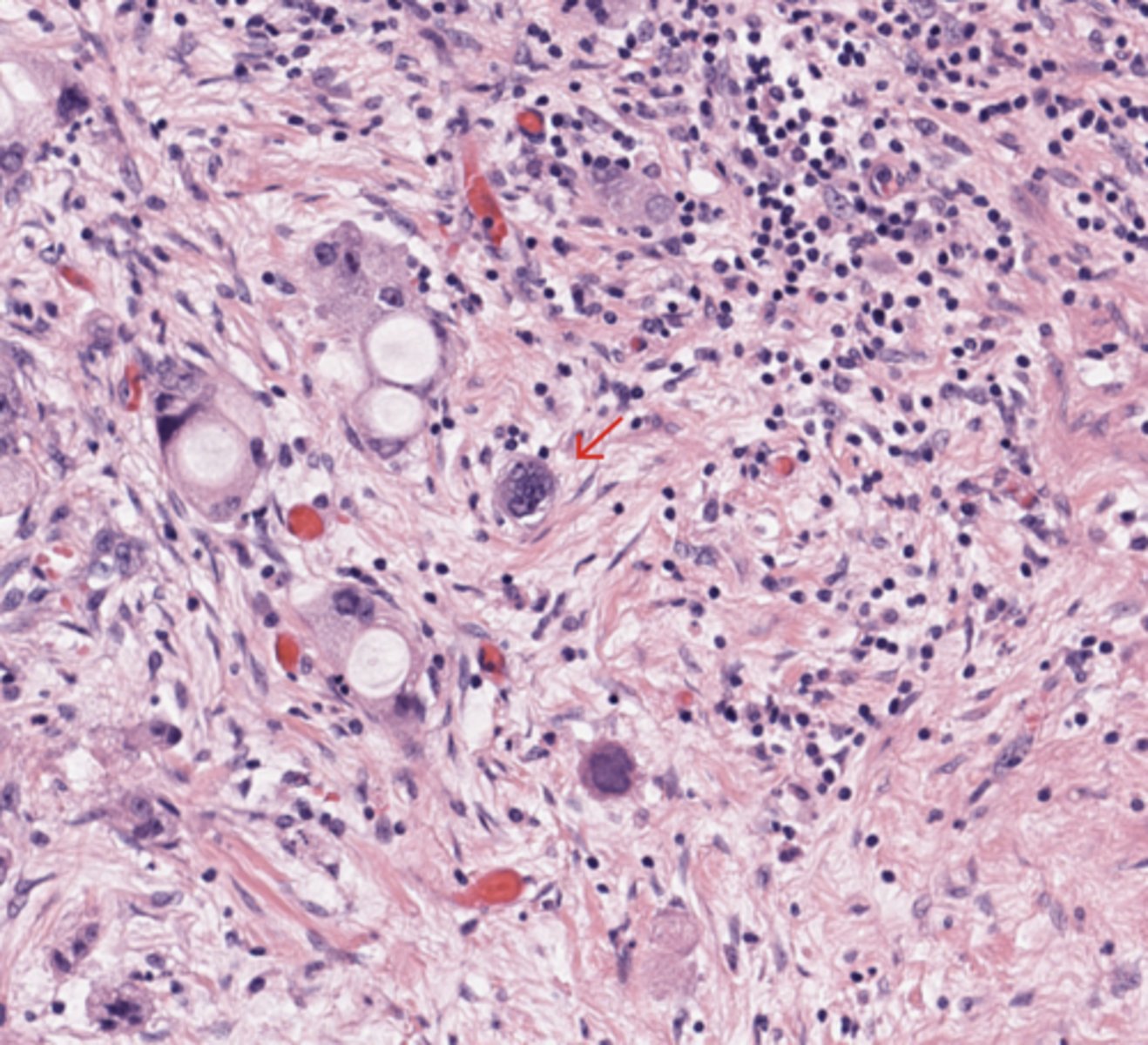
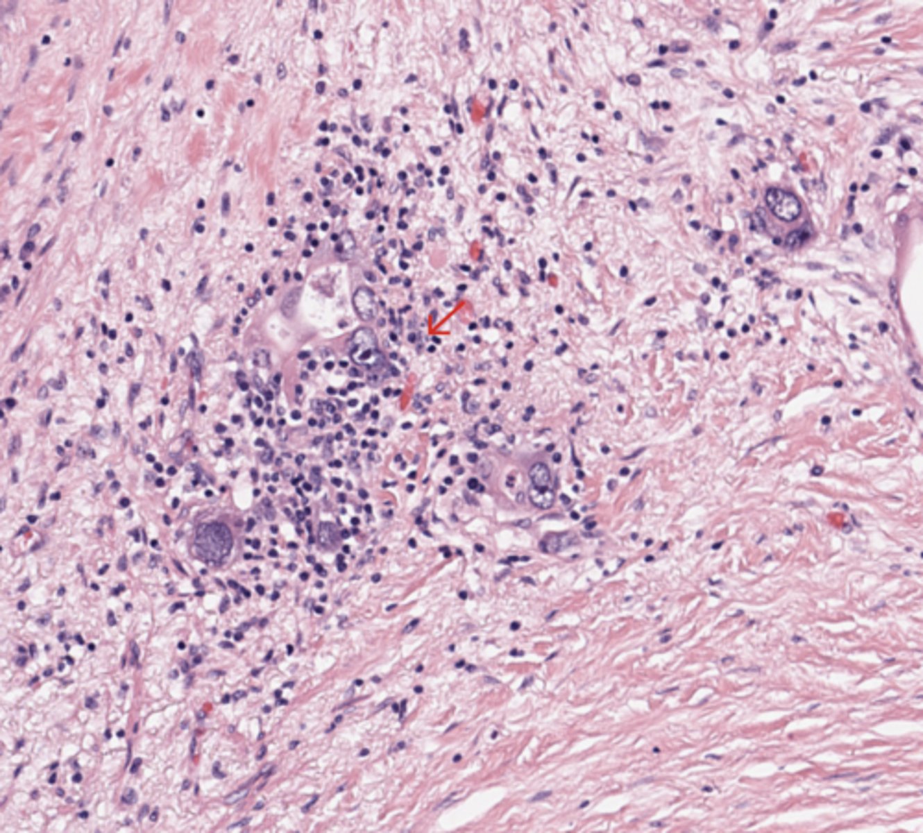
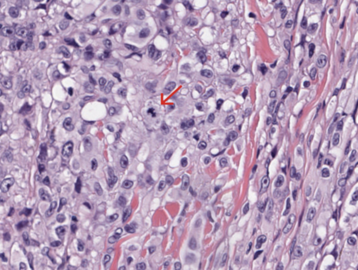
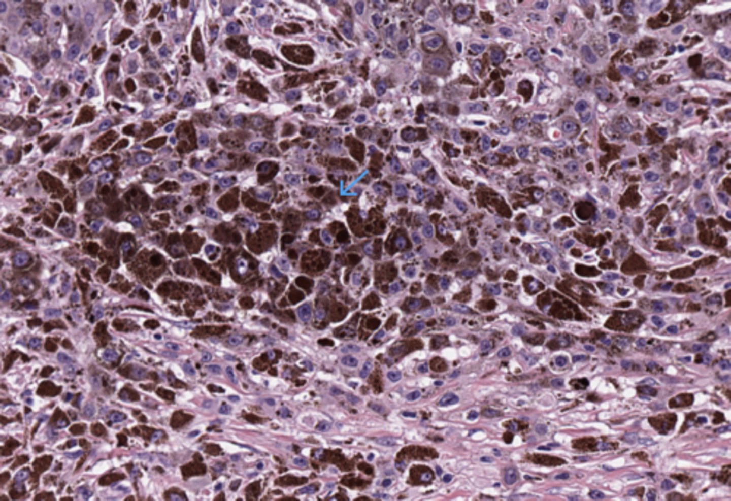
Pleomorphic cells
(Pancreas)
* My note - The red arrow points directly to a large, atypical pleomorphic cell nucleus.
What you see: This nucleus is significantly larger, darker (hyperchromatic), and more irregularly shaped than the small, uniform, round nuclei of the surrounding background lymphocytes.
Significance: Pleomorphism represents a high degree of variation in nuclear size and shape, which is a core histological hallmark of malignancy.
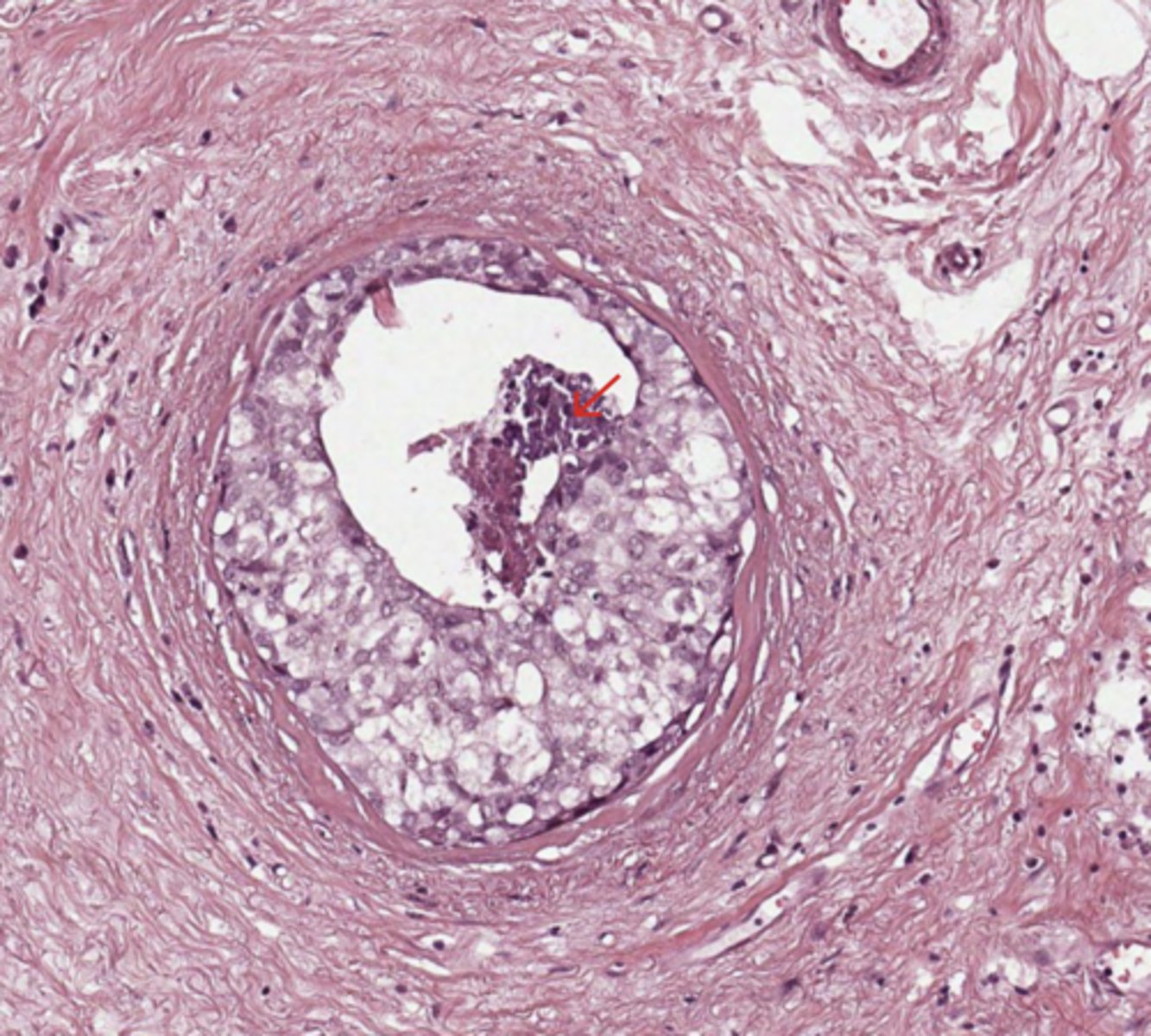
Pancreas
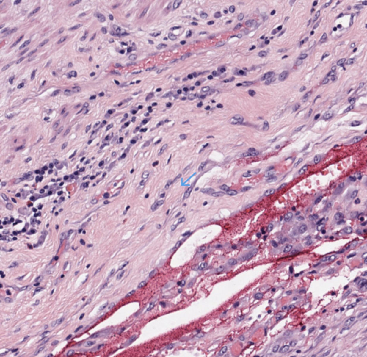
Vascular invasion
(Pancreas)
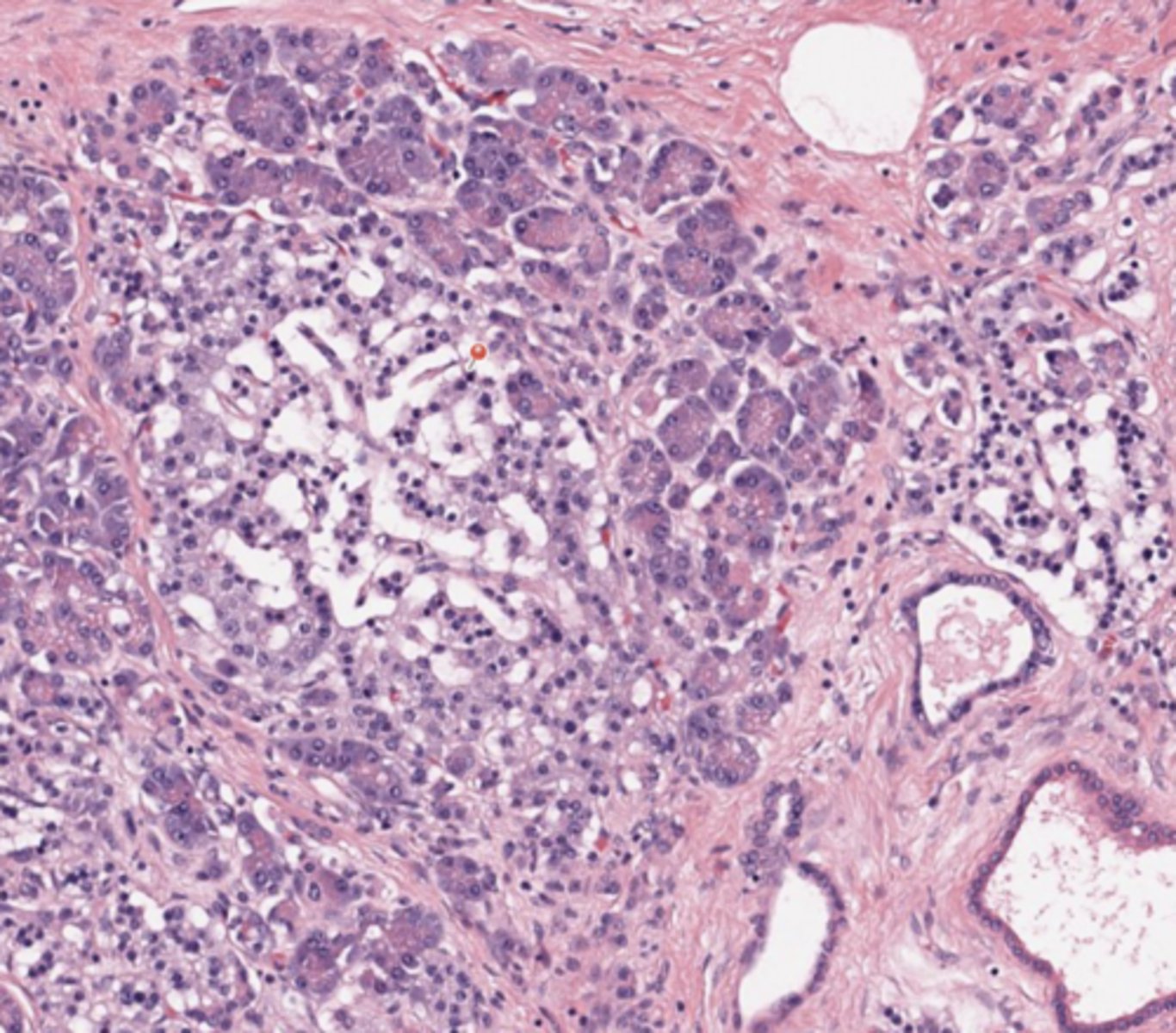
Pancreas
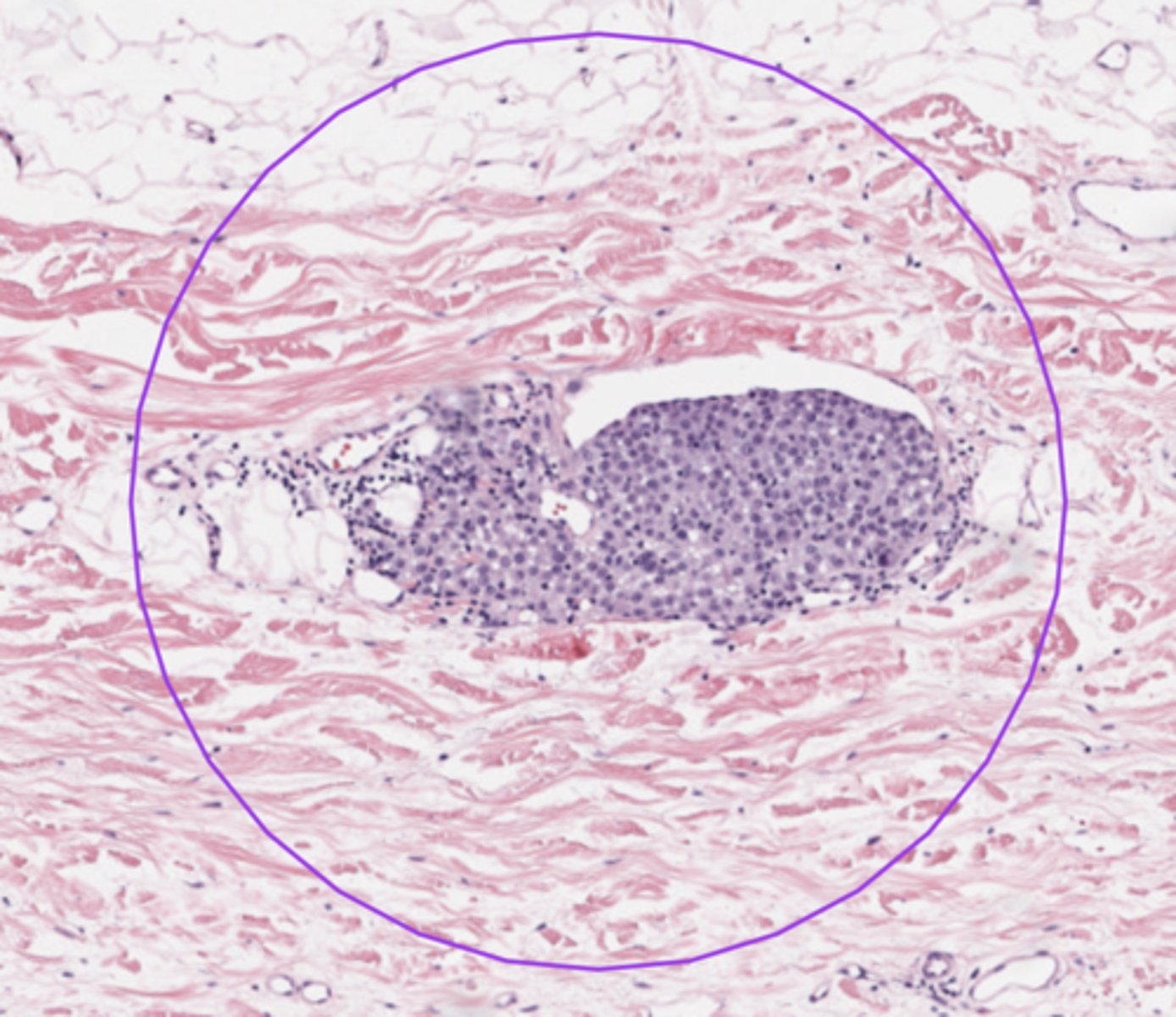
Islets of Langerhans
The islets of Langerhans represent the endocrine part of the pancreas. The cells are lighter than those of the pancreatic acini. Depending on the cell type, they secrete certain hormones into surrounding fenestrated capillaries. Beta cells (70%): Insulin Alpha cells (20%): Glucagon Delta cells (5%): Somatostatin PP cells (2%): Pancreatic polypeptide.
(Pancreas)
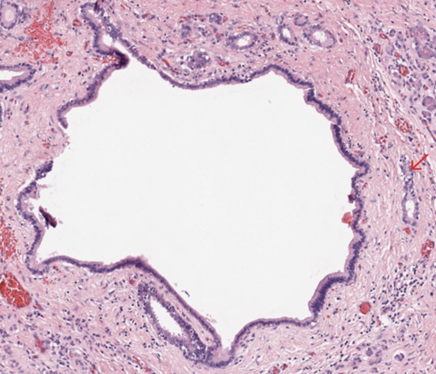
Pancreas
Pancreatic duct (big lumen)
(Pancreas)
* My note - the small lumen are probably interlobular ducts.
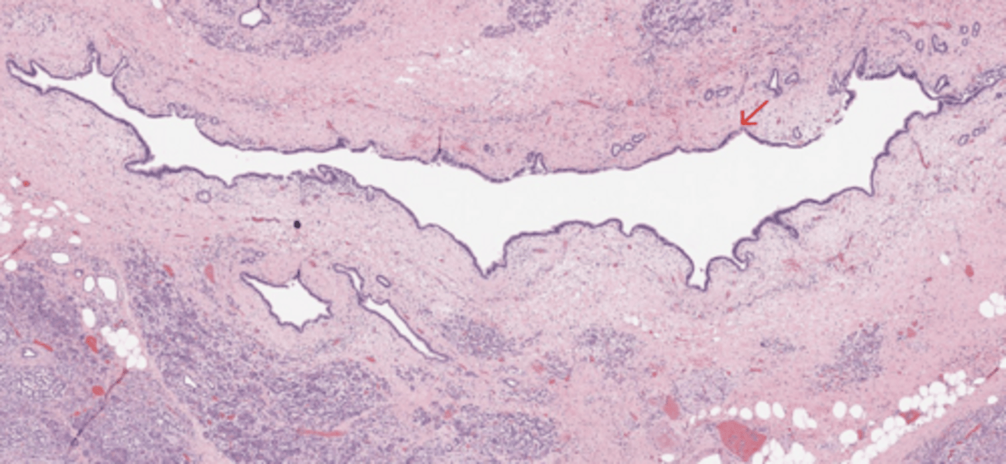
Pancreas
Pancreatic duct (duct of Wirsung)
(Pancreas)
Lung
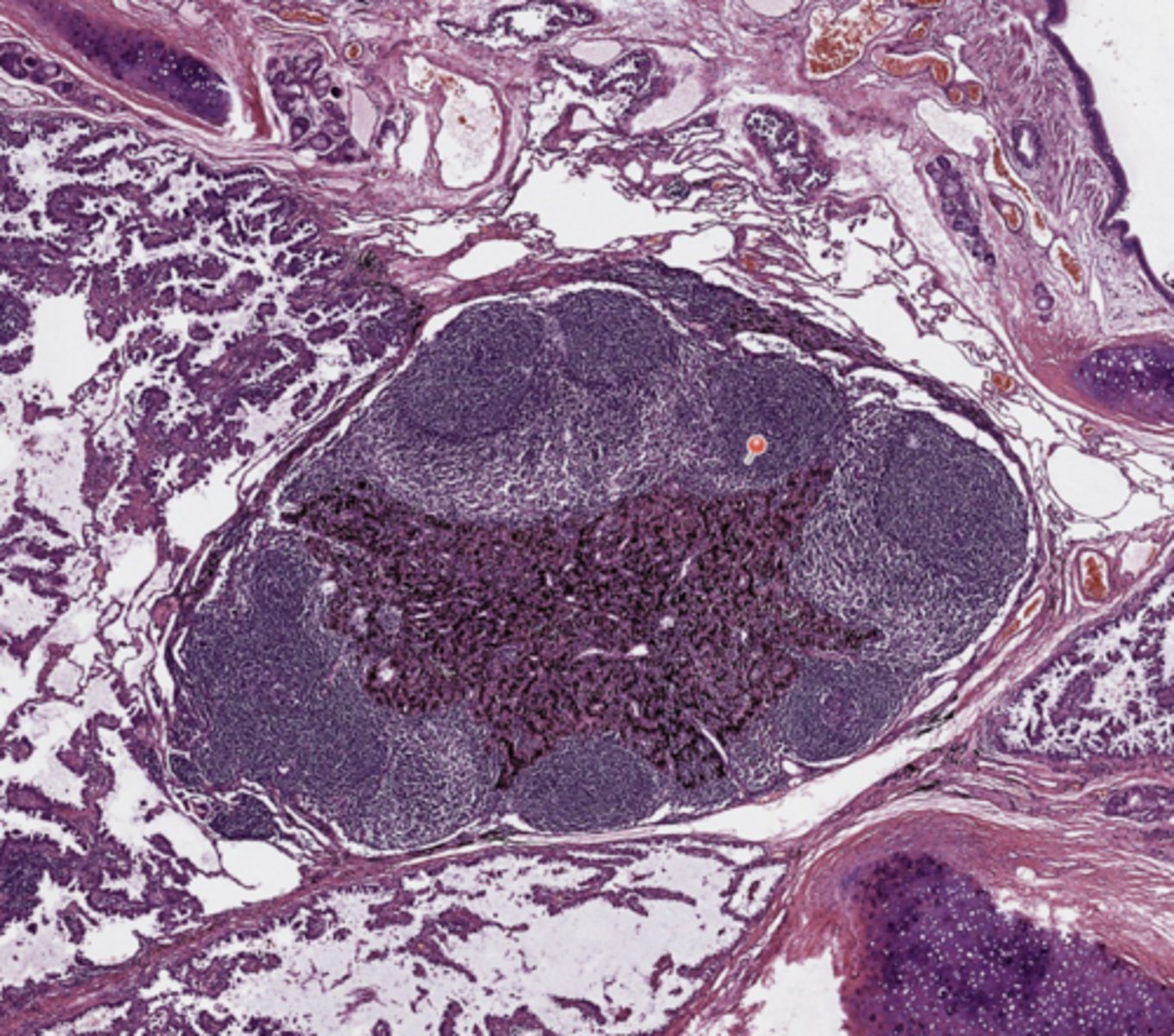
Lymphnode with marked antracosis
(Lung)
Lung
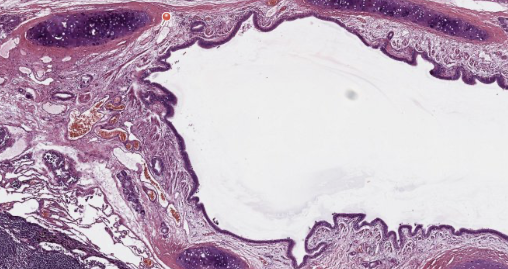
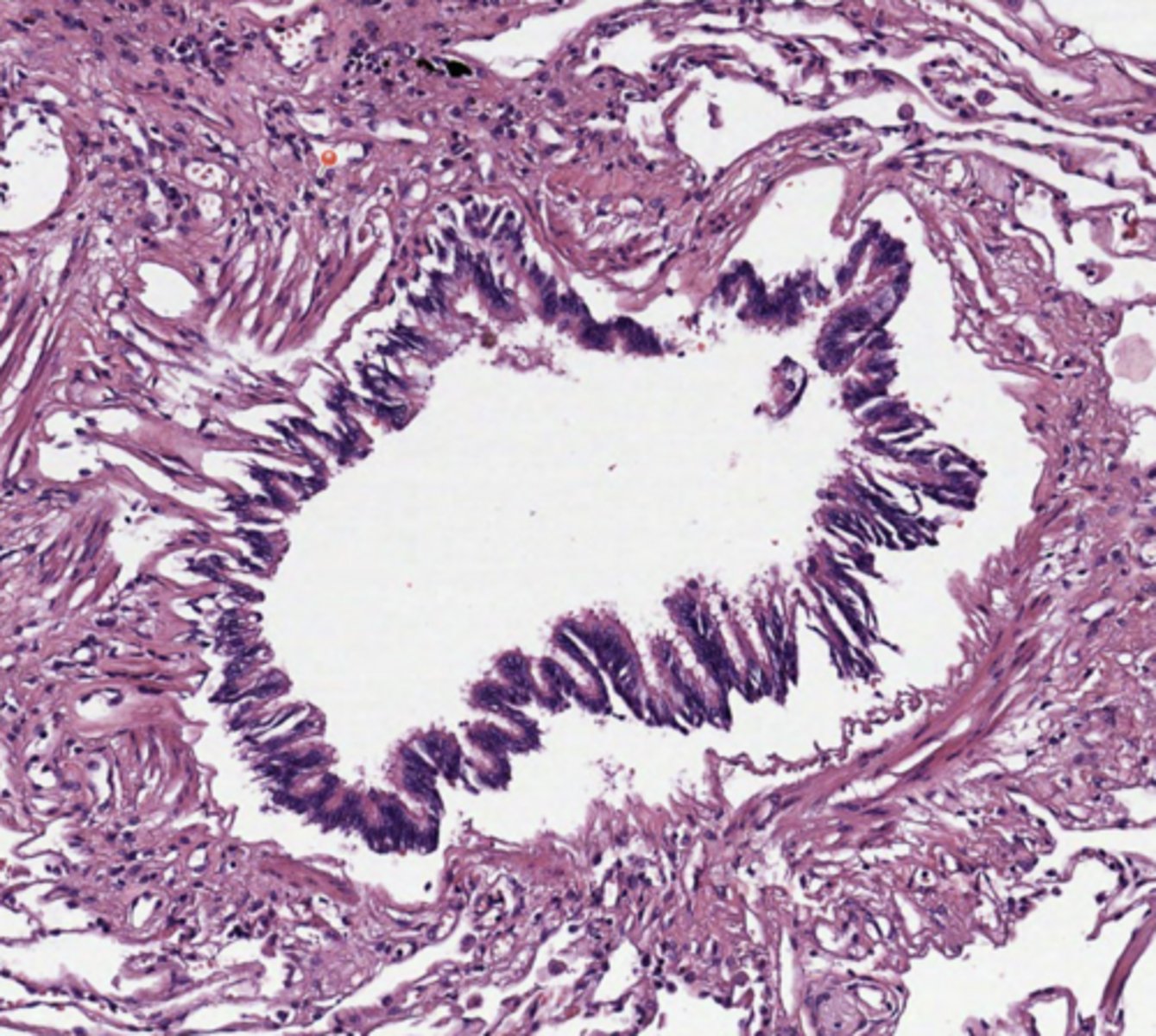
Main broncus
Primary Bronchi
Respiratory Epithelium - composed of a pseudostratified columnar epithelium.
Basement Membrane - thick, eosinophilic band beneath the epithelium.
Lamina Propria - dense irregular connective tissue.
Bronchial Cartilage
Peri-bronchiolar Glands
Smooth Muscle
(Lung)
Lung
Primary (Muscular) Bronchioles
Epithelium changes from pseudostratified columnar to simple, ciliated columnar epithelium as they decrease in diameter.
Club Cells - dome-shaped secretory cells.
Smooth Muscle - variable amounts present.
(Lung)
Lung
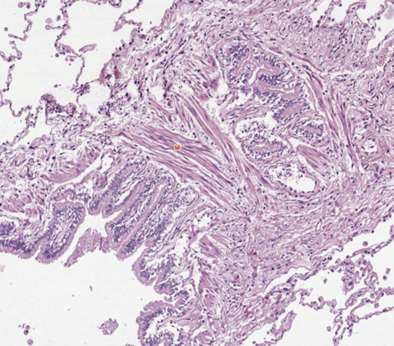
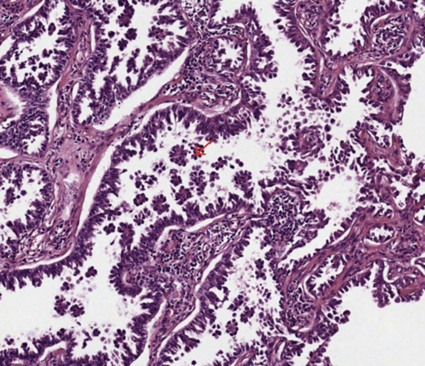
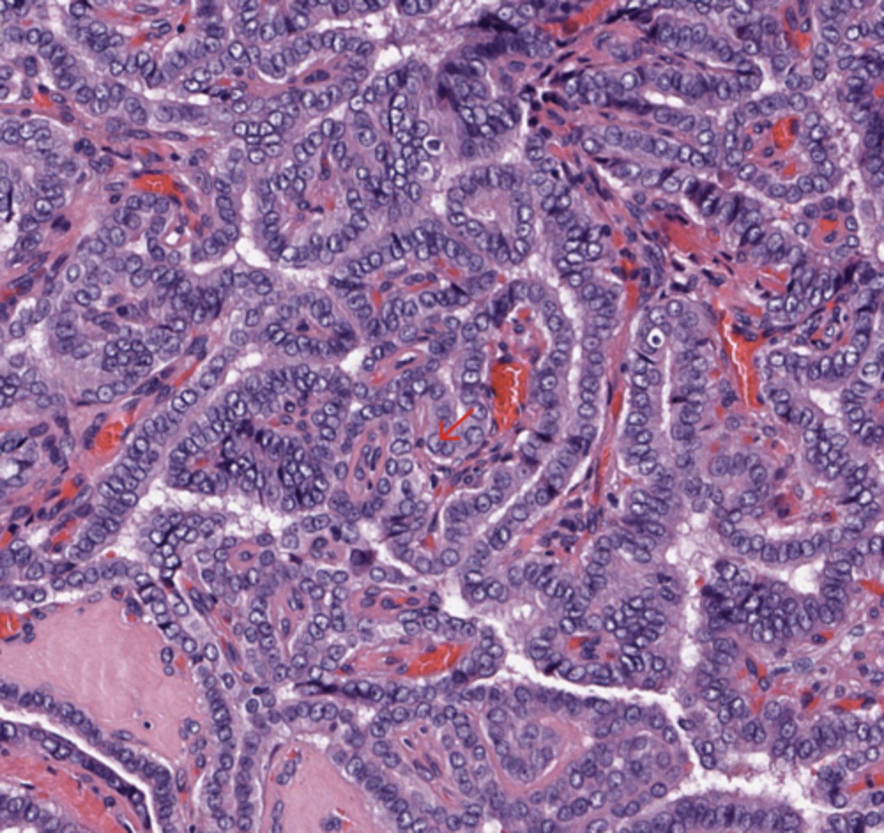
Neoplastic Papillae - Papillary growth pattern
This tumour consists of neoplastic glandular cells growing along the surface of fibrovascular cores
(Lung)
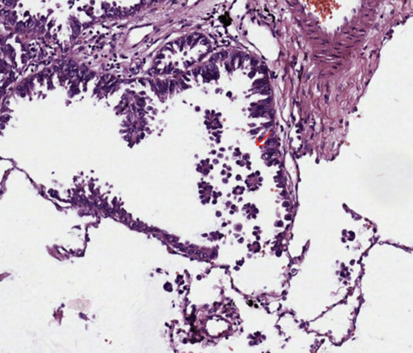
Lung
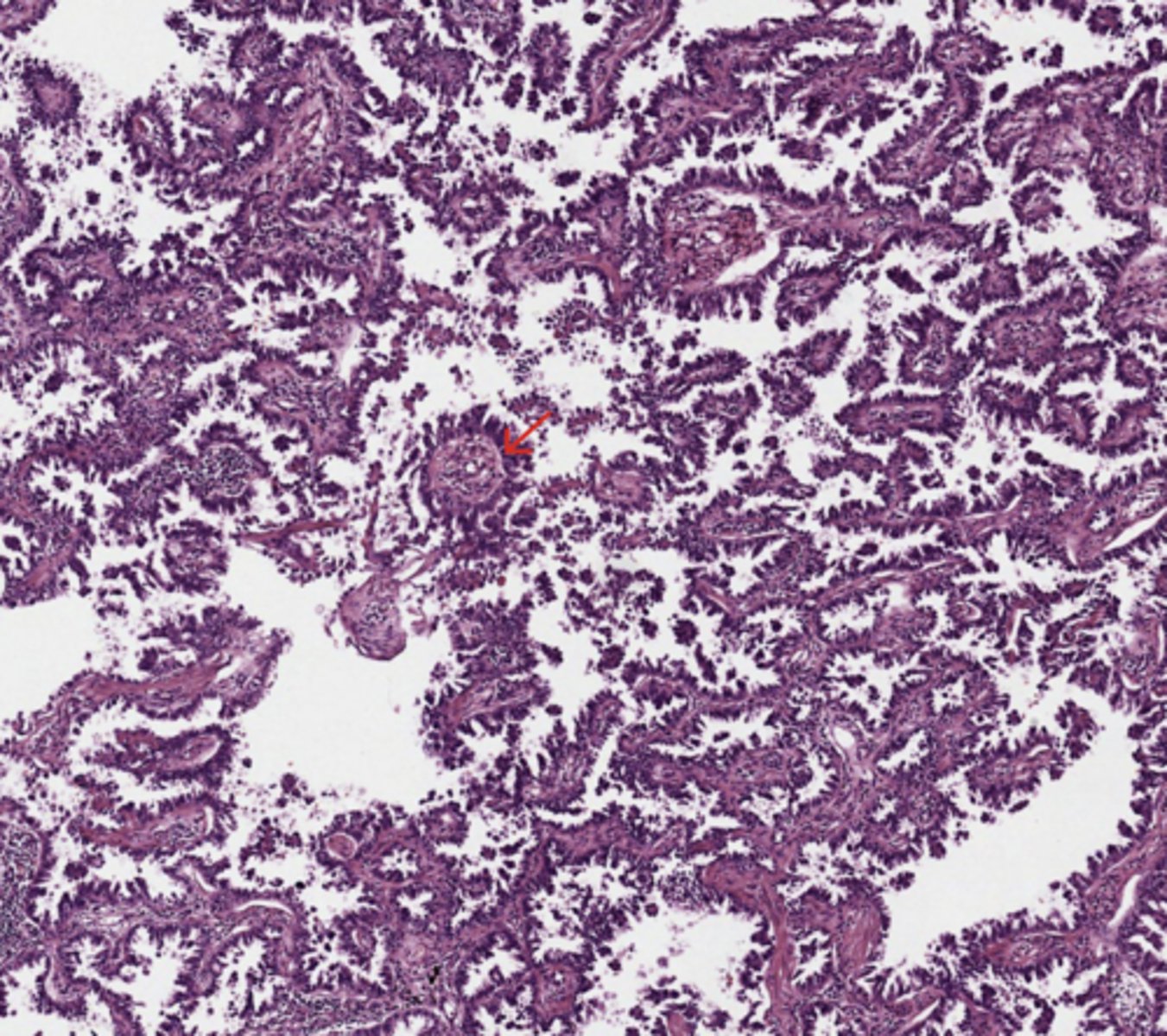
Micropapillae
Micropapillary architecture within the airspaces, without fibrovascular core.
(Lung)
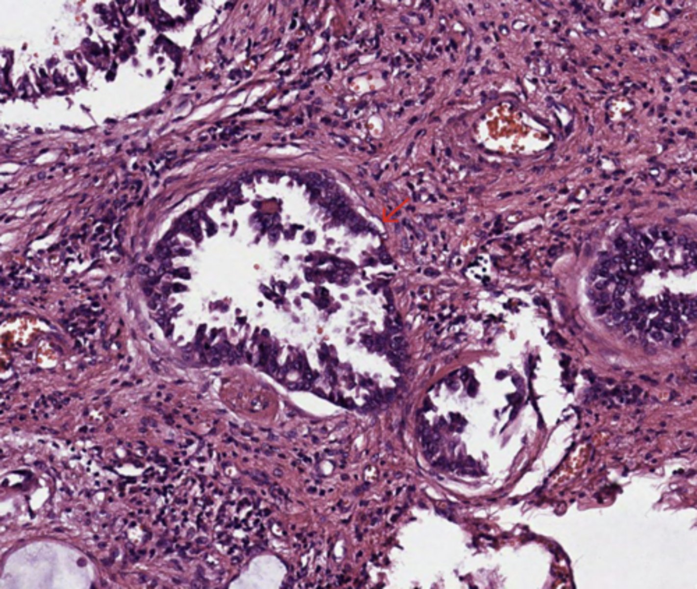
Lung
STAS
Spread through airspaces in adenocarcinoma
Many airspace clusters of tumour cells spread far beyond the tumour edge in a continuous manner , forming the lesion of spread through airspaces
(Lung)
Lung
Lymphovascular invasion
(Lung)
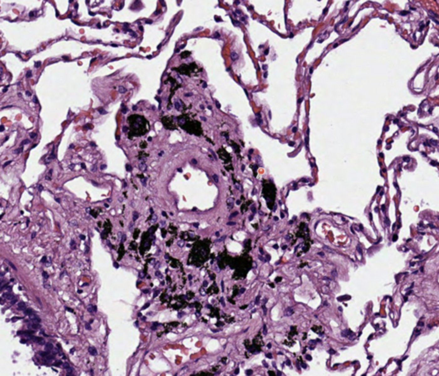
Lung
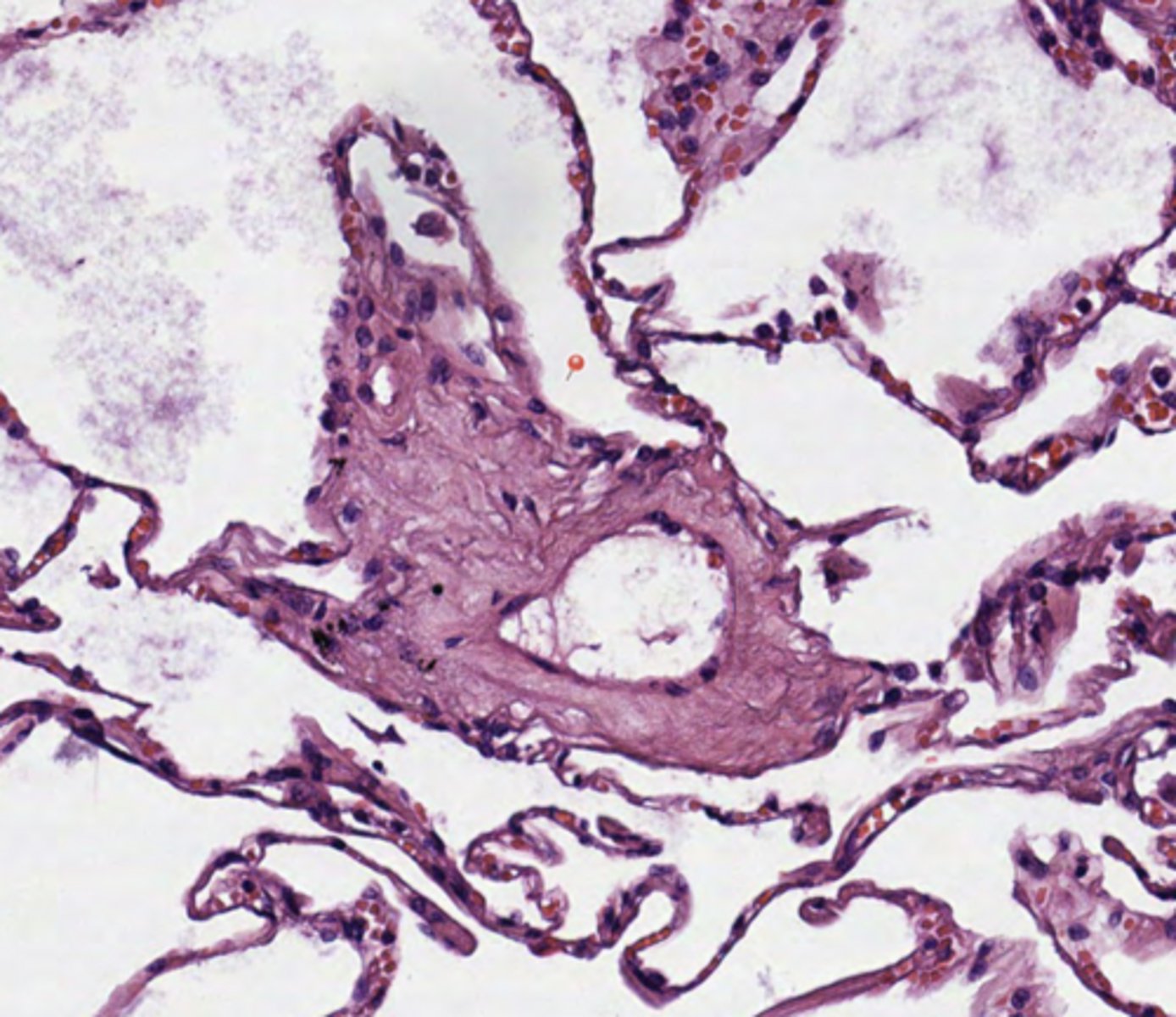
Antracosis
(Lung)
Lung
Secondary bronchus
(Lung)
Lung
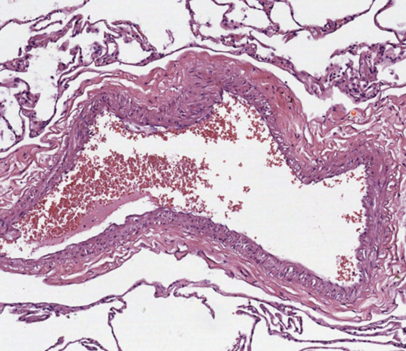
Artery
(Lung)
Lung
Vein
(Lung)
Thyroid
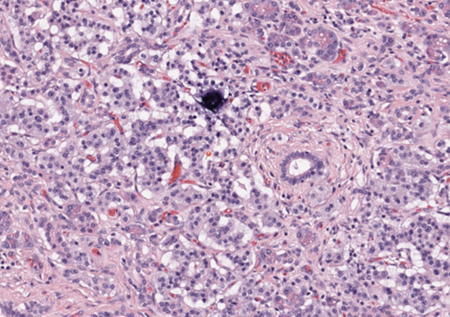
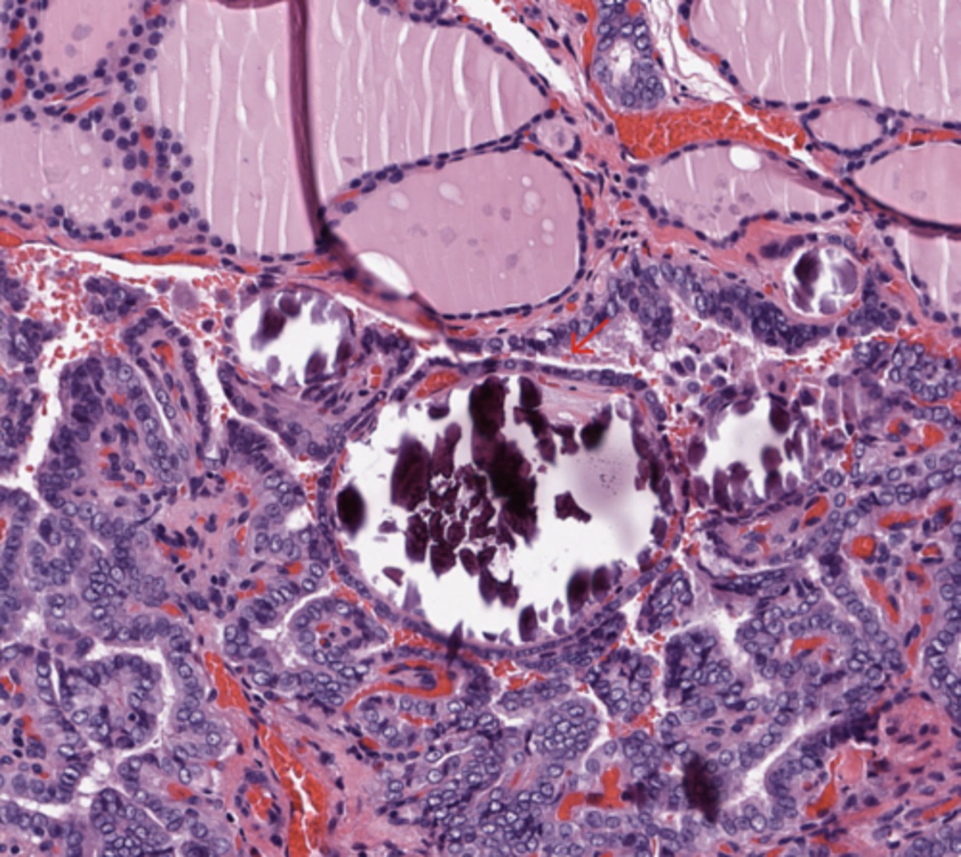
Nuclear features in PTC (Papillary Thyroid Carcinoma)
Change of nuclear size and shape, nuclear enlargement, elongation and overlapping. - Chromatin characteristics: chromatin clearing / optically clear chromatin, chromatin margination, glassy / ground glass nuclei, Orphan Annie eye (transparent) nuclei - Nuclear membrane irregularity: irregular nuclear contour, nuclear grooves and nuclear pseudoinclusions (represent cytoplasmic invaginations).
(Thyroid)
Thyroid
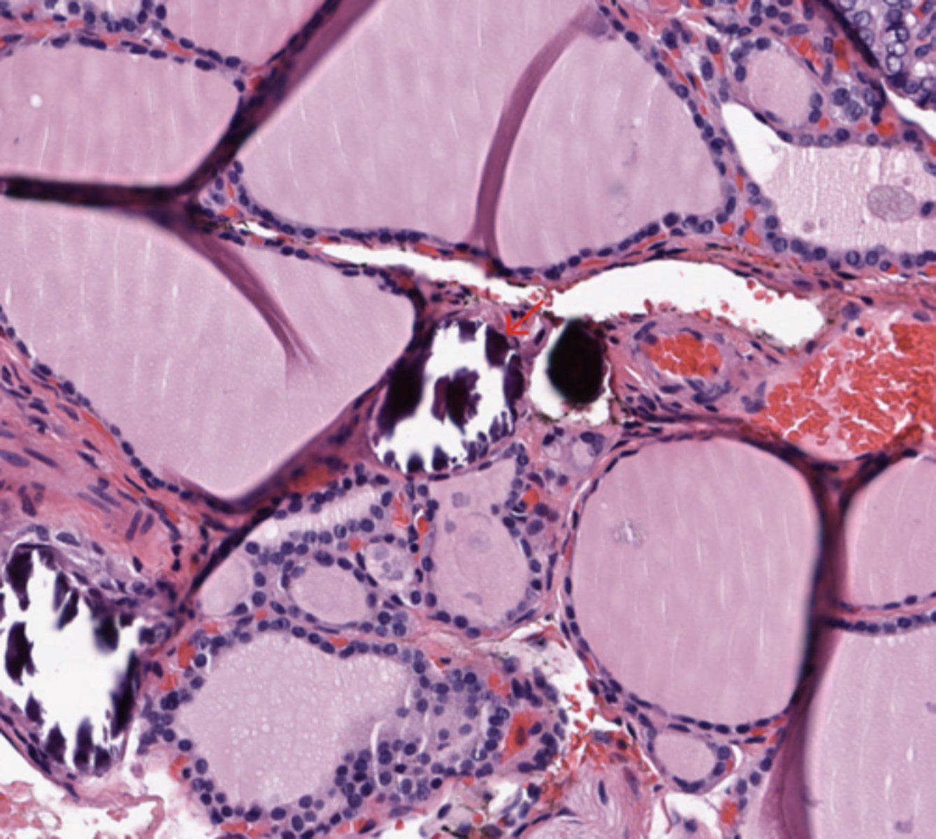
Psammoma Body
Psammoma bodies defined as laminated microcalcification are frequently associated with classic, tall cell, hobnail variants; it is postulated that psammoma bodies are formed in the hyalinized core / stalk of papillae.
(Thyroid)
Thyroid
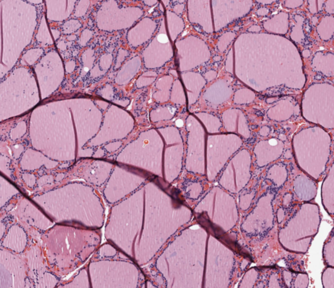
Colloid
Follicular cells normally synthesize thyroglobulin and secrete it into follicular lumen
Thyroid peroxidase, found in apical membrane of thyroid follicular cells, catalyzes iodination of tyrosine residues on thyroglobulin molecule and coupling of iodotyrosyl residues to form T4 (thyroxine) and T3, which are still bound to thyroglobulin, making them inactive; they are then stored as colloid.
In response to TSH, follicular cells pinocytose colloid, release the thyroglobulin, and secrete now active T4 and T3 into bloodstream
(Thyroid)
Thyroid
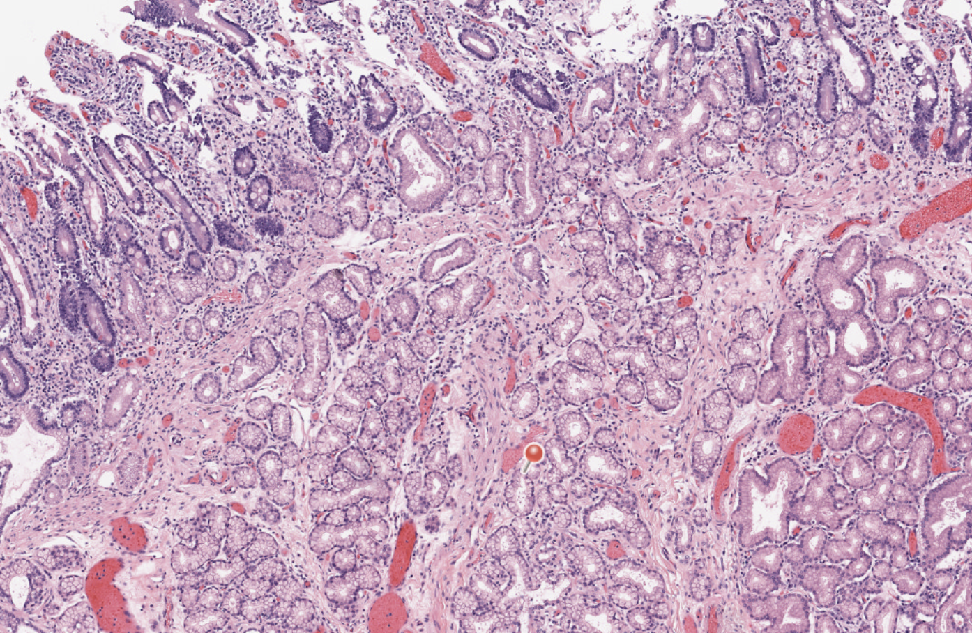
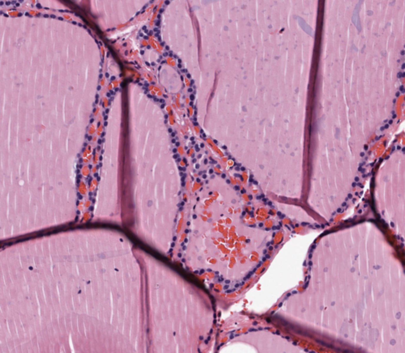
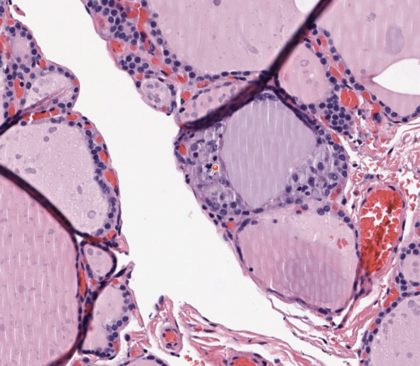
Normal thyroid follicules
Normal thyroid follicules are lined by a flat/ cuboidal epithelium. Its function is the production and secretion (into the blood) of thyroid homones.
(Thyroid)
Thyroid
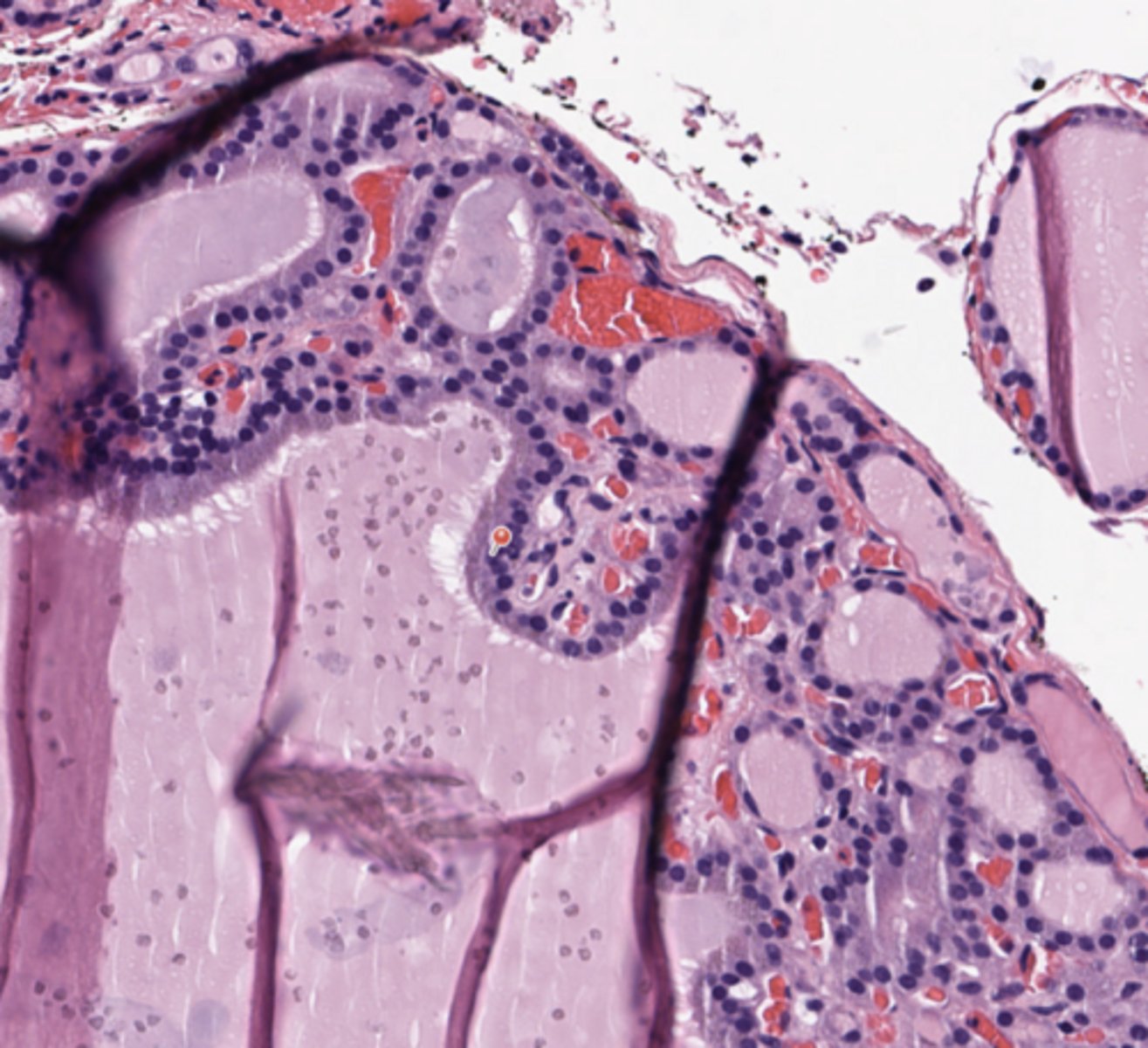
Papillary modification in benign hyperfunctioning nodule
In an hyperfunction setting, some hyperplastic thyroid nodules may exhibit papillary structures, named Sanderson polsters. They lack the typical cytological atypia and features of PTC.
(Thyroid)
Thyroid
Parafollicular C Cells
C cells represent 0.1% of gland, produce calcitonin.
(Thyroid)
Thyroid
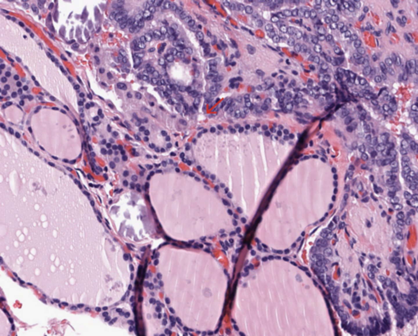
Invasive front in PTC
PTC usually shows infiltrative borders, which represent a key morphological feature distinguishing it from thyroid follicular adenoma, which is typically encapsulated.
(Thyroid)
Thyroid
Psammoma bodies interstitial spaces
Psammoma bodies in interstitial soaces may represent a sign of lymphatic invasion.
(Thyroid)
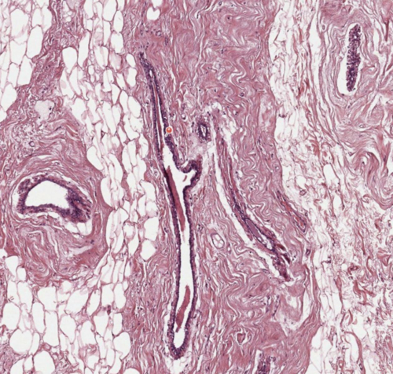
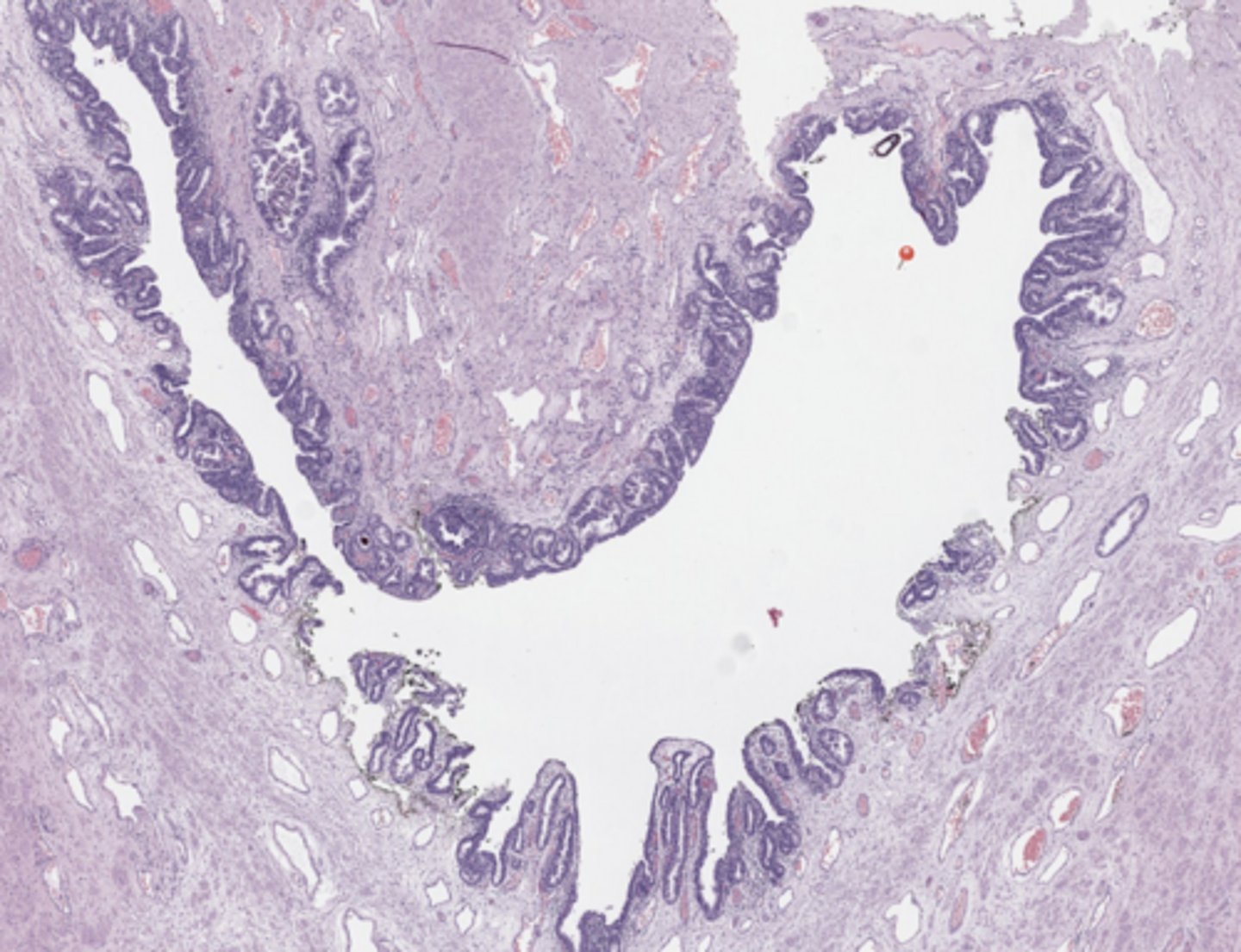
Prostate
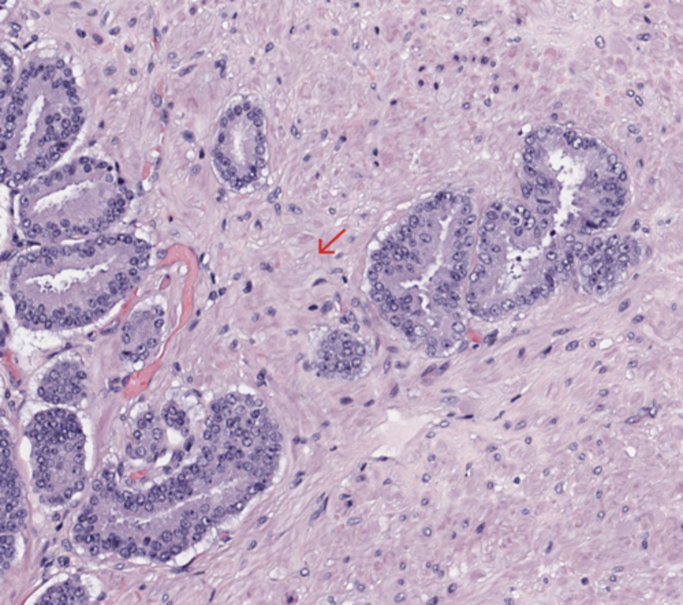
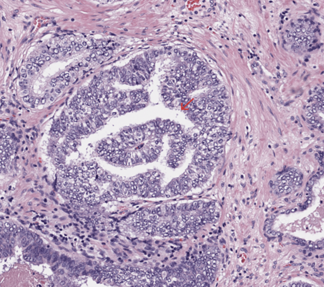
Infiltrative growth pattern
The infiltrative growth pattern typically consists of small atypical glands scattered between larger, more complex, and frequently paler benign glands.
(Prostate)
Prostate
Crowded glands
The presence of crowded glands in a linear arrangement spanning the width of the biopsy core should raise concern for malignancy. Single cells, cells arranged in cords, and fused and cribriform glands are less common patterns of invasion that are characteristic of high-grade prostatic carcinoma.
(Prostate)
Prostate
Prominent nucleoli
Prominent nucleoli are a common finding in prostate cancer; nuclear enlargement is often a helpful criterion, but its diagnostic value is not as high as that of the presence of prominent nucleoli.
(Prostate)
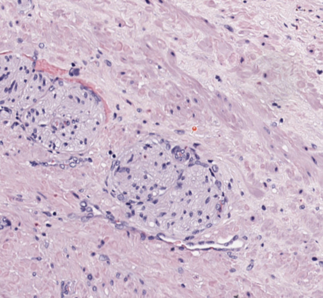
Prostate
Nerve
(Prostate)
Prostate
Glomerulations
they are a feature of prostate adenocarcinoma and are not seen in benign mimics. They are characterized by dilated glands containing intraluminal structures, which have a single point of attachment and which resemble renal glomeruli.
(Prostate)
Prostate
Prostatic urethra
(Prostate)
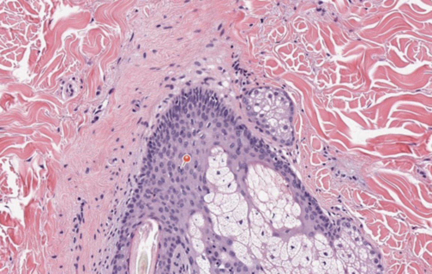
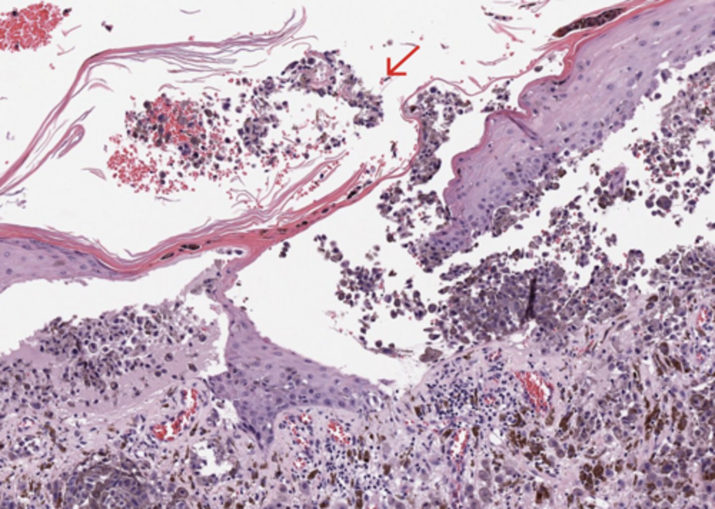
Skin
UV damage
Exposure to UV radiations damages the DNA in skin cells, triggering mutations (C>T substitutions) that cause melanocytes—the pigment-producing cells—to proliferate "without control", infiltrate, and metastatize
(Skin)
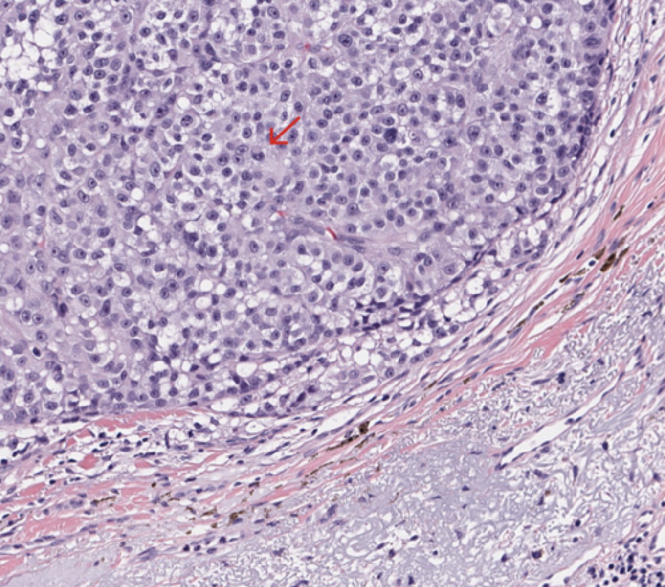
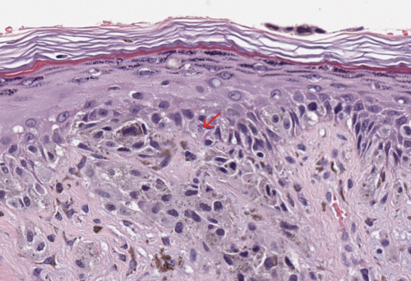
Skin
Epithelioid melanocytes
(Skin)
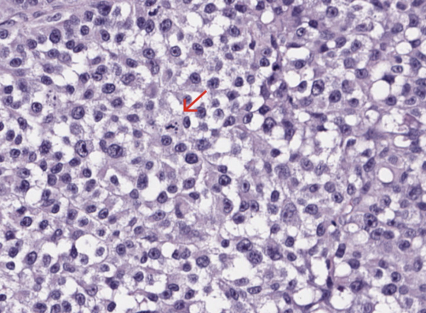
Skin
Mitosis
(Skin)
Skin
Mitosis
(Skin)
Skin
Mitosis
(Skin)
Skin
Deep infiltration (hypodermis)
(Skin)
Skin
Micro-satellitosis
(Skin)
Skin
Lympho-vascular invasion
(Skin)
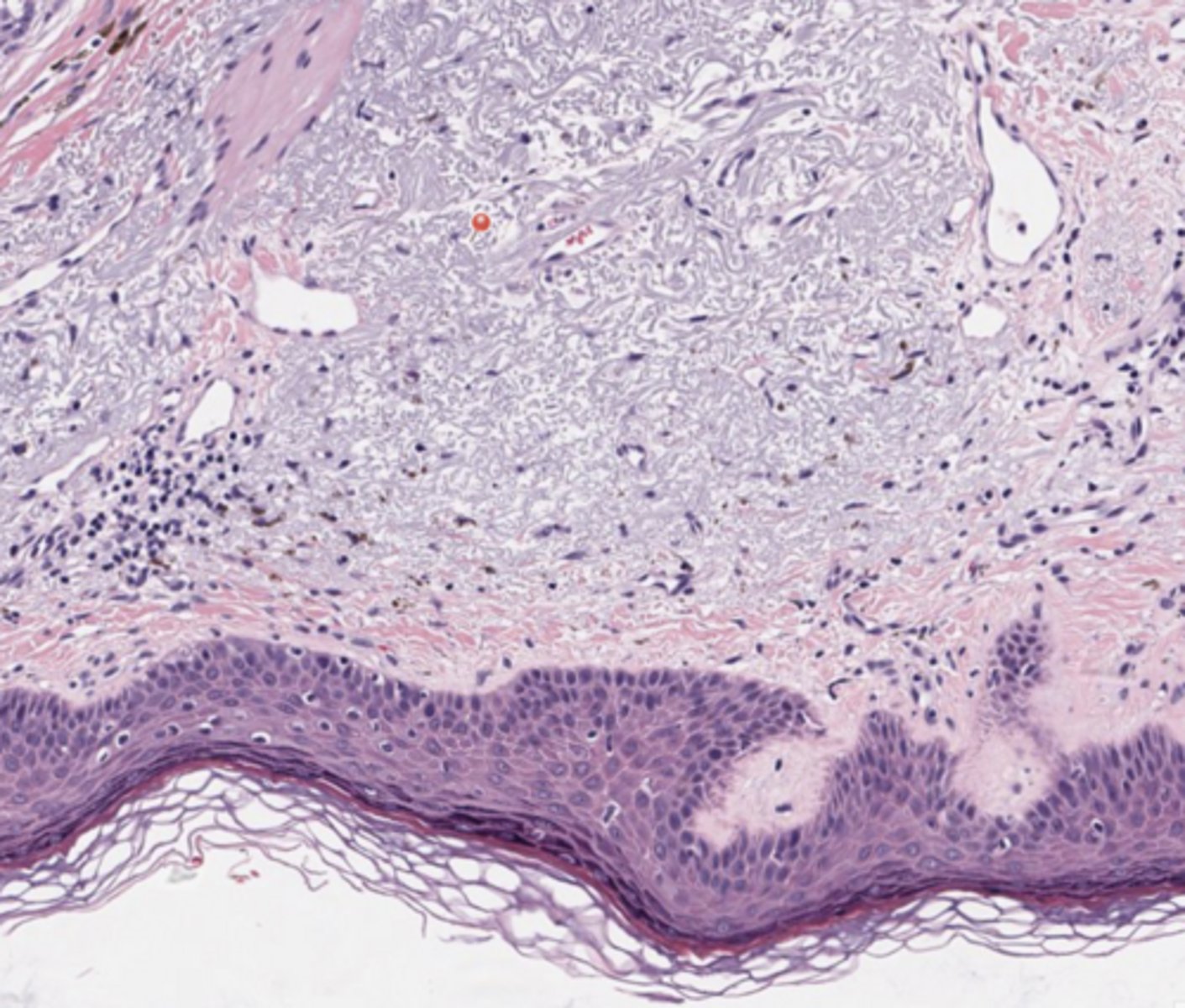
Skin
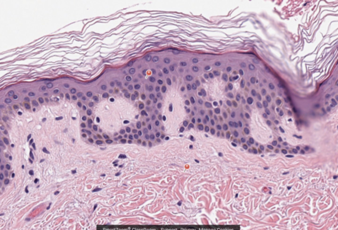
Upper pin - Adjacent epidermis
Lower pin - Papillary dermis
(Skin)
Skin
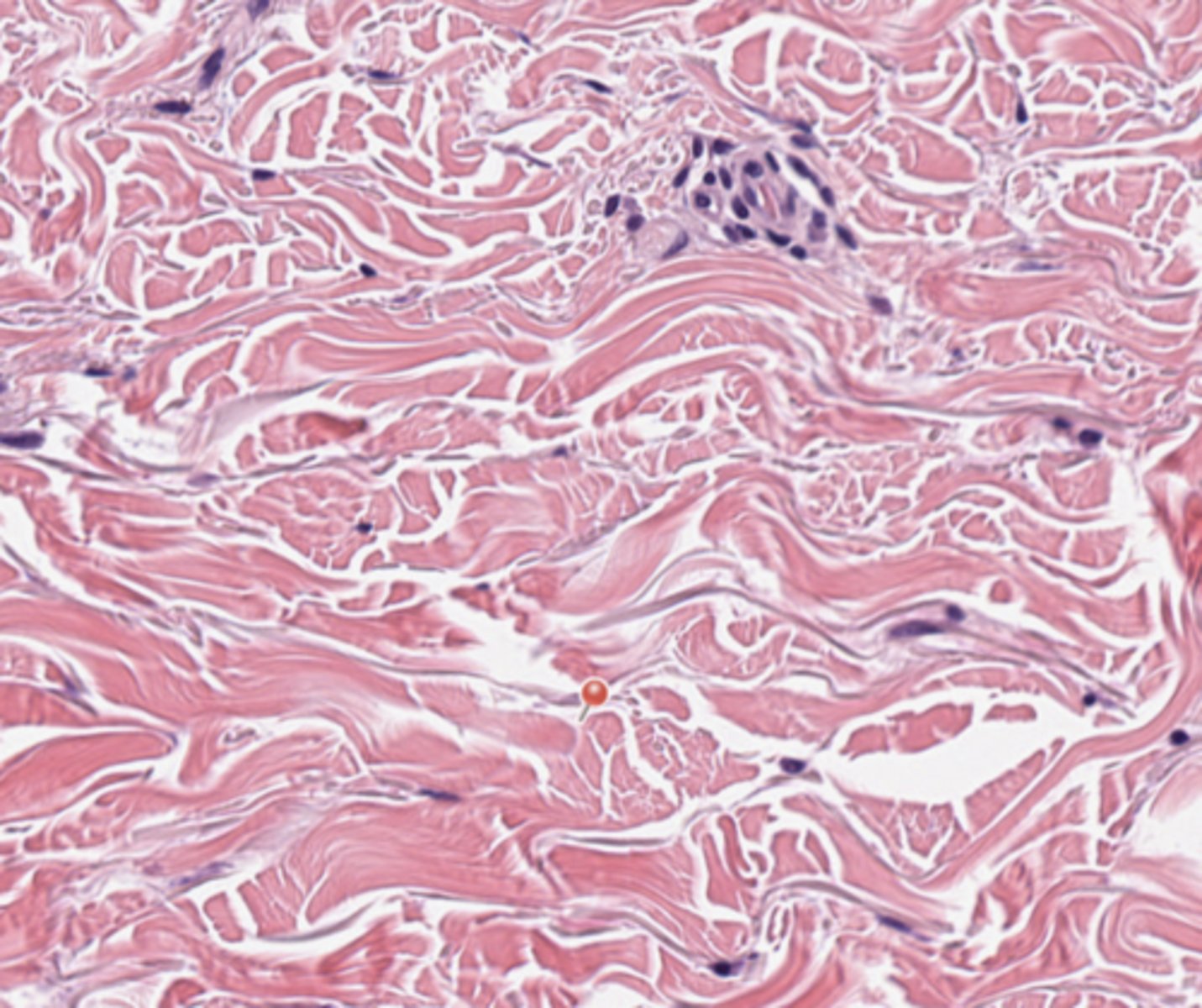
Reticular dermis
(Skin)
Skin
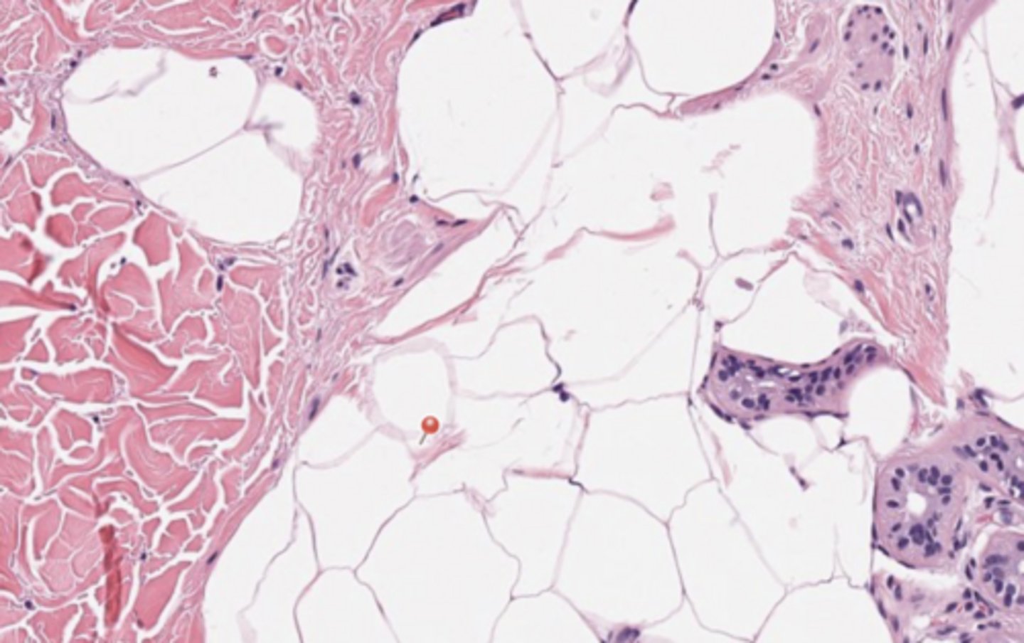
Hypodermis
(Skin)
Skin
Adnexal structure
(Skin)
Skin
Radial growth phase
(Skin)
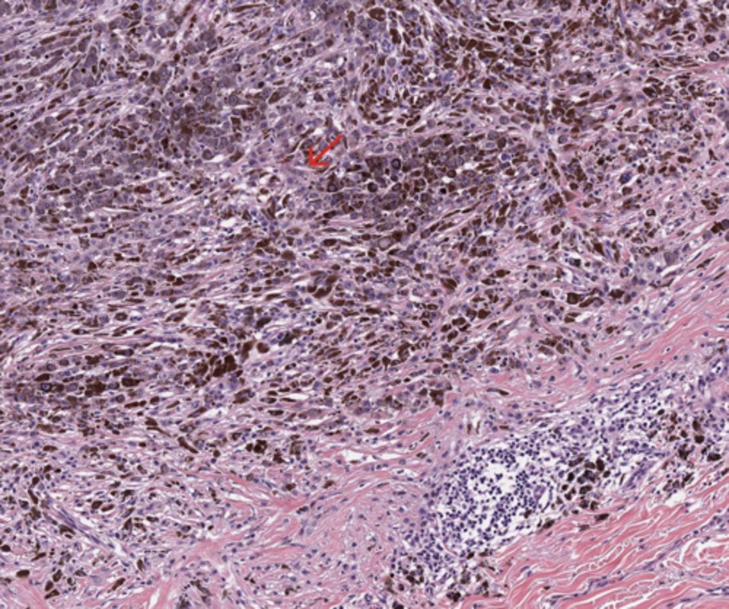
Skin
Vertical growth phase
(Skin)
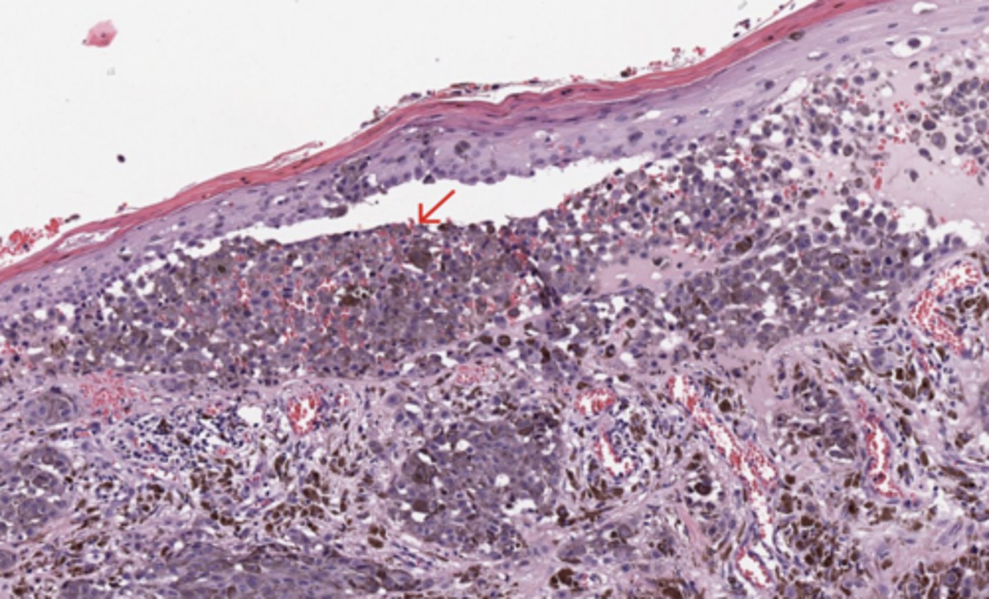
Skin
Epidermal effacement
(Skin)
Skin
Melanin pigment
(Skin)
Skin
Ulceration
(Skin)
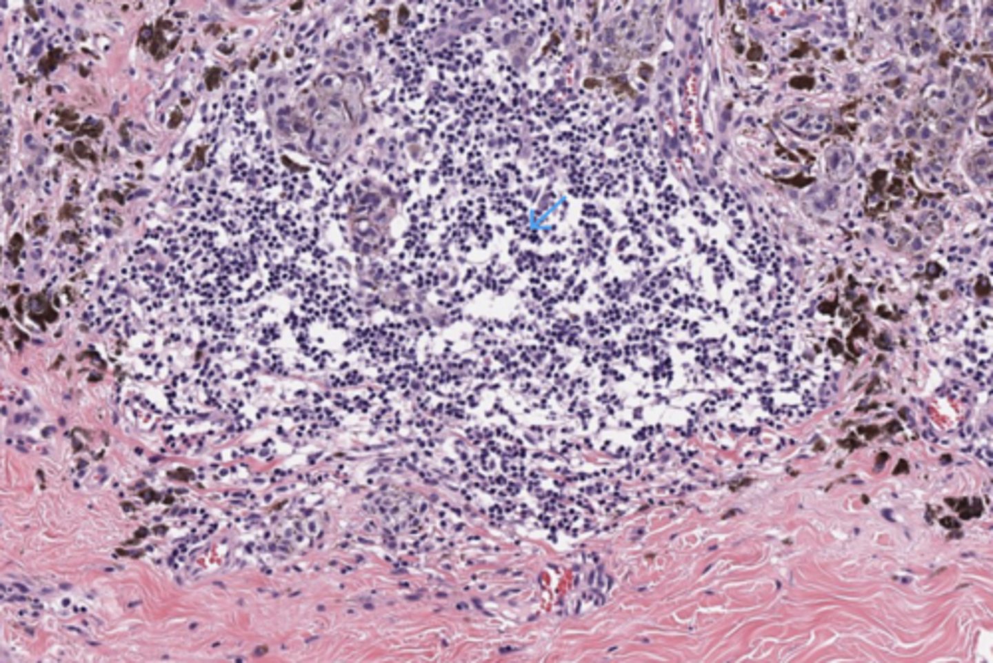
Skin
TILs
(Skin)
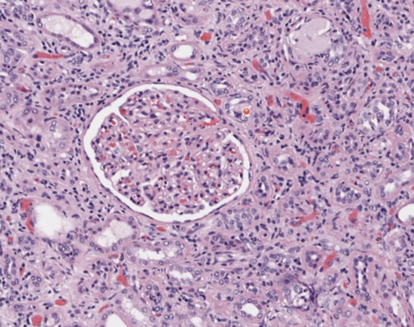
Urothelial tract
Renal glomerulus
(Urothelial tract)
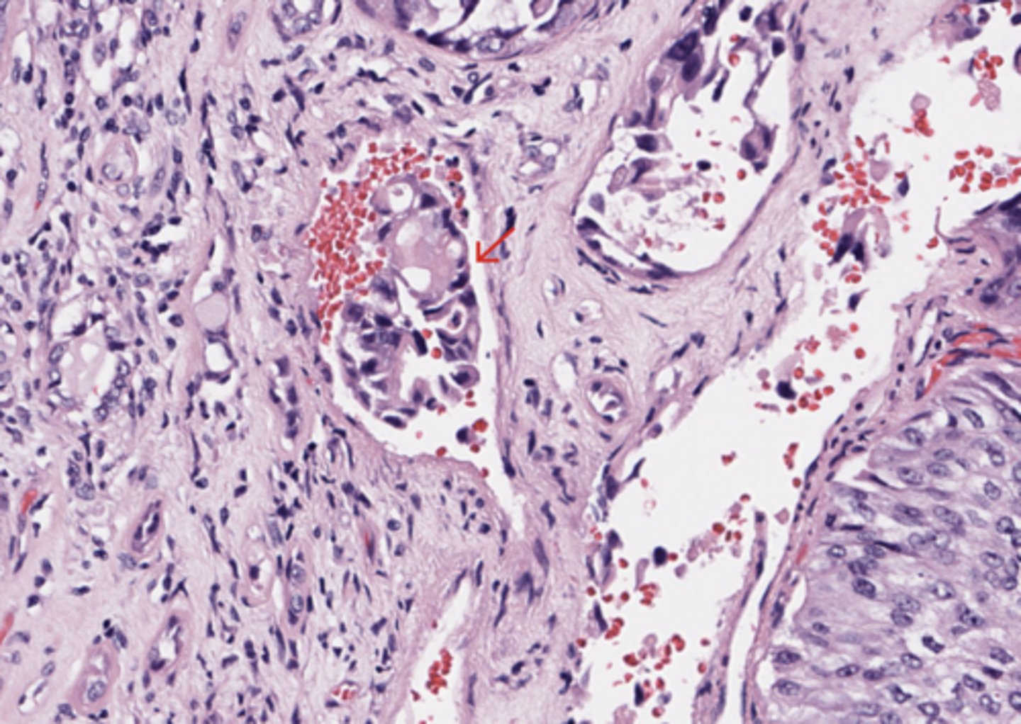
Urothelial tract
Lymph vascular invasion
(Urothelial tract)
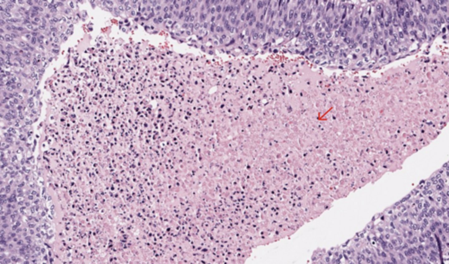
Urothelial tract
Necrosis
(Urothelial tract)
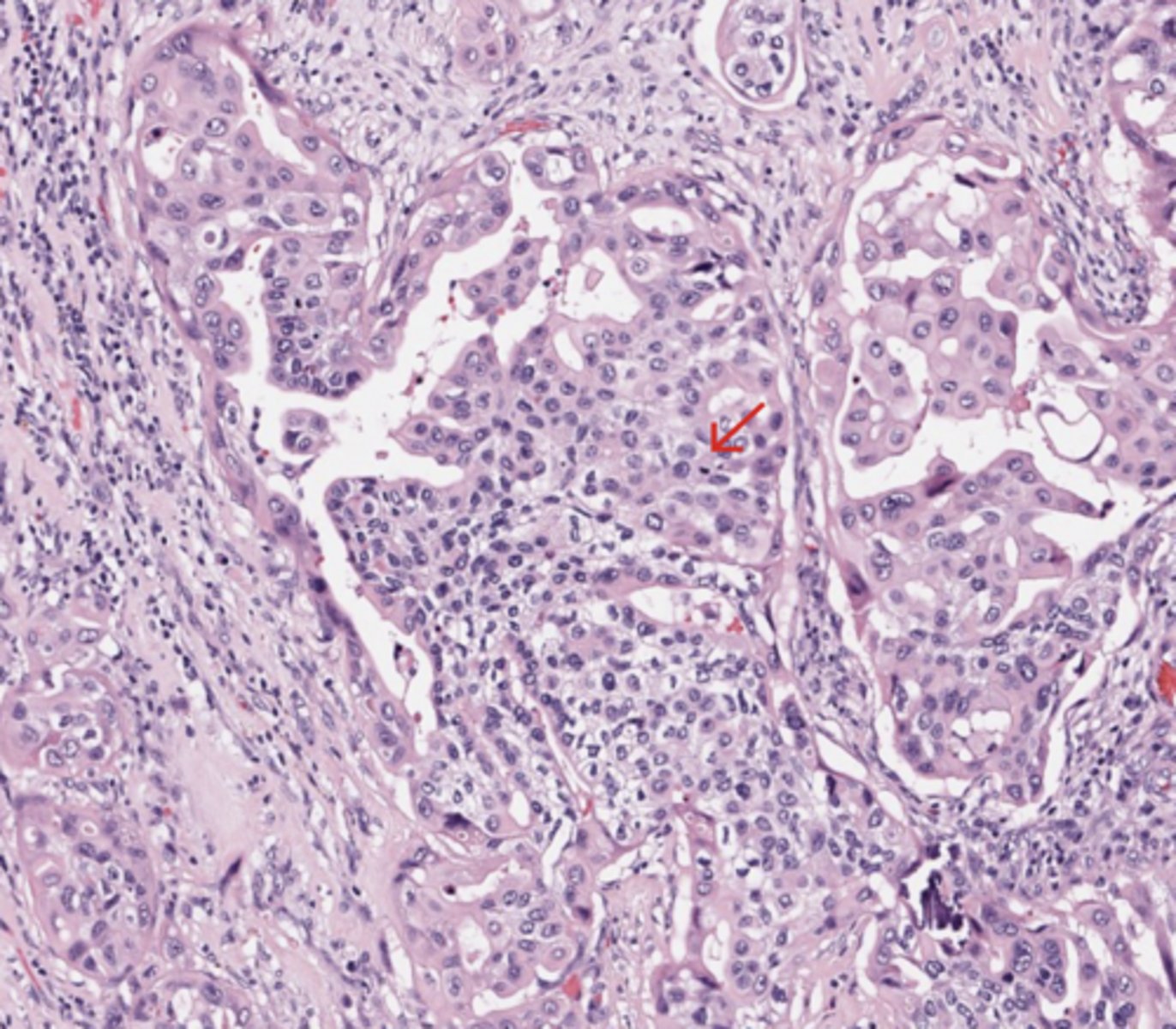
Urothelial tract
High Grade component
(Urothelial tract)
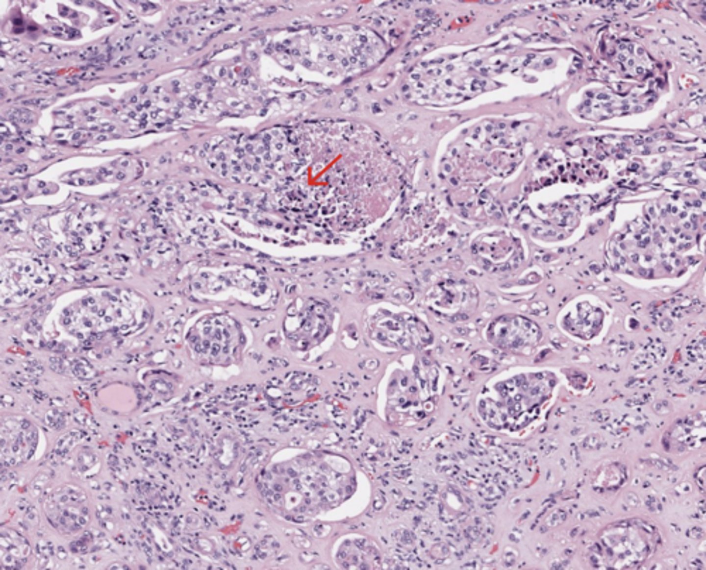
Urothelial tract
Invasion of renal parenchyma
T3 (Renal pelvis) Tumour invades beyond muscularis into peripelvic fat or renal parenchyma
(Urothelial tract)
Urothelial tract
Peripelvic Fat
(Urothelial tract)
Urothelial tract
Ureter
(Urothelial tract)