Dev Bio Final
1/66
There's no tags or description
Looks like no tags are added yet.
Name | Mastery | Learn | Test | Matching | Spaced | Call with Kai |
|---|
No analytics yet
Send a link to your students to track their progress
67 Terms
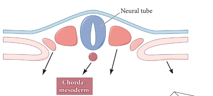
Identify the four main types of mesoderm
Chordamesoderm
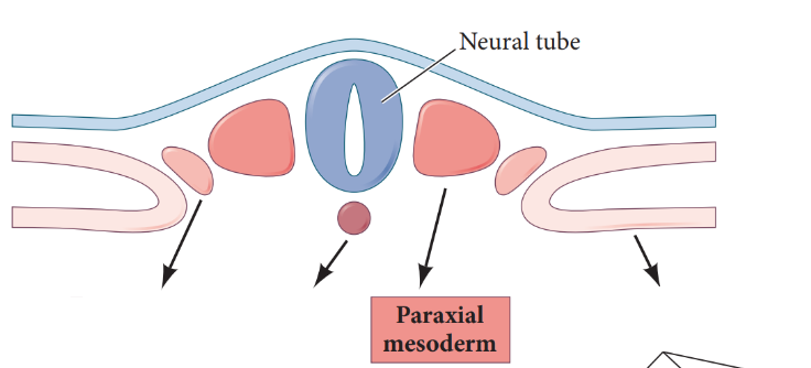
Paraxial mesoderm
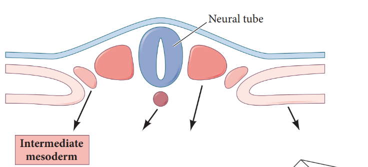
Intermediate mesoderm
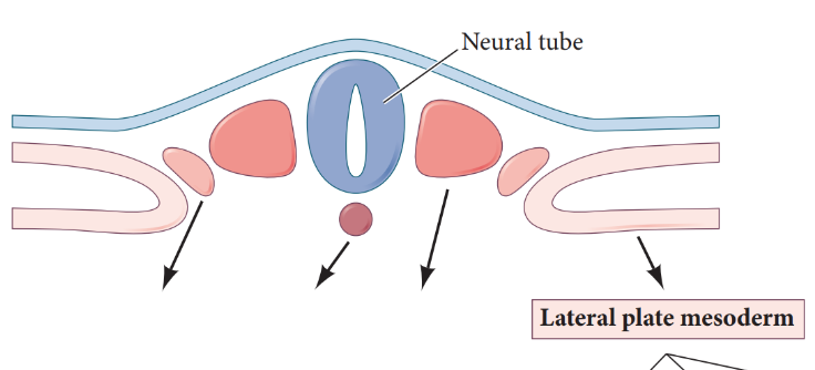
Lateral plate mesoderm
Chordamesoderm
forms the mesoderm
most central
Paraxial mesoderm
forms the somites; anteriormost paraxial mesoderm does not segment → becomes the head mesoderm
on both side of the notochord
Intermediate mesoderm
forms the urogenital system → kidneys and gonads
directly lateral to paraxial mesoderm
Lateral plate mesoderm
forms the heart, blood vessels, blood cells, linings of body cavity, pelvic and limb skeleton
farthest from notochord
Somitogenesis
the formation of somites, involving the periodic creation of an epithelial block by the mesenchymal cells of the presomitic mesoderm
Pre-somitic mesoderm
subset of paraxial mesoderm that is unsegmented and is located on each side of neural tube immediately behind the forming somites → precursor to somites
segments periodically in an anterior to posterior manner to form somites
What happens to the number of somites as development proceeds?
number of somites increases
How are somites built?
pre-somitic mesoderm only contains mesenchymal cells → mesenchymal to epithelial transition
become a compact epithelial tissue as fibronectin matrix deposition seals the new somite outer wall to form somite block
each somite separates from the pre-somitic mesoderm at the anterior end forming somite pairs
What are the 2 big questions regarding somitogenesis?
WHEN should a somite form → controlled by the clock
WHERE should the boundary form so somites are always the same size → controlled by wavefront
Describe the “clock and wavefront” model.
Describe the “clock and wavefront” model
In this model, two converging systems interact to regulate (1) where a boundary will be capable of forming (the wavefront) and (2) when epithelial boundary formation should occur (the clock).
Outline the clock and wavefront process
wavefront (determination front) is established by gradient of antagonistic retinoic acid and FGF
RA is high in anterior and decreases posteriorly
FGF is high in posterior and decreases anteriorly
high FGF signaling maintains PSM cells in immature state → cells that receive low RA and FGF concentration will become competent to form a boundary to create a somite
determination front is where RA and FGF overlap → cells are competent to Notch signaling to build somite
cyclic expression (turn on + off in cycles) of Notch genes (Lfng, Hairy1) → Notch turns on these genes which are expressed throughout PSM from posterior to region last somite formed
high Notch wave hits determination front to induce MET to create new somite boundary
Can cells form somites while exposed to high FGF?
NO
What happens if the clock is sped up?
formation of numerous, smaller somites
What happens if clock is slowed down?
formation of fewer, larger somites
Identify factors that influence somite identity/specification along the A-P axis
as somites form they are exposed to different Hox gene combinations depending on their position along A-P axis
each somite receives unique Hox code
specify somites to receive cervical, thoracic, lumbar, or sacral identities
What happens if Hox genes are not expressed in discrete regions?
overexpression leads to structural abnormalities
e.g. if thoracic Hox gene expression expands into cervical region → cervical vertebrae may develop ribs
What are the cells the somites give rise to?
Somites are transient and as they mature they split into compartments that give rise to specific tissue lineages:
sclerotome cells → contribute to cartilage
syndetome cells → contribute to tendons
myotome cells → contribute to skeletal muscle
endothelial cells → contribute to dorsal aorta
dermatome cells → contribute to dermis + skeletal muscle
Describe the different regions that the somite subdivides into
sclerotome: made up of migratory mesenchymal cells
dermomyotome: made up of remaining epithelial cells
How does the sclerotome region form?
EMT occurs in the ventromedial portion of the somite forming the sclerotome
Sclerotome cells give rise to
vertebrae along A-P axis, cartilage, tendons, meninges of spinal cord
What cells are within the sclerotome (ventromedial) division of the somite?
sclerotome cells
syndetome cells
endothelial cells
How does the dermomyotome form?
remaining epithelial structure in the dorsolateral region of the somites
does NOT undergo EMT
What two regions in the dermomyotome subdivided into?
Dermatome → precursor to dermis of the skin
Myotome → skeletal muscle precursor
How is the myotome region formed?
myoblasts (muscle precursor cells) migrate beneath the dermomyotome to produce the myotome
What are the two myotome regions?
Primaxial myotome/muscle
Abaxial myotome/muscle
Primaxial myotome
myoblasts in myotome closest to neural tube form the centrally located primaxial myotome
gives rise to: intercostal muscles of the ribs + deep muscles of the back
Abaxial myotome
myoblasts in the myotome farthest from the neural tube form the abaxial myotome
give rise to: body wall, limbs, tongue
Define the paracrine factors that form the different regions of the somite
Sclerotome: high Shh from floor plate + notochord
Dermatome: Neurotrophin-3 + Wnt1 from dorsal neural tube
Myotome:
Primaxial: low Shh, Wnt1 + Wnt3 from floor plate + notochord
Abaxial: BMP4 + Fgf5 from lateral plate mesoderm, Wnt from epidermis
Outline the process from somite to vertebrae formation
notochord induces surrounding mesenchyme cells to secrete epimorphin
epimorphin attracts sclerotome cells to region around notochord and neural tube where they condense and differentiate into cartilage
as nerves from the spinal cord migrate they split the sclerotome into an anterior and posterior segment
as motor neurons grow to innervate newly forming muscles the anterior segment of each sclerotome recombines with the posterior segment of the next anterior sclerotome to form new vertebrae → re-segmentation
Resegmentation
the primary segments established by somites reorganize by half a segment to form the definitive vertebral column
by shifting the skeletal segments by half a unit, each muscle block (myotome) directly straddles an intervertebral joint
muscle anchors to two successive vertebrae, providing the leverage necessary for lateral bending and movement of the spine
Intermediate mesoderm
forms the urogenital system consisting of the kidneys, gonads, and their associated ducts and outer portion of adrenal gland
located in trunk/posterior of body → in between paraxial and lateral plate mesoderm
Describe how scientists determined how location of the intermediate mesoderm was important for kidney induction
scientists separated intermediate mesoderm from paraxial mesoderm
intermediate mesoderm couldn’t turn on
determined that intermediate mesoderm must have contact with paraxial mesoderm to form kidneys
What are the 3 stages of mammalian kidney development
Pronephros → transient in humans; fish/amphibian larvae gain functional kidney at this stage
Mesonephros → transient
Metanephros → mammals get functional kidney at this stage
Pronephros
pronephric duct arises in intermediate mesoderm ventrolateral to anterior somites
cells migrate caudally and anterior region of duct induces adjacent mesenchyme to form pronephros of developing kidney
in mammals, pronephros degenerate but more caudal portions of pronephric duct persist to become nephric duct
central component of excretory system
Mesonephros
nephric duct induces adjacent mesenchyme to form mesonephros
as more tubules are induced caudally the anterior mesonephric tubules begin to regress through apoptosis
mesonephros is the main source for hematopoietic stem cells necessary for blood cell development; some become epididymis and vas deferens in males
Metanephros
permanent kidney of amniotes
formed through reciprocal induction between ureteric bud and metanephrogenic mesenchyme
Describe the use of reciprocal induction during kidney development
kidney is formed from 2 intermediate mesoderm populations:
ureteric bud and metanephrogenic mesenchyme
metanephrogenic mesenchyme becomes committed in posterior regions of intermediate mesoderm where it induces epithelium of ureteric bud to branch from each paired nephric duct
forms collecting ducts and ureters
when ureteric buds emerge from nephric ducts they enter the metanephrogenic mesenchyme where the epithelium at the tips of the branches induces the mesenchyme to aggregate
forms the nephrons and tubules through division
Explain how paracrine factors are critical for reciprocal induction
Wnt9 and Wnt6 are released from ureteric bud to induce the surrounding metanephorgenic mesenchyme to form the tubular epithelium (nephrons)
these paracrine factors induce Wnt4 which acts in an autocrine manner to complete MET transition
Explain bladder development and kidney connection
initially ureter empties into a cloaca which is lined by endoderm
maintained in adult birds, reptiles, amphibians and functions as waste receptacle for both intestine and kidney
in mammals, a urogenital septum forms separating the rectum from the urogenital sinus → forming the bladder
Cloaca
a single rear opening that serves as the common chamber for intestinal, urinary, and reproductive tracts replacing separate urinary and anal openings
Lateral plate mesoderm
forms the circulatory system consisting of the heart, blood cells, and blood vessels
located on the lateral side of each of the two bands of intermediate mesoderm
What does the lateral plate mesoderm subdivide into?
somatic mesoderm (body cavity) → dorsal layer
splanchnic mesoderm (circulatory system) → ventral layer
What is the space between the two layers?
coelom → becomes the body cavity by stretching from the future neck region to the posterior of the body
What does the somatic mesoderm divide into
somatic mesoderm underlies the ectoderm → together form the somatopleure
What does the somatopleure give rise to?
limb bones
pelvis
What does the splanchnic mesoderm divide into?
splanchnic mesoderm overlies the endoderm → together form the splanchnopleure
What does the splanchnopleure give rise to?
circulatory system
What does the coelom divide into?
pleural cavity → space surrounding thorax
pericardial cavity → space surrounding heart
peritoneal cavity → space surrounding abdomen
What is the first functional organ in the body?
the heart
How are the heart fields formed?
vertebrate heart arises from two regions of splanchnic mesoderm—one on each side of the body—that interact with adjacent tissue to become specified for heart development
cardiogenic mesoderm are cells within the heart field that form the heart
heart field is divided into 2 regions:
first field: progenitor cells form the primary heart tube but these cells have limited proliferative ability and will generate only the major portion of the left ventricle
second field: progenitor cells add cells to both anterior and posterior of heart tube → produce atria at posterior end and produce right ventricle and outflow tracts at anterior end
What are the cell types of the heart generated from the heart fields?
Endocardial endothelial cells: line/cushion the heart
Atrial myocyte: fill in atrial cavity to give musculature
Ventricular myocyte: muscle or Purkinje fibers that coordinate heartbeat
Describe the key signals that are needed to specify the cardiogenic vs hemangiogenic mesoderm
Cardiogenic mesoderm:
cells near anterior of body do not receive Wnt → blocked by inhibitors secreted by anterior endoderm
cells receive BMP + Fgf8 to convert LPM into cardiogenic mesoderm
Hemangiogenic mesoderm:
cells near posterior of body receive Wnt signals
converts LPM into hemangiogenic mesoderm
In the center of the embryo Noggin + Chordin signals from the notochord block BMP → cardiogenic + hemangiogenic fields do not form in the center of the embryo
Cardiogenic mesoderm
Precursor to the heart fields
Hemangiogenic mesoderm
Precursor to blood, blood vessels
Describe the process of heart tube formation starting with cardiogenic mesoderm and finishing with a single tube
cardiogenic mesoderm cells migrate from splanchnopleure creating two populations:
one on the right + left side of the neural tube
each side has its own first and second heart fields → each side forms its own heart tube
two endocardial tubes form that migrate and fuse together to create one heart tube
What happens if the two heart tubes fails to migrate and fuse?
Cardia bifida: two separate hearts form, one on each side of the body
cannot sustain life
How is the heart formed?
formation of the foregut allows for formation of single tube
final endocardial tube consists of:
endocardium surrounded by myocardium (muscle)
Describe the process of heart looping
at 21 days of development the human heart is a single chamber tube → must loop to become a two chambered tube: atrium to receive blood and ventricle to pump blood out
pressure from blood flow helps drive looping to completion
when looping is complete portion of the heart tube destined to become the atria lies anterior to the portion that becomes ventricles
the septa forms from migrating cardiac neural crest cells
What does the heart need in order to pump blood after it has formed?
vascular system must establish its circulatory loops → blood vessels
Outline vasculogenesis
network of blood vessels is created from scratch from the splanchnic mesoderm of the lateral plate
combination of BMP, Wnt, and Notch signaling activates Etv2 transcritption factor in LPM cells converting them to hemangioblasts
Low Notch signaling causes hemangioblasts to become endothelial (blood vessels)
High Notch signaling causes hemangioblasts to become blood cells
Outline angiogenesis
blood vessel network is remodeled into veins, arteries, and capillaries
What are the sites of vasculogenesis?
In amniotes, extraembryonic vasculogenesis occurs in “blood islands” of the yolk sac
formed by the hemangioblasts and give rise to early vasculature needed to feed the embryo
intraembryonic vasculogenesis forms the dorsal aorta