Unit 2: Brain and Neurones
1/24
There's no tags or description
Looks like no tags are added yet.
Name | Mastery | Learn | Test | Matching | Spaced | Call with Kai |
|---|
No analytics yet
Send a link to your students to track their progress
25 Terms
stimuli
change in the enviroment
Sensory neurone
carries electrical impulses from receptors to Central nervous system
Motor neurone
Carries electrical signal from CNS to effector
relay neurone
within the CNS and coordinates a response
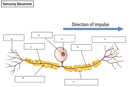
A) Dendrites
B) Nucleus of Schwann Cell
C) Dendron
D) Nucleus
E) Cell body
F) Schwann Cell - makes up myelin sheath - insulates dendron and axon - speeds up electrical impulses
G) Axon
H) Axon terminals
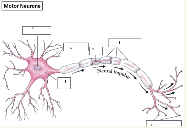
A) Nucleus
B) Cell body
C) Dendrites
D) Axon
E) Myelin sheath (also schwann cells)
F) Axon terminal
How is electrical impulse transferred across the synapse
Electrical impulse arrives at axon terminal, triggering vesicles containing neurotransmitters (chemical messengers) to move to the membrane of neurone.
Vesicles fuse with cell membrane and release neurotransmitters which diffuses across synapse (gap between two neurones)
Neurotransmitters binds to receptors on the next neurone initiating a new electrical impulse - neurotransmitters are complimentary to specific receptors
Function of the reflex arc
to prevent harm
Structure of Reflex arc
Nervous system carries fast electrical impulse
Stimuli is detected by receptors
impulses initially travels along sensory neurone which transmits the impulses from the sense organs to the CNS
Here, the impulses travels along the spinal cord to the brain which coordinates a response
The electrical impulse then travels back along the motor neurone to the effectors (muscles or glands) to carry out a response
relay neurone
cell body at the start
no myelin sheath - don’t need it
shorter than other neurones
surrounded by billions of interconnected neurones
What is the structure and main function of the spinal cord?
It acts as the main communication highway linking the brain to the peripheral nervous system. It also independently coordinates rapid reflex actions (via the reflex arc).
Structure: A long, cylindrical column of nervous tissue extending from the brain down through the spine.
What are the cerebral hemispheres (cerebrum) and what do they control?
Controls conscious thought, memory, language, intelligence, sensory processing (sight/hearing), and initiates voluntary movement.
Where is the cerebellum located and what is its function?
Structure: A smaller, folded structure located at the lower back of the brain (underneath the cerebrum).
Function: Controls balance, posture, and muscle coordination. It ensures voluntary movements are smooth and precise
What is the medulla oblongata and why is it vital for survival?
Structure: A tube-like structure located at the very base of the brain, directly connecting it to the spinal cord.
Function: Controls unconscious/involuntary life-support functions, such as regulating heart rate and breathing rate, as well as swallowing reflexes.
Why is investigating and treating brain tissue physically inside the skull so difficult and risky?
Physical barrier: The brain is fully enclosed by the hard bone of the skull (cranium), making physical access highly invasive.
High risk of damage: The brain is incredibly complex and delicate; any accidental surgical damage can cause permanent loss of vital functions (e.g., speech, movement, life support).
No regeneration: Central nervous system neurones do not easily repair or regenerate.
How does a CT (Computed Tomography) scan work to investigate the brain?
It fires a series of X-ray beams from many different angles around the head.
A computer processes these X-rays to build up a detailed 3D, cross-sectional image of the brain's physical structure.
What type of information do CT scans give a doctor, and what are they used to diagnose?
They show structural information only (a static picture of the physical tissues).
Used to identify abnormal structures like brain tumours, internal bleeding (hemorrhages), blood clots, or brain damage from a stroke.
How does a PET (Positron Emission Tomography) scan work to investigate the brain?
The patient is injected with a radioactive tracer attached to a biological molecule (usually glucose).
The scanner detects the radiation emitted by the tracer to see where it travels in the body.
What type of information do PET scans give a doctor, and what are they used to diagnose?
They show functional / metabolic information in real-time.
Active brain cells respire faster and use more glucose, taking up more tracer.
Used to study real-time brain activity during tasks, or to identify conditions where brain activity changes (e.g., Alzheimer's disease, epilepsy).
What is the fundamental difference between a CT scan and a PET scan when investigating the brain?
CT scan = Structure. It shows the physical shape, location, and anatomy of tissues (a static map).
PET scan = Function. It shows active cellular metabolism and which parts of the brain are working in real-time (a dynamic map).
Why can't the body naturally repair damage to the brain or spinal cord?
No mitosis/regeneration: Mature neurones in the central nervous system (CNS) do not readily divide by mitosis.
Permanent damage: Because they cannot replicate or replace themselves, any cell death or structural damage is usually permanent.
What is the physical consequence of a spinal injury, and why can't surgery fix it?
Consequence: A severed or crushed spinal cord cuts off electrical impulses between the brain and the rest of the body, leading to paralysis.
Why it can't be fixed: There is currently no medical way to successfully rewire or splice millions of microscopic nerve fibres back together across the damaged area.
What is the blood-brain barrier, and how does it limit the chemical/drug treatment of brain diseases?
What it is: A specialized, highly selective filtering mechanism in the capillaries supplying the brain.
The limitation: It is designed to keep toxins out, but it also blocks many useful medications (like standard chemotherapy drugs or antibiotics) from physically entering brain tissue.
Why is physically removing a brain tumour surgically so difficult and limited?
Inaccessibility: Tumours are often buried deep within the brain or surrounded by vital structures.
Collateral damage: Removing the tumour risks accidentally cutting or damaging the healthy, surrounding brain tissue, which could destroy essential functions like speech, memory, or movement.
If a patient undergoes treatment for a brain tumour (like radiotherapy or surgery), what limitations/risks remain?
The treatment itself can cause permanent damage to surrounding healthy neurones.
This can lead to long-term neurological problems, personality changes, or cognitive difficulties that cannot be reversed.