Anatomy of the Female Pelvis
1/28
There's no tags or description
Looks like no tags are added yet.
Name | Mastery | Learn | Test | Matching | Spaced | Call with Kai |
|---|
No analytics yet
Send a link to your students to track their progress
29 Terms
What does the bony pelvis consist of?
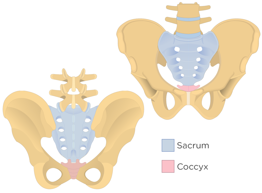
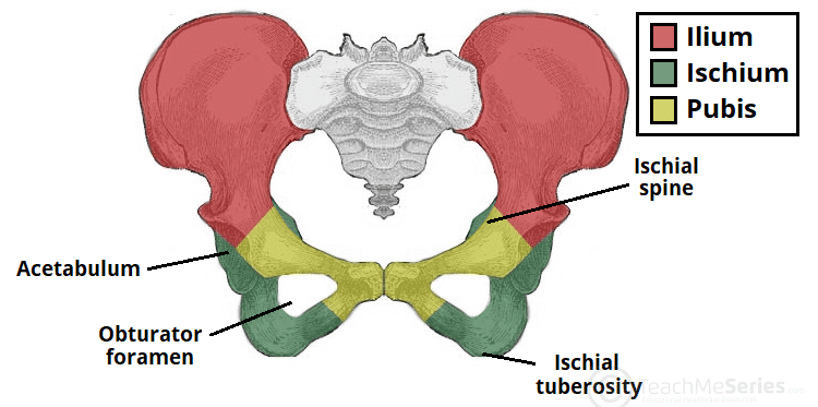
Sacrum, coccyx, and innominate bones
What are the innominate bones?
Ilium, ischium, and pubic symphysis
What are the markers of the posterior border of the pelvic cavity?
Sacrum and coccyx
What are the boundaries of the female pelvis?
From iliac crest to the pelvic diaphragm at the base of the pelvis
The pelvis is divided into what?
True (lesser) pelvis and false (major/greater) pelvis
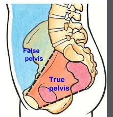
Where and what does the true (Lesser) pelvis consist of?
Inferior portion of pelvis that contains uterus, ovaries, fallopian tubes, urinary bladder, small bowel, sigmoid colon and rectum
Where and what does false (major/greater) pelvis consist of?
Superior portion of pelvis that is pelvic by virtue of its bony boundaries (iliac crest)
What imaginary line separates the true (lesser) pelvis from false (major/greater) pelvis?
Linea terminalis
Which female pelvic muscles may be shown sonographically? AKA 5 true pelvic muscles
Rectus abdominis muscles, the iliopsoas muscles, obturator internus muscles, piriformis muscles, and a group of muscles AKA pelvic diaphragm.
What is the pelvic diaphragm and where is it located sonographically?
Hammock shaped muscles (levator ani and coccygeus) that gives support to pelvic organs,
Location: transverse plane at level of vagina, posterior to bladder, vagina, and rectum.
What are the O.P.I muscles and where are they located sonographically?
Opturator internus (lateral to bladder)
Piriformis (posterolateeral)
Iliopsoas (anterolateral)
Location: transverse plane while scanning adnexas. They are adjacent/lateral to bladder/ovaries,uterus
What are the uterine ligaments?
The broad ligaments, round ligaments, suspensory ligaments of the ovary, and cardinal ligaments
What are the broad ligaments?
A double-layered fold of peritoneum connecting lateral sides of uterus to walls and floor of pelvis pelvic organs. This is the only ligament ever visualized on sono, only when there is pelvic ascites
What are the round ligaments?
They support the fundus of the uterus and extend from uterine cornua to labia majora between the folds of the broad ligaments
What are the suspensory ligaments of the ovaries (infundibulopelvic)?
They support the ovaries and tubes and extend from ovaries to pelvic side walls
What are the cardinal ligaments?
They support the cervix and extend from lateral surface of cervix to the lateral fornix of the vagina
Where is the vesicouterine pouch or anterior cul-de-sac located?
Anterior to the uterus and posterior to bladder
Where is the rectouterine pouch located?
Between rectum and uterus. AKA posterior cul-de-sac, pouch of douglas, or rectovaginal. It is also the most likely place for fluid to collect in the pelvis
Between the anterior wall of bladder and symphysis pubis lies what?
The space of Retzius aka retropubic space which contains extraperitoneal fat
What supplies blood to female genitalia?
Abdominal aorta
The aorta branches into what?
Paired common iliac arteries (bifurcation near umbilicus)
The common iliac arteries then divide into what
External iliac arteries and internal iliac arteries.
What are the uterine arteries?
Branches of internal iliac arteries
What are the branches of the uterine artery?
Arcuate arteries, along lateral aspect of myometrium. Which progress into the radial arteries which supply blood to deeper laters of myometrium. The radial arteries then divide into straight and spiral arteries which is in the layers of the endometrium. Straight feeds basal layers and spiral feeds decidual/functional layers of endo.
The ovarian arteries originate from where?
Lateral to aorta AKA gonadal arteries
The ovaries have dual blood supply what arteries is it?
Each ovary receives nourishment from an ovarian artery and a branch of uterine artery
All venous structures mirror their arterial counterparts with exception of what vein?
Left ovarian vein which drains directly into left renal vein
Uterine veins drain or return to what?
Internal iliac veins
What does the right ovarian vein drain into?
IVC