Eye and Ear Microscopic Anatomy
1/51
There's no tags or description
Looks like no tags are added yet.
Name | Mastery | Learn | Test | Matching | Spaced | Call with Kai |
|---|
No analytics yet
Send a link to your students to track their progress
52 Terms
What are the accessory structures of the eye and their main functions?
Eyebrows, eyelashes, eyelids → Block debris
Lacrimal glands → Moisten, clean, lubricate
Conjunctiva → Protective mucous membrane
Know this picture!
What is conjunctiva?
A specialized stratified columnar epithelium with goblet cells that lines the eye and eyelids
What are the two regions of the conjunctiva?
Ocular conjunctiva – covers sclera
Palpebral conjunctiva – lines eyelids
What is this?
Conjunctiva
Do the conjunctiva cover the cornea?
NO!
What are tarsal (Meibomian) glands?
Sebaceous glands in the eyelids that prevent tear evaporation and lubricate the ocular surface
What is the lacrimal apparatus responsible for?
Producing, collecting, and draining tears
Reduce friction
Continually cleanses and moistens
Contains lysozyme
What epithelium lines the lacrimal drainage system?
Canaliculus: stratified squamous
Lacrimal sac: pseudostratified ciliated
What is the flow of lacrimal fluid?
Lacrimal glands → Across eye → Lacrimal puncta → Lacrimal caruncle → Lacrimal canaliculi → Lacrimal sac → Nasolacrimal duct → Nasal cavity
What are the three layers of the eye wall?
Fibrous tunic → Sclera and cornea
Vascular tunic → Choroid, ciliary body, iris
Retina → Pigmented layer, neural layer
Communities with cerebrum through optic n. (CN 2)
What are the two cavities of the eye?
Anterior cavity → Aqueous humor
Posterior cavity → Vitreous humor
Includes the lens
What is the hyaloid canal?
Remnant of the embryonic hyaloid artery in the vitreous
What is the sclera composed of?
Dense irregular CT
Provide shape and protection
What is the corneoscleral junction?
The limbus
Where cornea merges with sclera
Most common site for melanoma
What is the relationship between the cornea and sclera?
Cornea → Most anterior portion of the eye
Sclera → Superficial posterior layer of the eye; The “white” of the eye
What are the components of the vascular tunic?
Choroid
Ciliary body
iris
What can be found in the vascular tunic?
Extensive array of blood vessels
Lymphatics
Intrinsic muscles of the eye
What is the function of the choroid?
Provides nutrients/oxygen to retina
Melanin absorbs stray light (melanocytes)
What are the two layers of the choroid?
Choroidocapillary lamina — Microvasculature provides nutrition for rental layers
Bruch’s membrane — Basal complex made of elastin and collagen fibers (collagen - elastin - collagen)
What is the function of the ciliary body?
Ciliary muscle → Lens accommodation
Circular bands of smooth muscle
Ciliary processes → Secrete aqueous humor
Folds of epithelium that line the muscle
Ciliary zonule → Suspensory ligaments extend from the ciliary body to the lends
What muscles control pupil size?
Sphincter pupillae → Constriction
Dilator pupillae → Dilation
What are the two layers of the retina?
Pigmented layer
Neural layer
What is the function of the pigmented layer?
Provide vitamin A
Transport nutrients
Remove waste
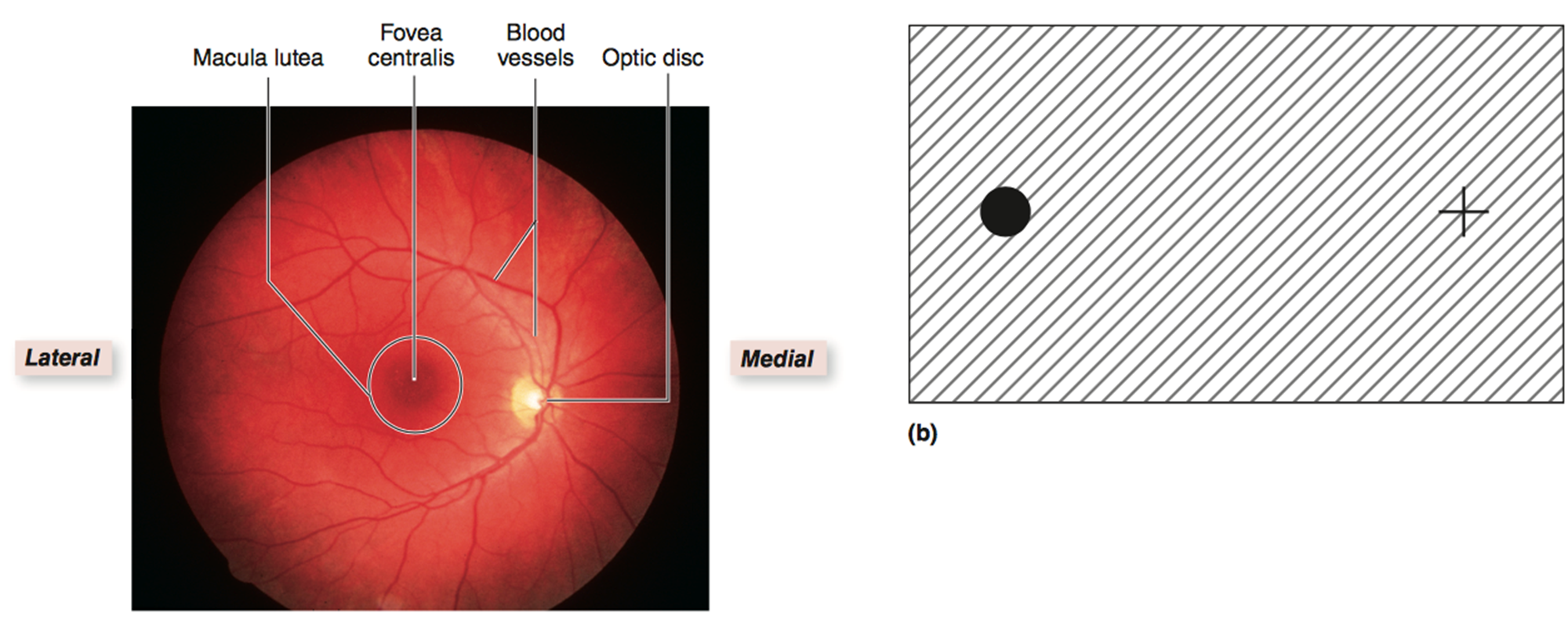
What is the fovea centralis?
Pit in the macula lutea with highest cone density → Sharpest vision
Light focus area for vision
What does the iris regulate?
The amount of light that enters the eye
List the five layers of the cornea (superficial → deep)
Corneal epithelium – non‑keratinized stratified squamous
Bowman membrane
thick (corneal) stroma – collagen + proteoglycans
Descemet membrane
Corneal endothelium – simple squamous
Which layer of the cornea maintains corneal transparency?
Stroma (regular collagen attachment)
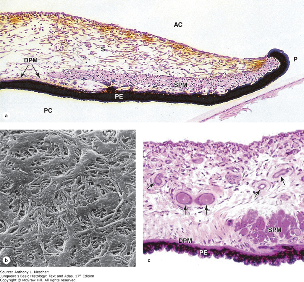
What is this?
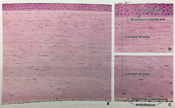
Five layers of the cornea
What is this?
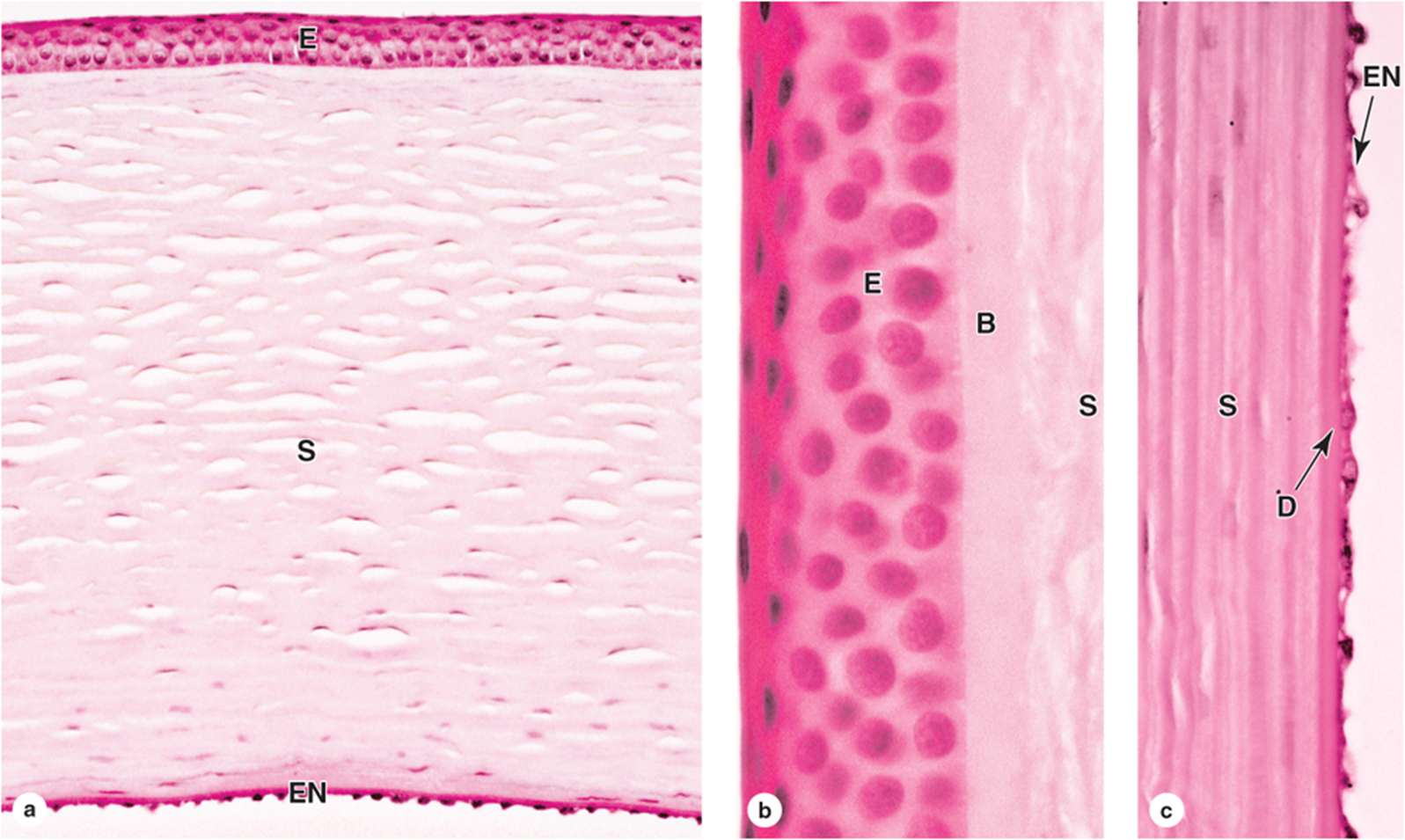
Five layers of the cornea
What is this?
Layers of the vascular tunic
What histologic features identify conjunctiva?
Stratified columnar epithelium
Goblet cells
Highly vascular
What is the iris?
Colored portion of the eye
Center of iris → Pupil
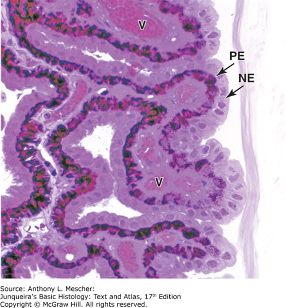
What is this?
Iris
PE - Posterior pigment epithelium
DPM - Dilator pupillae muscle
SPM - Sphincter pupillae muscle
What are the principal components of the lens?
Lens capsule
Lens epithelium — Simple cuboidal epithelium
Lens fibers
What is the ora serrata?
The junction between the retina and the ciliary body
What are the two photoreceptors?
Rods → Contrast dark and light tones
Cones → Color (Red, Green, Blue)
Where is the macula lutea located?
Lateral to the optic disc
What separates the two cavities of the eye?
The lens
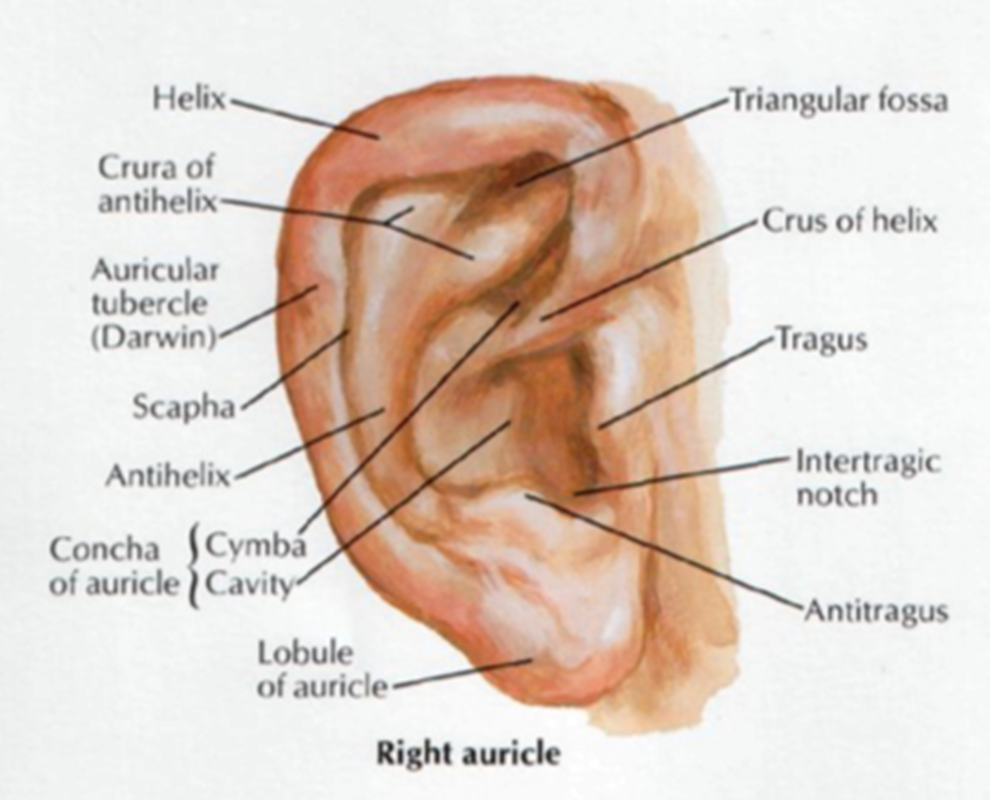
What structures make up the external ear?
Auricle
External acoustic meatus
Tympanic membrane
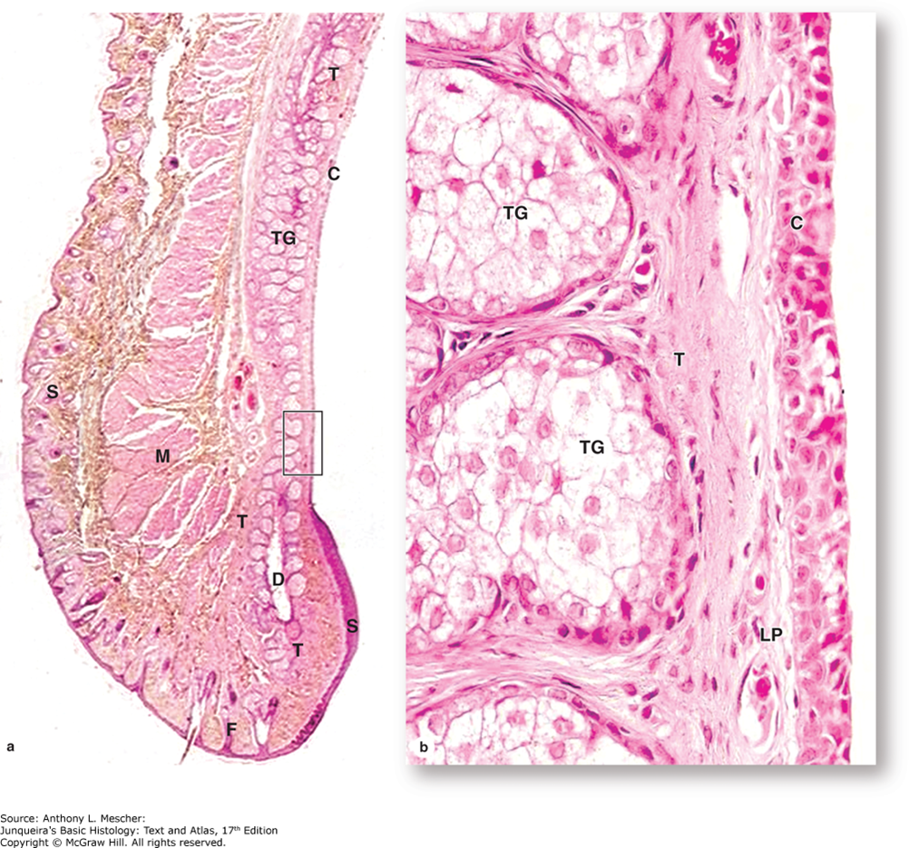
What do ceruminous glands produce?
Cerumen (ear wax)
What are the boundaries of the middle ear?
Lateral: tympanic membrane
Medial: oval & round windows
Inferior tube leads to the nasopharynx via the eustachian tube
What epithelium lines the middle ear?
Non‑keratinized stratified squamous epithelium
What are the auditory ossicles?
Malleus
Incus
Stapes
What is the function of the ossicles?
Amplify sounds waves to the oval window
What muscles protect the ear from loud sounds?
Stapedius
Tensor tympani
What is the bony labryinth?
Cavities in temporal bone housing membranous labyrinth
What are the two major functional divisions of the inner ear?
Vestibular complex – balance
Cochlea – hearing
What do semicircular canals detect?
Positional changes of the head (X, Y, Z planes)
What nerve carries vestibular and auditory signals in the inner ear?
CN VIII (Vestibulocochlear nerve)
What can be found within the middle ear?
Tympanic cavity
Auditory ossicles
How much larger is the tympanic membrane compared to the oval window?
20x larger