forensic practical
1/109
There's no tags or description
Looks like no tags are added yet.
Name | Mastery | Learn | Test | Matching | Spaced | Call with Kai |
|---|
No analytics yet
Send a link to your students to track their progress
110 Terms
A body is discovered in a locked room at 10:00PM. The environmental temperaturehasbeenasteady25°C. Upon examination, the forensic team finds that rigor mortis is present in the jaw, neck, and upper limbs, but the lower limbs are still flaccid. During transport, the body is roughly handled , and the stiffness in the arms is lost
1-According to the "Rule of 12," how long does it take rigor mortis to fully develop
2-Based on the distribution described (jaw, neck, and arms rigid, while legs are flaccid), twhat is the estimated post mortem time
3-If the stiffness in the arms was lost due to physical disruption during transit, is it going to redevelop ?
Answer: It takes approximately 12 hours to complete all over the body (from head to feet).
Answer: Rigor mortis follows a proximal-distal distribution. Since it has reached the upper limbs but not yet the lower limbs, the estimated PMI is between 2 and 12 hours, likely around 8–12 hours.
Answer: If rigor is broken by mechanical force while it is still developing, it does not redevelop and may be misleading in estimating the PMI. This is known as "broken rigor mortis."
Case
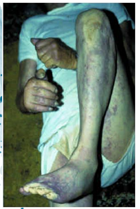
A body is found in an open field. The forensic examiner observes greenish discoloration of the whole skin, blisters (bullae), and bloating of the features.
Questions
What is the first external sign of putrefaction, and when does it appear after death in winter?
If these advanced signs (whole-body greening and bloating) are present, estimate the PMI for both a summer and winter context.
Answers
Answer: The first sign is greenish discoloration in the right iliac fossa, appearing about 48 hours after death in winter.
Answer: In winter, these signs indicate a PMI of approximately one week. In summer, the process is accelerated and takes about half that time, suggesting a PMI of 3-4 days.
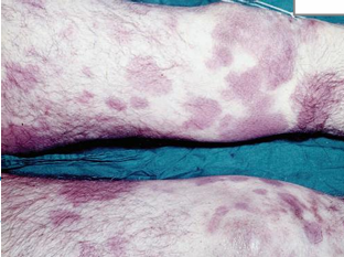
Case
During examination of a dead body, multiple-coloured patches are observed on the skin, as shown in the image.
Questions
What is the name of this postmortem change?
Describe the initial stage of this postmortem change.
What is the main differential diagnosis (D.D.) for this condition?
Answers
Answer: Postmortem lividity / Hypostasis
Answer: The initial stage of postmortem lividity is characterized by a reddish-purple patchy discoloration.
Answer: D.D. Bruises
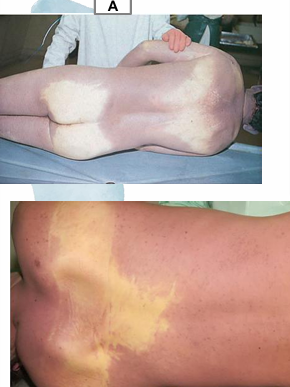
Case
Questions
What postmortem change is described by the confluent reddish-purple discoloration on the back?
What causes the contact flattening (contact pallor) observed over the scapula and buttocks?
Answers
Answer: Postmortem lividity / Hypostasis.
Answer: Contact flattening (contact pallor) is caused by pressure from the surface the body is resting on, which compresses the local capillaries and prevents blood from pooling in those specific areas (such as the scapula and buttocks).
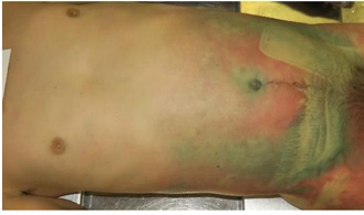
Questions
What postmortem change is characterized by a greenish discoloration of the skin on the anterior abdominal wall?
What is the typical timeframe for this specific postmortem change to appear?
Answers
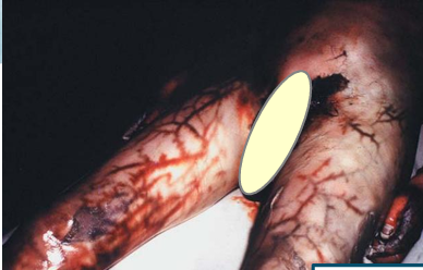
Answer: Putrefaction.
Answer: Time: 24–48 hours.
Questions
What postmortem change is characterized by the description provided?
What is the cause of the change in the skin?
What is the typical timeframe for this change to appear?
Answers
Answer: Putrefaction / Marbling of the skin.
Answer: Cause: Superficial vessels distended with putrefactive gases.
Answer: Time: 24–48 hours.
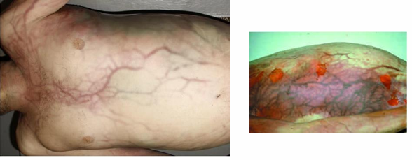
Questions
What postmortem change is characterized by a distinctive, web-like pattern on the skin?
What is the underlying cause of the change of the skin during putrefaction?
Answers
Answer: Putrefaction / Marbling of the skin.
Answer: Cause: Superficial vessels distended with putrefactive gases (bacterial breakdown of hemoglobin reacts with hydrogen sulfide to form sulfhemoglobin, staining the vessel walls).

Questions
What postmortem change is described by these advanced signs?
What are the specific features observed in this stage?
Answers
Answer: Advanced Putrefaction.
Answer: Describe: skin discoloration, marbling, and skin slippage.
Questions
What postmortem change is characterized by these features?
What are the specific features observed during this stage?
What is the typical timeframe for these changes to occur?
Answers
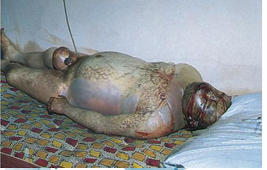
Answer: Advanced Putrefaction.
Answer: Describe: skin discoloration, marbling, and gaseous distension of the face, abdomen, and scrotum.
Answer: Time: After about a week.
Questions
What postmortem change is characterized by these features?
What is the typical timeframe for this change to appear?
What is the main differential diagnosis (D.D.) for this condition?
Answers
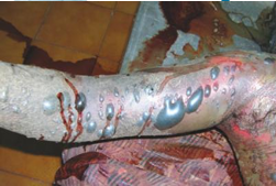
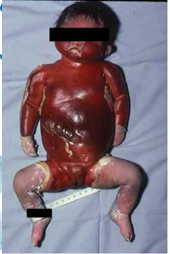
Answer: Postmortem blisters on the right arm / Putrefaction.
Answer: Time: After about a week.
Answer: D.D. Burn Bullae
Questions
What specific putrefactive changes are described by these features?
What is the typical timeframe for these changes to occur?
Answers
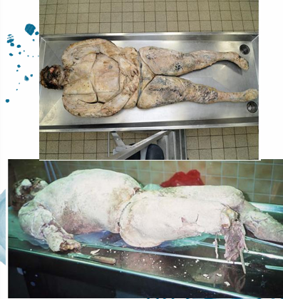
Answer: Putrefactive changes: Degloving and Destocking
Answer: Time: After about 2 weeks
Questions
What postmortem change is characterized by these skin features?
What are the specific physical and color changes observed in the skin during this process?
Answers
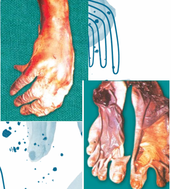
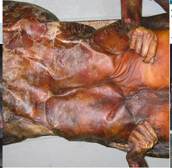
Answer: Mummification.
Answer: Describe: The skin is dry, hard, and leatherlike. Its color changes to blackish-brown.
Questions
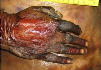
What postmortem change is shown affecting the hand?
What are the specific physical and color changes observed in the skin during this process?
Answers
Answer: Mummification of the hand.
Answer: Describe: The skin is dry, hard, and leatherlike. Its color changes to blackish-brown.S
Questions
What postmortem change is characterized by these features?
What are the specific physical changes observed in the fat tissue during this process?
Answers
Answer: Saponification / Adipocere.
Answer: Describe: Subcutaneous and intra-abdominal fat is replaced by greyish-white, soap-like waxy substance.
S
Questions
What postmortem change is characterized by these features?
What is the medicolegal (ML) importance of this condition?
Answers
Answer: Maceration.
Answer: ML. importance: Its presence is indicative of death in the uterus.
Questions
What postmortem change is characterized by this sudden onset?
What is the typical time of occurrence for this change?
Answers
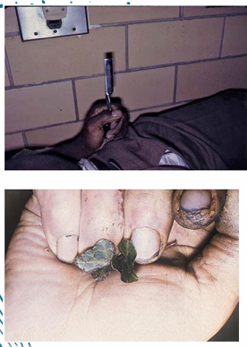
Answer: Cadaveric Spasm
Answer: Time of occurrence: Immediately at the moment of death
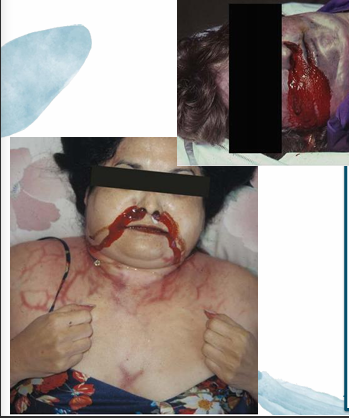
Questions
What postmortem change is characterized by these features?
How is the fluid from the mouth and nostrils described during this process?
What is the differential diagnosis (D.D.) for this fluid?
Answers
Answer: Putrefaction with postmortem purge and marbling of the skin
Answer: Describe: blood-stained, thick, coarse, offensive frothy fluid from the mouth and nostrils during putrefaction.
Answer: D.D: Antemortem bleeding or drowning froth.
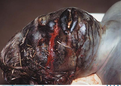
Questions
What postmortem change is characterized by these features?
What are the specific features observed during this process?
Answers
Answer: Decomposition with postmortem purge
Answer: Describe: Bloating (swelling) of the face and blood-tinged fluid from the nostrils and mouth (postmortem purge) during decomposition.
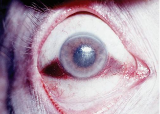
Questions
What postmortem change is characterized by the description provided?
What is the cause of this specific ocular postmortem change?
Answers
Answer: Tache noire.
Answer: Cause: Postmortem drying of sclera of the eyes.
Questions
What postmortem change is characterized by these specific time intervals?
What is the onset time for this change?
What is the estimated time passed since death when this change is fully established?
Answers
Answer: Fixed Rigor mortis
Answer: Its onset: 2 hours after death
Answer: Time passed since death: 12 hours
Questions
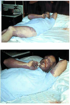
What is the position of the body at the time of death based on the findings?
What evidence supports the conclusion that the body was moved after livor mortis had fixed?
Answers
Answer: The lividity pattern is consistent with the man being on his face in the bed. The fluid in the nose matches up with the stained area in the bed.
Answer: The pattern of the bedding on the leg suggests the decedent was lying on his face, and he had been moved after the livor mortis had fixed.
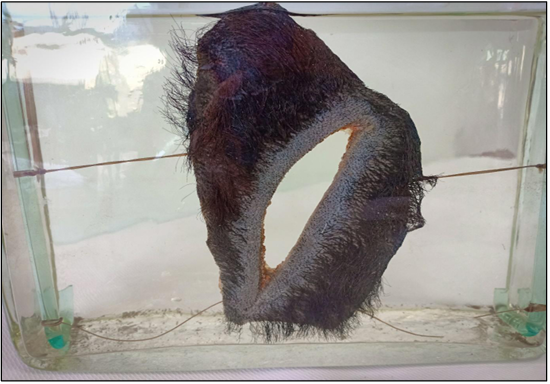
Questions
What is the specimen being examined?
What are the descriptive features of the injury on the scalp?
What is the likely causative instrument for this type of injury?
How is the time of death estimated in relation to the injury?
What are the possible causes of death?
What is the final diagnosis for this specimen?
Answers
Answer: The specimen is a piece of the scalp.
Answer: Description of Injury: The injury is a cut wound characterized by regular edges, acute angles, and is about 8cm in length.
Answer: Causative Instrument: It is caused by a sharp-edged instrument like a knife.
Answer: Time of Death: The victim died shortly after the injury due to the absence of signs of surgical interference, healing, or sepsis.
Answer: Cause of Death: The possible causes of death are hemorrhage, shock, or brain injuries.
Answer: Diagnosis: Antemortem Cut Wound in The Scalp.
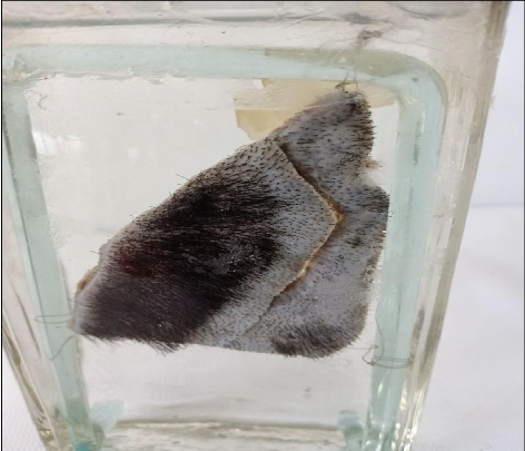
Questions
What is the specimen being examined?
What are the descriptive features of the injury on the scalp?
What is the likely causative instrument for this type of injury?
How is the time of death estimated in relation to the injury?
What are the possible causes of death?
What is the final diagnosis for this specimen?
Answers
Answer: The specimen is a piece of the scalp.
Answer: Description of Injury: The injury is a cut wound formed of parts characterized by regular edges and acute angles in each part, with a curved overall shape, and is about 6 cm in length.
Answer: Causative Instrument: It is caused by the edges of broken glass.
Answer: Time of Death: The victim died shortly after the injury due to the absence of signs of surgical interference, healing, or sepsis.
Answer: Cause of Death: The possible causes of death are hemorrhage, shock, or brain injuries.
Answer: Diagnosis: Antemortem Cut Wound in The Scalp.
Questions
What is the specimen being examined?
What are the descriptive features of the injury on the scalp?
What is the likely causative instrument for this type of injury?
How is the time of death estimated in relation to the injury?
What are the possible causes of death?
What is the final diagnosis for this specimen?
Answers
Answer: The specimen is a piece of the scalp.
Answer: Description of Injury: The injury is a contused wound characterized by irregular edges, multiple angles, a linear shape, and is about 5 cm in length.
Answer: Causative Instrument: It is caused by a heavy blunt instrument like a stick.
Answer: Time of Death: The victim died shortly after the injury due to the absence of signs of surgical interference, healing, or sepsis.
Answer: Cause of Death: The possible causes of death are hemorrhage, shock, or brain injuries.
Answer: Diagnosis: Antemortem Contused Wound in The Scalp.
Questions
What is the specimen being examined?
What are the descriptive features of the injury on the scalp?
What is the likely causative instrument for this type of injury?
How is the time of death estimated in relation to the injury?
What are the possible causes of death?
What is the final diagnosis for this specimen?
Answers
Answer: The specimen is a piece of the scalp.
Answer: Description of Injury: The injury is a contused wound characterized by irregular edges, multiple angles, an irregular shape, and is about 5 cm in length.
Answer: Causative Instrument: It is caused by a heavy blunt instrument like a big stone.
Answer: Time of Death: The victim died shortly after the injury due to the absence of signs of surgical interference, healing, or sepsis.
Answer: Cause of Death: The possible causes of death are hemorrhage, shock, or brain injuries.
Answer: Diagnosis: Antemortem Contused Wound in The Scalp.
Questions
What is the specimen being examined?
What are the descriptive features of the injury on the skin?
What is the likely causative instrument for this type of injury?
How is the time of death estimated in relation to the injury and medical intervention?
What are the possible causes of death?
What is the final diagnosis for this specimen?
Answers
Answer: The specimen is a piece of the skin.
Answer: Description of Injury: The injury is a contused wound characterized by irregular edges, multiple angles, a linear shape, and is about 10 cm in length.
Answer: Causative Instrument: It is caused by a heavy blunt instrument like a stick.
Answer: Time of Death: The victim died shortly after surgical interference due to the absence of signs of healing or sepsis.
Answer: Cause of Death: The possible causes of death are hemorrhage, shock, or other injuries.
Answer: Diagnosis: Antemortem Contused Wound in The Skin.
Questions
What is the specimen being examined?
What are the descriptive features of the injury on the skin?
What is the likely causative instrument for this type of injury?
How is the time of death estimated in relation to the injury?
What are the possible causes of death?
What is the final diagnosis for this specimen?
.
Answers
Answer: The specimen is a piece of the skin.
Answer: Description of Injury: The injury is a stab wound characterized by regular edges, one acute angle, one rounded angle, and is about 2 cm in length.
Answer: Causative Instrument: It is caused by a sharp-pointed, single-bladed instrument like a knife.
Answer: Time of Death: The victim died shortly after the injury due to the absence of signs of surgical interference, healing, or sepsis.
Answer: Cause of Death: The possible causes of death are hemorrhage, shock, or injury to a vital organ.
Answer: Diagnosis: Antemortem Stab Wound in The Skin
Questions
What is the specimen being examined?
What are the descriptive features of the injury on the skin?
What is the likely causative instrument for this type of injury?
How is the time of death estimated in relation to the injury?
What are the possible causes of death?
What is the final diagnosis for this specimen?
Answers
Answer: The specimen is a piece of the skin.
Answer: Description of Injury: The injury is a stab wound characterized by regular edges, two acute angles, and is about 2 cm in length.
Answer: Causative Instrument: It is caused by a sharp-pointed, double-bladed instrument like a dagger.
Answer: Time of Death: The victim died shortly after the injury due to the absence of signs of surgical interference, healing, or sepsis.
Answer: Cause of Death: The possible causes of death are hemorrhage, shock, or injury to a vital organ.
Answer: Diagnosis: Antemortem Stab Wound in The Skin.
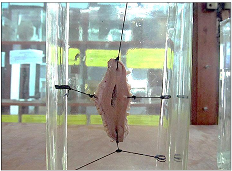
Questions
What is the specimen being examined?
What are the descriptive features of the injury on the heart?
What is the likely causative instrument for this type of injury?
How is the time of death estimated in relation to the injury?
What are the possible causes of death?
What is the final diagnosis for this specimen?
Answers
Answer: The specimen is a heart of an adult human being.
Answer: Description of Injury: The injury is a longitudinal penetrating stab wound and is about 2 cm in length in the left ventricle.
Answer: Causative Instrument: It is caused by a sharp-pointed / sharp-edged instrument like a dagger or knife.
Answer: Time of Death: The victim died shortly after the injury due to the absence of signs of surgical interference, healing, or sepsis.
Answer: Cause of Death: The possible causes of death are hemorrhage, shock, or cardiac tamponade (collection of the blood in the pericardial sac).
Answer: Diagnosis: Antemortem Stab Wound in The Heart.
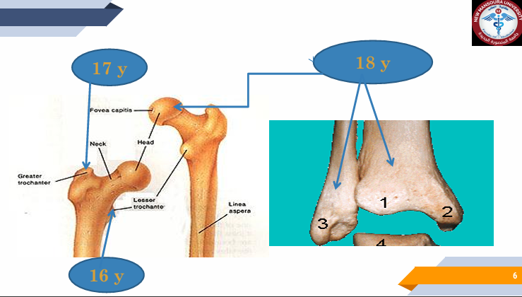
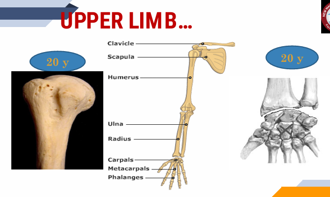
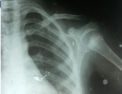
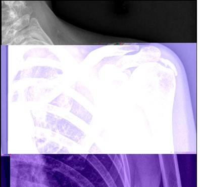
X-ray on the shoulder joint : Age less than 20 years old in male
ps. انا معنديش فكره هي كدا طبيعيه ولا ال لاب اتهبل
X-ray on the shoulder joint : Age more than 20 years old in male
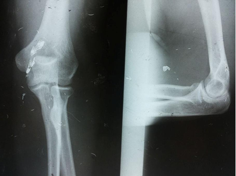
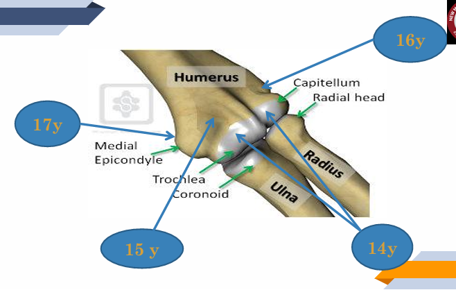
X-ray on the elbow joint : Age more than 17 years old in male
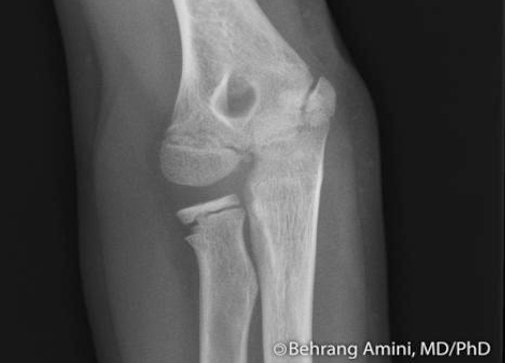
X-ray on the Elbow joint : Age less than 14 years old maleٍ

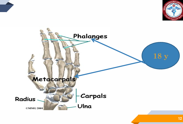
X-ray on the Wrist joint : Age less than 18 years old in male
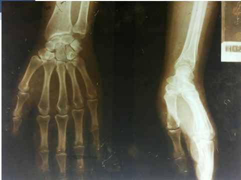
Wrist joint : Age more than 18 years old in male
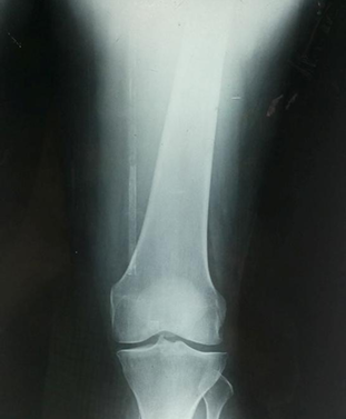
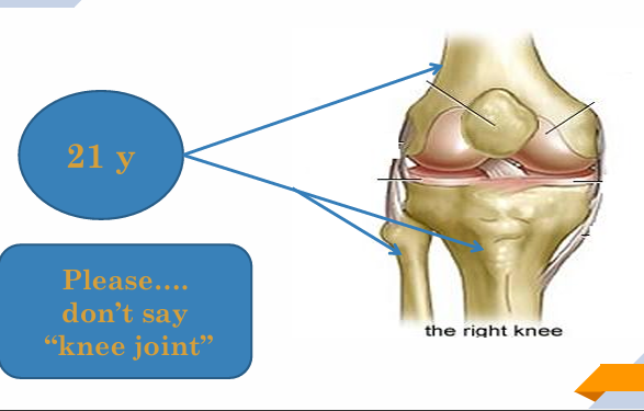
X-ray on the Knee joint : Age more than 21 years old in maleٍ
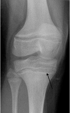
X-ray on the Knee joint : Age less than 21 years old in male
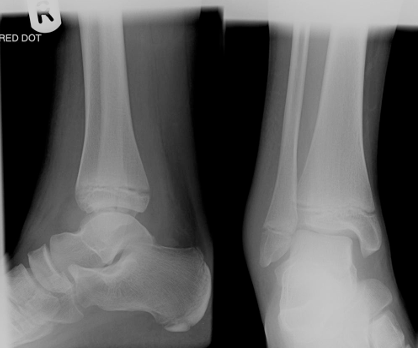
Ankle joint : Age less than 18 years old in male
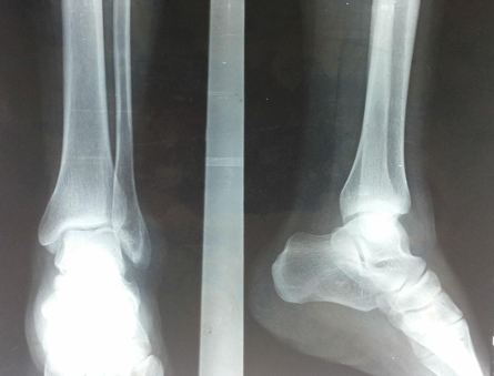
X-ray on ankle joint : Age more than 18 years old in male
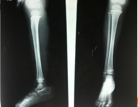
X-ray on the Knee and ankle joints : Age less than 18 years old in male
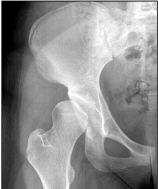
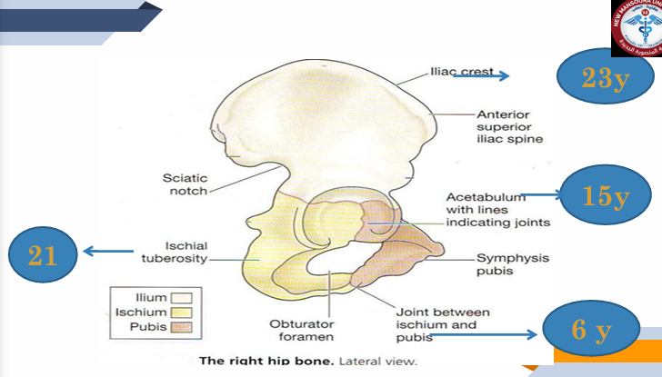
X-ray on the Hip joint : Age less than 23 years old in maleٍ
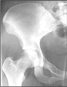
X-ray on the Hip joint : Age more than 23 years old in male
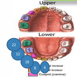
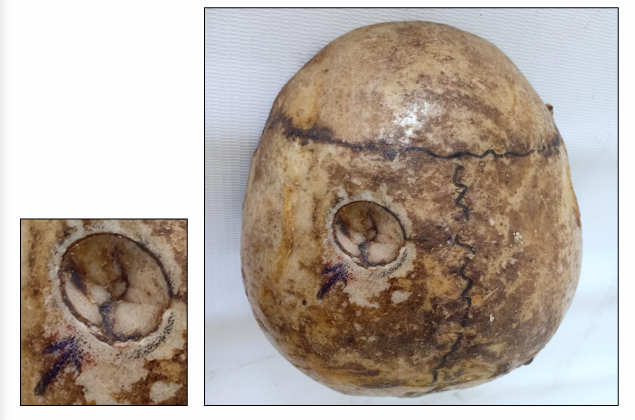
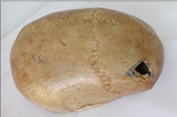
child skull 6-7 yo
Skull aged more than 12 and less than 18-25 years old
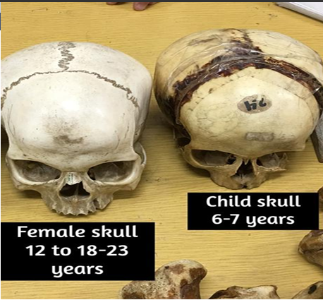
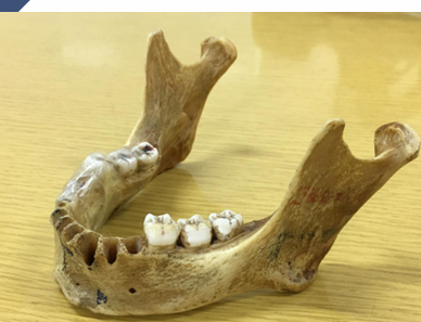
adult mandible more than 18 years (in the middle age)
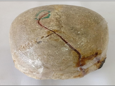
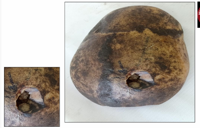
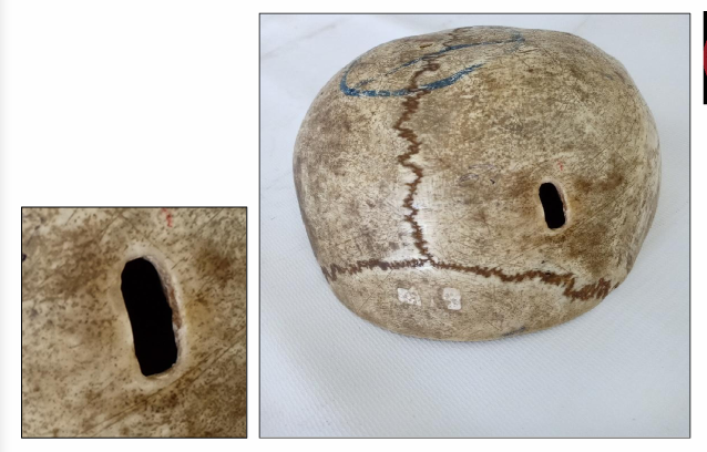
Questions
What is the specimen being examined?
What are the types and descriptive features of the fracture shown on the skull?
What type of causative instrument and force mechanics produce this injury?
How is the time of death estimated?
What are the possible causes of death?
What is the final diagnosis for this specimen?
Answers
Answer: The specimen is vault of skull.
Answer: Type & Description of Injury: Fissure fracture starting from the middle of the lower part of the occipital bone and running into the sagittal suture, leading to its separation (Diastatic Fracture). It is linear, about 10 cm in length, and runs in an irregular course.
Answer: Causative Instrument: Heavy blunt instrument with a wide striking surface and low momentum of force (e.g., Iron Bar or Heavy Stick of Wood).
Answer: Time of Death: Shortly after injury due to the absence of signs of surgical interference, healing, or sepsis, and the sharp edges of the fracture.
Answer: Cause of Death: Severe concussion or compression.
Answer: Diagnosis: Diastatic Fissured Fracture in The Occipital Bone of The Vault of The Skull.
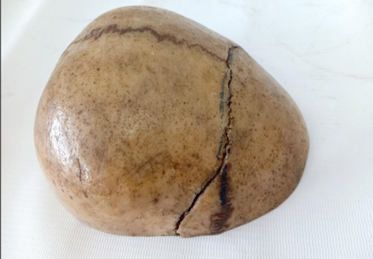
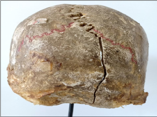
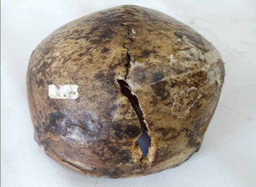
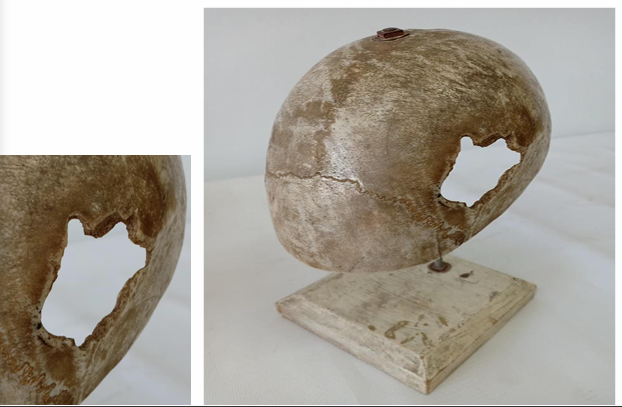
Questions
What is the specimen being examined?
What are the types and descriptive features of the fracture?
What type of causative instrument and force mechanics produce this injury?
How is the time of death estimated?
What are the possible causes of death?
What is the final diagnosis for this specimen?
Answers
Answer: The specimen is vault of skull.
Answer: Type & Description of Injury: Fissure fracture extending from the lower part of the parietal bone passing upwards and to the left into the coronal suture, leading to its separation (Diastatic Fracture). It is linear, about 5 cm in length, and runs in an oblique course.
Answer: Causative Instrument: Heavy blunt instrument with a wide striking surface and low momentum of force (e.g., Iron Bar or Heavy Stick of Wood).
Answer: Time of Death: Shortly after injury due to the absence of signs of surgical interference, healing, or sepsis, and the sharp edges of the fracture.
Answer: Cause of Death: Severe concussion or compression.
Answer: Diagnosis: Diastatic Fissured Fracture in The Parietal Bone of The Vault of The Skull.
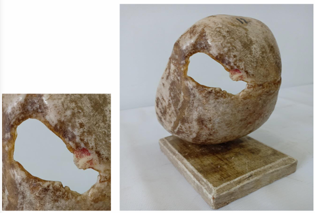
Questions
What is the specimen being examined?
What are the type and descriptive features of the fracture?
What type of causative instrument and force mechanics produce this injury?
How is the time of death estimated?
What are the possible causes of death?
What is the final diagnosis for this specimen?
Answers
Answer: The specimen is vault of skull.
Answer: Type & Description of Injury: Fissure fracture running an oblique course, extending upwards from the lower part of the occipital bone on the right side towards the left part of the lambdoid suture. It is linear, about 15 cm in length, and runs in an oblique course.
Answer: Causative Instrument: Heavy blunt instrument with a wide striking surface and low momentum of force (e.g., Iron Bar or Heavy Stick of Wood).
Answer: Time of Death: Shortly after injury due to the absence of signs of surgical interference, healing, or sepsis, and the sharp edges of the fracture.
Answer: Cause of Death: Severe concussion or compression.
Answer: Diagnosis: Fissured Fracture in The Vault of The Skull.
Questions
What is the specimen being examined?
What are the type and descriptive features of the fracture?
What type of causative instrument produces this injury?
How is the time of death estimated?
What are the possible causes of death?
What is the final diagnosis for this specimen?
Answers
Answer: The specimen is vault of skull.
Answer: Type & Description of Injury: Cut fracture extending from the posterior part of the right parietal bone to the squamous part of the occipital bone, crossing the right part of the lambdoid suture. It is linear, about 7 cm in length, with sharp regular edges.
Answer: Causative Instrument: Heavy sharp-edged instrument (e.g., Butcher’s Knife).
Answer: Time of Death: Shortly after injury due to the absence of signs of surgical interference, healing, or sepsis, and the sharp edges of the fracture.
Answer: Cause of Death: Brain laceration or compression.
Answer: Diagnosis: Cut Fracture in The Vault of The Skull.
Questions
What is the specimen being examined?
What are the types and descriptive features of the injuries and surgical interventions on the skull?
What type of causative instrument produces the primary injury?
How is the time of death estimated?
What are the possible causes of death?
What is the final diagnosis for this specimen?
Answers
Answer: The specimen is vault of skull.
Answer: Type & Description of Injury:
Cut Fracture: Located in the right parietal bone lying transversely, cutting the outer table only. It is 10 cm posterior to the coronal suture, close to the sagittal suture at its medial end, linear in shape, with a sharp edge, and 4 cm in length.
Trephine decompression operation gap: Located behind the cut fracture, rounded in shape, and 1 cm in diameter.
Answer: Causative Instrument: Heavy sharp-edged instrument (e.g., Butcher’s Knife).
Answer: Time of Death: Shortly after injury due to the absence of signs of healing or sepsis, and the sharp edges of the gap.
Answer: Cause of Death: Brain lacerations, compression, or surgical interference.
Answer: Diagnosis: Cut Fracture of The Vault of The Skull with Trephine Operation.
Questions
What is the specimen being examined?
What are the types and specific descriptive features of the two injuries on the skull?
What type of causative instrument and angle of impact produce these injuries?
How is the time of death estimated?
What are the possible causes of death?
What is the final diagnosis for this specimen?
Answers
Answer: The specimen is vault of skull.
Answer: Type & Description of Injury:
1st Cut Fracture: Lies transversely in the left parietal bone, 3 cm lateral to the sagittal suture and 4 cm posterior to the coronal suture, with the outer table of the bone being separated.
2nd Cut Fracture: Lies obliquely about 1 cm behind the first fracture, 4 cm lateral to the sagittal suture and 5 cm posterior to the coronal suture. Its lower edge is straight, regular, and slanting, while its upper edge is shelved up.
Answer: Causative Instrument: Heavy sharp-edged instrument with high momentum of force (e.g., Grubber or Fas) hitting the bone in an oblique direction (causing shelving of the bone and slanting of the edges).
Answer: Time of Death: Shortly after injury due to the absence of signs of surgical interference, healing, or sepsis, and the sharp edges of the fracture.
Answer: Cause of Death: Severe concussion or compression.
Answer: Diagnosis: Cut Fractures in The Left Parietal Bone of The Vault of The Skull.
Questions
What is the specimen being examined?
What are the type and specific descriptive features of the fracture, including its directional depth indicators?
What type of causative instrument and force mechanics produce this injury?
How is the time of death estimated?
What are the possible causes of death?
What is the final diagnosis for this specimen?
Answers
Answer: The specimen is vault of skull.
Answer: Type & Description of Injury: Rounded localized depressed fracture in the right parietal bone (2 cm lateral to sagittal suture & 7 cm posterior to coronal suture). It is 2 cm in diameter, shows a depressed external table with a fractured inner table, and is deeper in its left anterior part (indicating the assailant hit from the front using their right hand).
Answer: Causative Instrument: Heavy blunt instrument with a small rounded striking surface and high momentum of force (e.g., head of a Hammer).
Answer: Time of Death: Shortly after injury due to the absence of signs of surgical interference, healing, or sepsis, and the sharp edges of the fracture.
Answer: Cause of Death: Severe concussion, brain lacerations, or compression.
Answer: Diagnosis: Localized Depressed Fracture in The Right Parietal Bone of The Vault of The Skull.
Questions
What is the specimen being examined?
What are the type and specific descriptive features of the fracture, including its directional depth indicators?
What type of causative instrument and force mechanics produce this injury?
How is the time of death estimated?
What are the possible causes of death?
What is the final diagnosis for this specimen?
Answers
Answer: The specimen is vault of skull.
Answer: Type & Description of Injury: Rounded localized depressed fracture in the left parietal bone (2 cm lateral to sagittal suture & 2 cm posterior to coronal suture). It is 2 cm in diameter, shows a depressed external table with a fractured inner table, and is deeper in its left posterior part (indicating the assailant hit the victim from the left side and slightly from backward using his right hand).
Answer: Causative Instrument: Heavy blunt instrument with a small rounded striking surface and high momentum of force (e.g., head of a Hammer).
Answer: Time of Death: Shortly after injury due to the absence of signs of surgical interference, healing, or sepsis, and the sharp edges of the fracture.
Answer: Cause of Death: Severe concussion, brain lacerations, or compression.
Answer: Diagnosis: Localized Depressed Fracture in The Left Parietal Bone of The Vault of The Skull.
Questions
What is the specimen being examined?
What are the type and specific descriptive features of the primary fracture and its radiating components?
What type of causative instrument and force mechanics produce this injury?
How is the time of death estimated?
What are the possible causes of death?
What is the final diagnosis for this specimen?
Answers
Answer: The specimen is vault of skull.
Answer: Type & Description of Injury: Rounded depressed comminuted fracture in the left parietal bone (7 cm lateral to sagittal suture & 6 cm posterior to coronal suture). It shows fragmentation of the bone ("spiderweb fracture"), depression into the cranial cavity, and two radiating fissure fractures:
1st radiating fracture: 5 cm in length, extending from the fracture site to the left coronal suture.
2nd radiating fracture: 2 cm in length, extending downward.
Answer: Causative Instrument: Heavy blunt instrument with a wide striking surface and very high momentum of force (e.g., Large piece of wood or Iron bar).
Answer: Time of Death: Shortly after injury due to the absence of signs of surgical interference, healing, or sepsis, and the sharp edges of the fracture.
Answer: Cause of Death: Severe concussion, brain lacerations, or compression.
Answer: Diagnosis: Depressed Comminuted Fractures of The Vault of The Skull.
Questions
What is the specimen being examined?
What are the type, specific descriptive features, and dimensions of the fracture?
What type of causative instrument and cross-sectional shape produce this injury?
How is the time of death estimated?
What are the possible causes of death?
What is the final diagnosis for this specimen?
Answers
Answer: The specimen is vault of skull.
Answer: Type & Description of Injury: Penetrating fracture in the right frontal bone (5 cm right to the frontal suture & 3 cm in front of the coronal suture). It is triangular in shape, with dimensions of 2 X 2 X 1.5 cm.
Answer: Causative Instrument: Heavy sharp-ended instrument with a triangular surface in cut section and a high momentum of force (e.g., a horn of an Ox).
Answer: Time of Death: Shortly after injury due to the absence of signs of surgical interference, healing, or sepsis, and the sharp edges of the fracture.
Answer: Cause of Death: Brain lacerations or compression.
Answer: Diagnosis: Penetrating Fracture in The Right Frontal Bone of The Vault of The Skull.
Questions
What is the specimen being examined?
What are the type, specific descriptive features, and dimensions of the fracture?
What type of causative instrument and cross-sectional shape produce this injury?
How is the time of death estimated?
What are the possible causes of death?
What is the final diagnosis for this specimen?
Answers
Answer: The specimen is vault of skull.
Answer: Type & Description of Injury: Penetrating fracture in the left parietal bone (4 cm lateral to sagittal suture, 1.5 cm in front of coronal suture). It is rectangular in shape with sharp edges, sizes 1 x 0.3 cm, and shows inward depression of both the outer and inner tables of the skull.
Answer: Causative Instrument: Heavy small sharp-ended instrument with a rectangular surface in cut section and a high momentum of force (e.g., a chisel).
Answer: Time of Death: Shortly after injury due to the absence of signs of surgical interference, healing, or sepsis, and the sharp edges of the fracture.
Answer: Cause of Death: Brain lacerations or compression.
Answer: Diagnosis: Penetrating fracture in the left parietal bone of the vault of the skull.
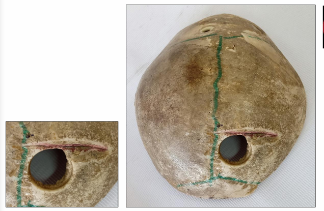
Questions
What is the specimen being examined?
What are the type, specific descriptive features, and dimensions of the surgical intervention on the skull?
What specific tools and techniques are evidenced by the features of the gap?
How is the time of death estimated?
What are the possible causes of death?
What is the final diagnosis for this specimen?
Answers
Answer: The specimen is vault of skull.
Answer: Type & Description of Injury: Decompression operation in the left parietal bone, presenting as a transverse oblong gap. It is located 5 cm lateral to the sagittal suture and 1 cm posterior to the coronal suture, measuring about 4 x 3 cm in size.
Answer: Evidence of Tools/Techniques:
Trephine operation: Evidenced by a smooth contour in the antero-medial part of the gap.
Bone nibbling forceps: Evidenced by marks of bone fragmentation at the edges of the gap, indicating widening of the surgical defect.
Answer: Time of Death: Shortly after injury due to the absence of signs of healing or sepsis, and the sharp edges of the gap.
Answer: Cause of Death: Brain lacerations, compression, or surgical interference.
Answer: Diagnosis: Decompression Operation of The Vault of The Skull.
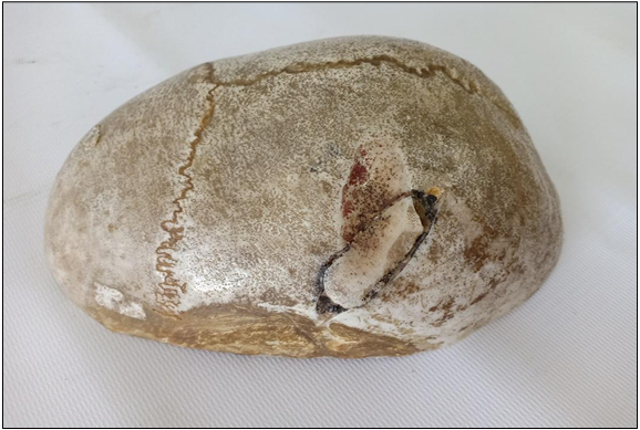
Questions
What is the specimen being examined?
What are the type, specific descriptive features, and dimensions of the gap on the skull?
How is the time of death estimated based on the healing signs of the bone and tissue?
What are the possible causes of death?
What is the final diagnosis for this specimen?
Answers
Answer: The specimen is vault of skull.
Answer: Type & Description of Injury: Decompression operation gap in the left side of the frontoparietal bone, located about 2 cm from the midline. It is transverse oblong in shape, measures 6 x 3 cm in size, and shows a smooth rounded edge with the beginning formation of a fibrous membrane.
Answer: Time of Death: The victim died 3 months after the operation, indicated by the smooth rounded edges of the bone gap and the beginning of fibrous membrane formation.
Answer: Cause of Death: The possible causes of death are minor trauma, sepsis, or other causes.
Answer: Diagnosis: Decompression Operation of The Vault of The Skull.
Questions
What is the specimen being examined?
What are the type, specific descriptive features, and locations of the findings on the skull?
What is the vascular origin of the primary pathology?
How is the time of death estimated in relation to the surgical procedure?
What is the possible cause of death?
What is the final diagnosis for this specimen?
Answers
Answer: The specimen is vault of skull.
Answer: Type & Description of Injury:
Surgical finding: 1 trephine operation in the vault.
Pathological finding: 2 big, oval-shaped hematomas measuring about 3x7 cm in size, located in the left parieto-temporal region between the inner table of the bone and the dura mater.
Answer: Vascular Origin: Rupture of the middle meningeal artery or one of its branches.
Answer: Time of Death: The victim died during the operation (before decompression) because the hematoma is still present.
Answer: Cause of Death: Brain compression.
Answer: Diagnosis: Extradural Haematoma & Trephine Operation.
Questions
What is the specimen being examined?
What are the four specific pathological and traumatic findings shown on the skull?
How is the antemortem nature of the injury determined?
What type of causative instrument is indicated by these collective findings?
How is the time of death estimated?
What are the possible causes of death?
What is the final diagnosis for this specimen?
Answers
Answer: The specimen is vault of skull.
Answer: Type & Description of Injury:
1. Small Rounded Penetrating Fracture: Located in the left parietal bone (2 cm lateral to sagittal suture & 4 cm posterior to coronal suture), rounded in shape, and 3 mm in diameter.
2. Sub-periosteal hemorrhage: Located on the posterior part of the left parietal bone and on the occipital bone.
3. Extradural hemorrhage: Located in the middle of the anterior part of the vault of the skull and in the right parietal bone, between the dura and the skull bone.
4. Subdural hemorrhage: Located on both sides of the falx cerebri.
Answer: Vital Reaction: It is an antemortem injury due to the presence of hemorrhages.
Answer: Causative Instrument: A blunt instrument with a sharp pointed object fixed to it (e.g., a piece of wood with a nail fixed to it).
Answer: Time of Death: Shortly after injury due to the absence of signs of surgical interference, healing, or sepsis.
Answer: Cause of Death: Brain laceration and brain compression.
Answer: Diagnosis: Penetrating Fracture In The Vault Of The Skull With Extradural, Subdural, Subperiosteal Hemorrhages.
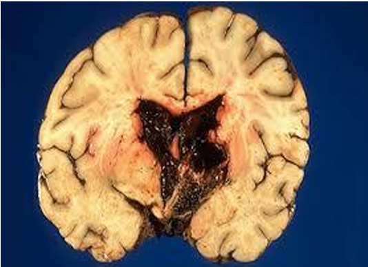
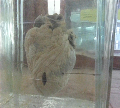
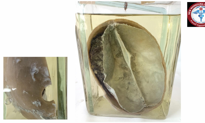
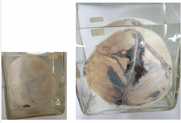
Questions
What is the specimen being examined?
What are the specific internal and external findings regarding the injury?
What is the possible cause of death?
What is the final diagnosis for this specimen?
Answers
Answer: The specimen is a transverse section in the brain.
Answer: Type & Description of Injury:
Internal: Pathological intracerebral hemorrhage, with the main bulk of the blood present inside the brain substance.
External: No laceration, congestion, or ulceration on the outer surface of the brain.
Answer: Cause of Death: Brain compression.
Answer: Diagnosis: Pathological Intracerebral Haemorrhage.
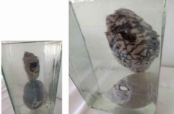
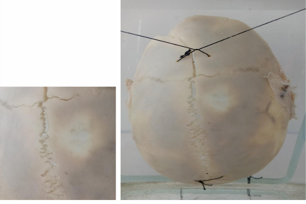
Questions
What is the specimen being examined?
What are the type, specific descriptive features, and dimensions of the skull fracture?
How is the antemortem nature of the injury determined?
What type of causative instrument and cross-sectional shape produce this injury?
How is the time of death estimated?
What are the possible causes of death?
What is the final diagnosis for this specimen?
Answers
Answer: The specimen is a vault of the skull with the underlying dura mater and the left cerebral hemisphere.
Answer: Type & Description of Injury: Traumatic penetrating fracture in the vault of the skull at the left fronto-parietal region, oval in shape, and about 1x2 cm in size.
Answer: Vital Reaction: The injury is antemortem due to evidence of subperiosteal, extradural, and intracerebral hemorrhage.
Answer: Causative Instrument: Tip of an Iron Bar (pointed end with a rounded cut section).
Answer: Time of Death: Shortly after injury due to the absence of signs of surgical interference, healing, or sepsis.
Answer: Cause of Death: Brain compression or brain laceration.
Answer: Diagnosis: Traumatic Intracerebral Haemorrhage.
Questions
What are the specific path, descriptive features, and sutures involved in the fracture?
How is the antemortem nature of the injury determined?
What type of causative instrument and force mechanics produce this injury?
How is the time of death estimated?
What are the possible causes of death?
What is the final diagnosis for this specimen?
Answers
Answer: Type & Description of Injury: Fissure fracture in the frontal bone extending from the anterior part of the left side to the midline, passing towards and running into the sagittal suture, completely separating the sagittal suture and the medial parts of the coronal and lambdoid sutures (Diastatic fissure fracture).
Answer: Vital Reaction: It is an antemortem injury due to the presence of subperiosteal hemorrhage in the right parietal bone.
Answer: Causative Instrument: Heavy blunt instrument with a wide striking surface and low momentum of force (e.g., Iron Bar or Heavy Stick of Wood).
Answer: Time of Death: Shortly after injury due to the absence of signs of surgical interference, healing, or sepsis, and the sharp edges of the fractures.
Answer: Cause of Death: Severe concussion or brain compression.
Answer: Diagnosis: Diastatic Fissure Fracture in The Frontal Bone with Subperiosteal Haemorrhage.
Case Summary
Scenario: 28-year-old male found suspended from a ceiling fan by a bedsheet, feet partially touching the ground.
Findings: Oblique, non-continuous ligature mark high in the neck; pale face, minimal petechiae; saliva dribbling from the angle of the mouth; no signs of struggle.
Questions
What is the most likely cause of asphyxia?
Is the manner of death suicidal, homicidal, or accidental?
Answers
Answer: Hanging (Incomplete hanging).
Answer: Suicidal. (Proven by antemortem saliva dribbling, oblique/non-continuous mark, and lack of struggle signs).
Case Summary
Scenario: 35-year-old female found dead at home. Suspicion of domestic violence.
Findings: Bruises, abrasions, and fingernail marks on the neck; congested face with petechial hemorrhages; fracture of the hyoid bone.
Questions
Type of asphyxia?
Manner of death?
Importance of hyoid fracture?
Answers
Answer: Manual strangulation (Throttling).
Answer: Homicidal.
Answer: Diagnostic of local neck compression: It proves heavy, direct pressure was applied to the neck (highly characteristic of manual strangulation in victims over 30–35 years old when the bone has ossified).
Scenario: Body found on the ground with a rope tightly wound around the neck.
Findings: Horizontal ligature mark completely encircling the neck, located low in the neck, showing a deep groove with underlying tissue damage, and corresponding to the ligature material.
Questions
Differentiate from hanging
Manner of death?
Answers
Differentiate from hanging: * This case (Ligature Strangulation): The mark is horizontal, continuous (completely encircling the neck), and located low down.
Hanging: The mark is typically oblique, non-continuous (interrupted at the knot), and located high up in the neck.
Manner of death: Homicidal.
Case Summary
Scenario: An infant was found dead in bed next to his mother with a history of co-sleeping.
Findings: No external injuries; cyanosis.
Questions
Type of asphyxia?
Possible manners?
Answers
Answer: Overlaying (a form of suffocation / accidental smothering).
Answer: Accidental (most common due to co-sleeping) or Homicidal (infanticide masked as an accident).
Scenario: 60-year-old man collapses while eating meat.
Findings: Piece of meat lodged in the airway; cyanosis and congestion; no trauma.
Questions
Mechanism of death?
Type of asphyxia?
Answers
Mechanism of death: Vagal inhibition (cardiac arrest from laryngeal nerve stimulation) or acute asphyxia.
Type of asphyxia: Choking (mechanical block of the air passages from within).
Scenario: Worker trapped under a heavy object.
Findings: Intense cyanosis of the face; petechiae in the eyes and skin; congested upper body.
Questions
Diagnosis?
Mechanism?
Answers
Diagnosis: Traumatic asphyxia (Crush asphyxia / Perthes' syndrome).
Mechanism: Mechanical fixation of the chest: Heavy pressure prevents respiratory movements, compressing the superior vena cava and forcing blood backward, which ruptures capillaries and causes intense upper-body congestion and petechiae.
Scenario: Body recovered from a canal.
Findings: Fine froth at the mouth and nostrils; water in the lungs; washerwoman’s skin.
Questions
Cause of death?
Possible time of death?
Answers
Cause of death: Drowning (Wet drowning).
Possible time of death: Estimated by the presence of washerwoman's skin, which typically develops after 12 - 18 hours of immersion in water.
Scenario: Victim found with signs of smothering and chest compression.
Findings: Petechiae; signs of suffocation; evidence of pressure on the chest.
Questions
Diagnosis and manner of death?
Answers
Diagnosis: Burking (a combination of homicidal smothering and traumatic/crush asphyxia).
Manner of death: Homicidal.
Case Summary
Scenario: 28-year-old male found dead in his room, suspended from a ceiling fan by a bedsheet.
Findings: Oblique, non-continuous ligature mark high up in the neck; pale face; non-protruded tongue; saliva dribbling from the angle of the mouth; no signs of struggle in the room.
Questions
What is the most likely cause of death?
Is this more consistent with suicidal or homicidal manner?
Is it AM or PM and why?
Answers
Most likely cause of death: Asphyxia due to hanging.
Manner of death: Suicidal. (Supported by the high, oblique, non-continuous mark, lack of struggle signs, and typical domestic venue).
AM or PM and why: AM (Antemortem). Proven by the saliva dribbling from the angle of the mouth, which is a vital reaction that can only occur if the victim was suspended while alive and secreting saliva under pressure.
Scenario: 2-year-old child brought dead after playing alone.
Findings: Cyanosis of the face; abrasions around the mouth and nose; congested conjunctiva with petechial hemorrhages; no foreign body in the airway.
Questions
What is the most likely type of asphyxia?
What findings support this diagnosis?
What is the possible manner of death?
Type of asphyxia: Smothering.
Supporting findings: Abrasions around mouth and nose + general asphyxia signs (petechiae, cyanosis) + clear airway.
Manner of death: Accidental or Homicidal.
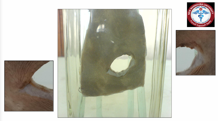
Questions
What is the specimen being examined?
What are the specific descriptive characteristics of the wound edges and diameter?
What distance of firing is indicated, and why?
How is the antemortem nature of this injury determined?
What type of causative instrument produced this injury?
How is the time of death estimated?
What are the possible causes of death?
What is the final diagnosis?
Answers
Answer: Piece of skin.
Answer: 2 mm diameter, circular hole with regular and inverted edges.
Answer: Far distance, due to the absence of powder marks.
Answer: Presence of subcutaneous hemorrhage.
Answer: Rifled weapon.
Answer: Shortly after injury (no surgery, healing, or sepsis signs).
Answer: Hemorrhage, shock, or injury of vital organs.
Answer: Typical Inlet in The Skin Caused by a Bullet.
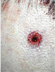
Questions
What is the specimen being examined?
What are the specific descriptive features of the wound edges and diameter?
What specific finding proves a contact range of firing, and what form does it take?
How is the antemortem nature of this injury determined?
What type of causative instrument and firing pattern produced this injury?
How is the time of death estimated?
What are the possible causes of death?
What is the final diagnosis?
Answers
Answer: Piece of skin.
Answer: 5 mm diameter, circular hole with regular, inverted, and burned edges.
Answer: Muzzle imprint in the form of a patterned abrasion.
Answer: Presence of subcutaneous hemorrhage.
Answer: Automatic rifled weapon in a contact pattern.
Answer: Shortly after injury (no surgery, healing, or sepsis signs).
Answer: Hemorrhage, shock, or injury of vital organs.
Answer: Contact Inlets in The Skin.
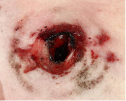
Questions
What is the specimen being examined?
What are the specific descriptive features of the wound edges and diameter?
How is the antemortem nature of this injury determined?
What type of causative instrument produced this injury?
How is the time of death estimated?
What are the possible causes of death?
What is the final diagnosis?
Answers
Answer: Piece of skin.
Answer: 1.2 cm diameter, circular hole with irregular and everted edges.
Answer: Irregular and everted edges with no surrounding powder marks make us sure that this is an exit wound , type shi
Answer: Presence of subcutaneous hemorrhage.
Answer: Rifled weapon.
Answer: Shortly after injury (no surgery, healing, or sepsis signs).
Answer: Hemorrhage, shock, or injury of vital organs.
Answer: Exit Wound in The Skin Caused by a Bullet.
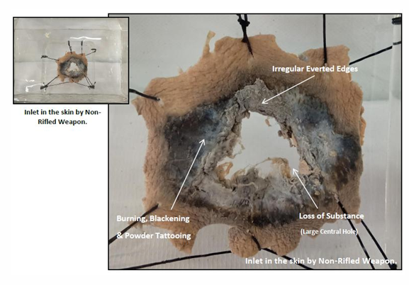
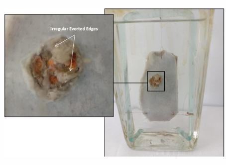
Questions
What is the specimen being examined?
What are the specific descriptive features of the wound edges, diameter, and dispersion?
What specific features indicate a "near firing" range?
How is the antemortem nature of this injury determined?
What type of causative instrument produced this injury?
How is the time of death estimated?
What are the possible causes of death?
What is the final diagnosis?
Answers
Answer: Piece of skin.
Answer: 2 cm diameter, large circular hole with irregular edges and no dispersion.
Answer: Presence of burning, blackening, and tattooing surrounding the hole.
Answer: Presence of subcutaneous hemorrhage.
Answer: Non-rifled weapon (firing shots).
Answer: Shortly after injury (no surgery, healing, or sepsis signs).
Answer: Hemorrhage, shock, or injury of vital organs.
Answer: Inlet in the skin by Non-Rifled Weapon.
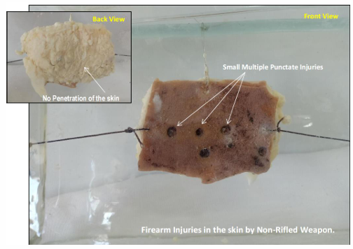
Questions
What is the specimen being examined?
What are the specific shape, count, diameter, and penetration characteristics of the injuries?
What characteristic of the wound edges is described?
How is the antemortem nature of this injury determined?
What type of causative instrument produced this injury?
How is the time of death estimated?
What is the possible cause of death?
What is the final diagnosis?
Answers
Answer: Piece of skin.
Answer: Multiple small punctate injuries, 3 mm in diameter, superficial, and not penetrating the skin.
Answer: Burned edges.
Answer: Presence of subcutaneous hemorrhage.
Answer: Non-rifled weapon (firing shots).
Answer: Shortly after injury (no surgery, healing, or sepsis signs).
Answer: Other associated injuries.
Answer: Firearm injury of the skin caused by shots from a non-rifled weapon.
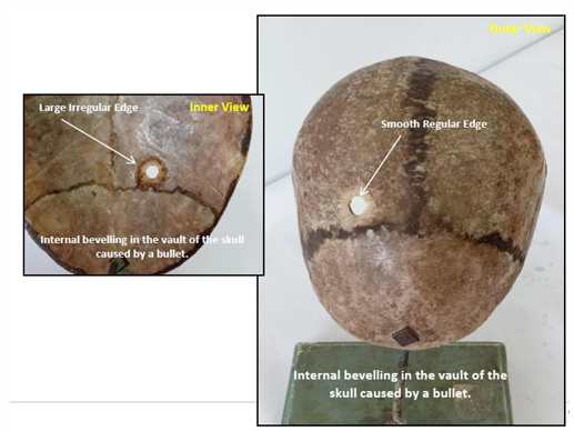
Questions
What is the specimen being examined?
What are the specific structural differences between the openings of the outer and inner tables of the skull?
What is the specific pathological term used for this type of skull bone involvement?
What are the precise anatomical measurements and landmark relations of the injury site?
What type of causative instrument produced this injury?
How is the time of death estimated?
What are the possible causes of death?
What is the final diagnosis?
Answers
Answer: Vault of skull.
Answer: Outer table: 5 mm diameter, smooth, and regular edges. Inner table: Larger opening with irregular edges.
Answer: Internal beveling (indicating a firearm inlet).
Answer: 5 mm diameter, 3 cm lateral to the sagittal suture, and 1 cm posterior to the coronal suture in the right parietal bone.
Answer: Rifled weapon (firing bullets).
Answer: Shortly after injury (no surgery, healing, or sepsis signs).
Answer: Intracranial hemorrhage or extensive brain lacerations.
Answer: Internal bevelling in the vault of the skull caused by a bullet.
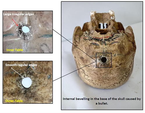
Questions
What is the specimen being examined?
What are the specific structural differences between the outer and inner table openings?
What is the precise anatomical location and diameter of the skull injury?
What type of causative instrument produced this injury?
How is the time of death estimated?
What are the possible causes of death?
What is the final diagnosis?
Answers
Answer: Vault of skull.
Answer: Outer table: Smooth and regular edges. Inner table: Larger opening with irregular edges (internal beveling).
Answer: 7 mm diameter, located in the center of the occipital bone.
Answer: Rifled weapon (firing bullets).
Answer: Shortly after injury (no surgery, healing, or sepsis signs).
Answer: Intracranial hemorrhage or extensive brain lacerations.
Answer: Internal bevelling in the vault of the skull caused by a bullet.
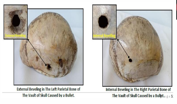
Questions
What is the specimen being examined?
What are the specific anatomical locations and measurements for both injuries?
How do the outer and inner tables differ structurally in the right parietal injury?
How do the outer and inner tables differ structurally in the left parietal injury?
What forensic terms describe the skull bone changes seen in the first and second injuries respectively?
What are the causative instrument and the specific trajectory of the missile?
How is the time of death estimated?
What are the possible causes of death?
What is the final diagnosis?
Answers
Answer: Vault of skull.
Answer:
Right parietal: 8 cm lateral to sagittal suture, 10 cm posterior to coronal suture.
Left parietal: 12 cm lateral to sagittal suture, 8 cm posterior to coronal suture.
Answer: Outer table: Smooth, regular edges (5 mm diameter). Inner table: Larger opening with irregular edges.
Answer: Outer table: 5 mm diameter with irregular edges. Inner table: Smooth, regular circular hole.
Answer: First injury shows internal beveling (inlet); second injury shows external beveling (exit).
Answer: Rifled weapon (firing a bullet); trajectory is from right to left, slightly downwards.
Answer: Shortly after injury (no surgery, healing, or sepsis signs).
Answer: Hemorrhage, shock, or injury of vital organs.
Answer: Internal Beveling & External Beveling of The Right & Left Parietal Bones of The Vault of Skull caused by a bullet.
identify specimen ?
characters ?
characters:
• Length: 16 cm •
Mouth & nose are separate openings
• Sex well differentiated at 4 months. “Male”
• Four limbs are well-formed showing finger & toes.
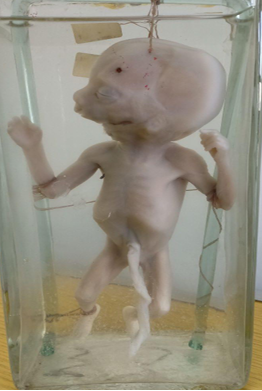
Specimen: • The specimen is a male foetus four months gestation
identify specimen ?
characters ?
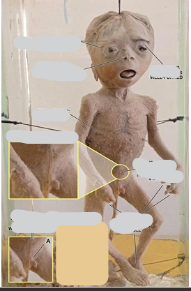
Male & Female Foetuses 6 Months Gestation:
Characters:
• Length: 30 cm
• Mouth & nose are separate openings
• Sex well differentiated: male
• Four limbs are well-formed, showing fingers and toes.
• Eyebrows & eyelashes are well formed “at six months.”
• Umbilical cord is attached above the pubis “at six months.” Specimen: Teeth
• The specimen is a male foetus at six months gestations.
identify specimen ?
characters ?
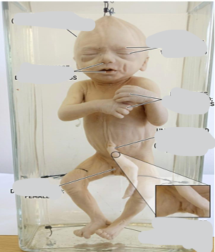
Specimen
Type: Complete female fetus
Gestational Age: 7 Months of gestation
Physical Characteristics & Measurements
Crown-Heel Length: 35 cm
Sexual Differentiation: Well-differentiated female genitalia
Facial Development: Mouth and nose are fully separate openings
Ocular Features: Eyebrows and eyelashes are distinct and well-formed
Extremities: Four well-formed limbs showing distinct fingers and toes
Umbilical Attachment: Attached low on the abdominal wall, directly above the pubis
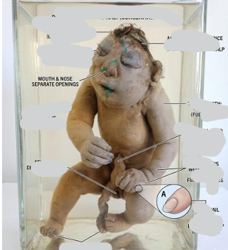
Specimen
Type: Complete female fetus showing congenital anomaly
Gestational Age: 9 Months of gestation (Full Term)
Physical Characteristics & Measurements
Crown-Heel Length: About 45–50 cm
Sexual Differentiation: Well-differentiated female genitalia
Facial & Ocular Features: Mouth and nose are fully separate openings; eyebrows and eyelashes are well-formed
Scalp & Nails: Scalp hair is 2–3 cm in length; nails extend beyond the tips of the fingers
Extremities: Four well-formed limbs showing distinct fingers and toes
Umbilical Attachment: Attached precisely at the middle of the body
Congenital Anomalies
Diagnosis: Anencephaly (absence of a major portion of the brain, skull, and scalp)
Questions
What organs are included in this specimen?
What is the estimated gestational age based on uterus size?
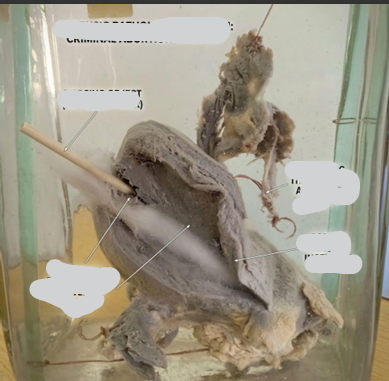
What finding inside the uterus proves a criminal abortion, and what is the causative instrument?
How is the time of death estimated?
What is the cause of death?
What is the final diagnosis?
Answers
Answer: Uterus, cervix, and adnexa.
Answer: 8–10 weeks of gestation (size of a moderate orange).
Answer: An elongated stick passing through the uterus and penetrating the fundus.
Answer: Immediately after the injury, due to the absence of vital reaction.
Answer: Severe hemorrhage.
Answer: Criminal Abortion.
Questions
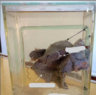
What organs are included in this specimen?
What is the estimated gestational age based on uterus size?
What three specific pathological signs are visible on the opened uterine wall?
How is the time of death estimated, and what finding supports this?
What is the cause of death?
What is the final diagnosis?
Answers
Answer: Uterus, cervix, and adnexa.
Answer: 10–12 weeks of gestation (size of a fetus head).
Answer: Very thick wall, signs of sepsis/necrotic tissues, and ulceration areas.
Answer: 4–6 weeks after induction, proven by the presence of vital reaction (sepsis).
Answer: Puerperal Sepsis.
Answer: Criminal Abortion.
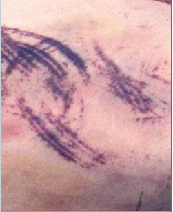
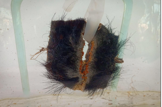
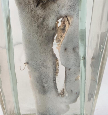
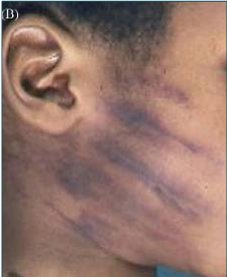
Mention the type of child abuse?
• Physical Abuse • Multiple tram-line bruises caused by a linear rod-like object, such as a stick.
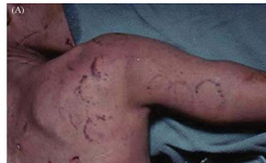
Mention the type of child abuse?
• Physical Abuse Patterned bruises: Bite marks
Mention the type of child abuse?
• Physical Abuse Patterned bruises caused by the imprint of the hand (hand slap)
Mention the type of child abuse?
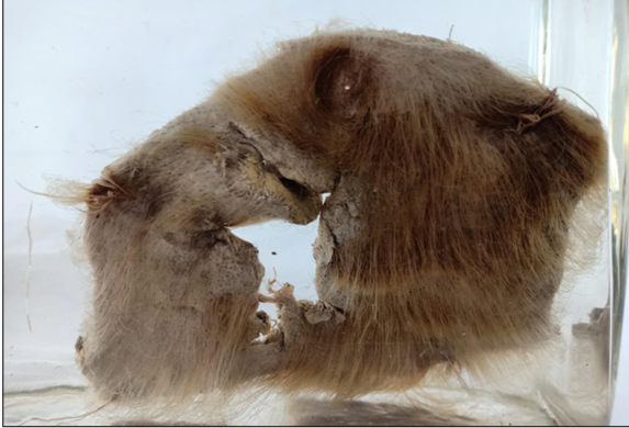
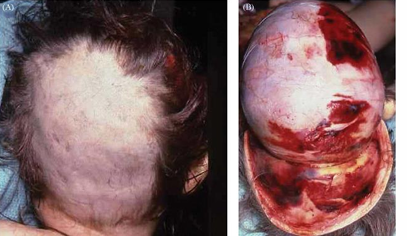
• Physical Abuse
External surface of the scalpwith alopecia (A). The internal surface shows diffuse subgaleal hemorrhage (B)
Mention the type of child abuse?
Neglect: A starved child.