Anatomy and Physiology
1/38
There's no tags or description
Looks like no tags are added yet.
Name | Mastery | Learn | Test | Matching | Spaced | Call with Kai |
|---|
No analytics yet
Send a link to your students to track their progress
39 Terms
Nasal Bridge
The saddle-shaped area of the nose that includes the nasal root and the lateral (side) aspects of the nose. It forms the upper bony portion of the nose.
Nasal Tip
The most anterior, projecting portion of the nose. Its position and projection are supported by the columella and underlying cartilage.

Columella
The tissue that separates the two nostrils externally. It acts like a supporting column for the nasal tip and should ideally be straight and supported by a straight nasal septum.
Naris (plural: nares) / nostril
The external openings of the nose through which air enters and exits. A single opening is called a naris, while both openings together are the nares.

Ala Nasi
The curved outer side ("wing") of a nostril.
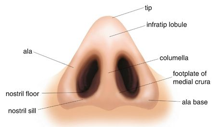
Alar Rims
The curved outer edges that surround the openings of the nostrils.

Alar Base
The area where the ala nasi joins the upper lip.

Nasal Sill
The base or floor of the nostril opening. It forms the lower border of the nostril.
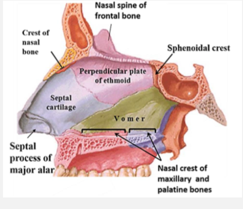
Vomer
Perpendicular structure that fits in median palatine suture groove, below septal cartilage and above hard palate
Turbinates (nasal conchae)
Curved, shelf-like bony structures inside the nasal cavity that are covered by mucous membrane.
They project from the lateral walls of the nose into the nasal cavity.
Their functions are to:
Filter inhaled air
Warm inhaled air
Humidify inhaled air
Direct airflow toward the olfactory region for smell
Superior Turbinate
The highest and smallest turbinate.
Located near the roof of the nasal cavity.
Helps direct airflow toward the olfactory (smell) receptors
Middle Turbinate
Located below the superior turbinate.
Forms part of the lateral wall of the nasal cavity.
Helps regulate airflow and drainage from several sinuses.
Inferior Turbinate
The largest and lowest turbinate.
Plays a major role in warming, humidifying, and filtering inspired air.
Meatuses
Passageways or spaces located directly beneath each turbinate.
Air flows through these spaces during breathing.
Superior Nasal Meatus
Space beneath the superior turbinate.
Allows airflow through the upper portion of the nasal cavity.
Middle Nasal Meatus
Space beneath the middle turbinate.
Important drainage pathway for several paranasal sinuses.
Inferior Nasal Meatus
Space beneath the inferior turbinate.
The largest nasal passageway for airflow.
Paranasal Sinuses
Air-filled cavities located within the bones of the face and skull.
Connected to the nasal cavity through small openings called ostia.
Four paired sinuses:
Frontal sinuses – in the forehead
Ethmoid sinuses – between the eyes
Maxillary sinuses – beneath the cheeks
Sphenoid sinuses – deep within the skull
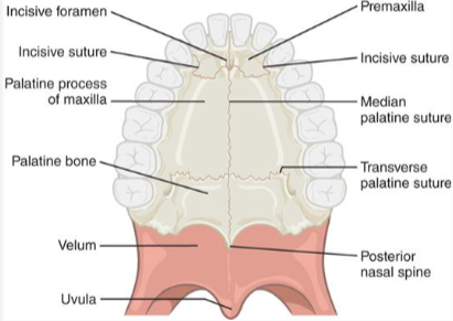
Incisive Foramen
an opening located at the junction between the premaxilla (the triangular bone at the very front of the hard palate) and the maxillary bones
the starting point of embryological development of lip and palate
It marks the boundary between two major developmental regions:
the primary palate (formed from the premaxilla)
the secondary palate (formed from the palatal shelves of the maxilla)
Sphenoid and Temporal Bones
Cranial bones that provide the structural base and attachment sites for muscles involved in velopharyngeal function.

Medial & Lateral Pterygoid Plates
Thin bony projections of the pterygoid process of the sphenoid bone
These plates form part of the posterior nasal and oral framework
Serve as attachment sites for muscles of the pharynx and soft palate, contributing to velopharyngeal movement.

Pterygoid Hamulus
A hook-like projection at the inferior end of the medial pterygoid plate
Acts as a pulley for the tendon of the tensor veli palatini muscle, allowing the muscle to change direction and assist in tensing the soft palate and opening the eustachian tube.
Adenoid Pad
A mass of lymphoid tissue located on the posterior pharyngeal wall in the nasopharynx, just behind the velum (soft palate)
Part of the immune system and help produce antibodies, especially in early childhood
in young children, often large and can assist with velopharyngeal closure, contributing to what is called velophadenoidal closure.
Cleft Risk for chronic otitis media
In individuals with a cleft palate, the muscles of the velopharyngeal mechanism (especially the tensor veli palatini) are abnormally positioned or function poorly, so the eustachian tube does not open effectively.
This leads to:
Poor ventilation of the middle ear
Fluid buildup (otitis media with effusion)
Increased risk of recurrent ear infections
Child risk for Ear Infection
Eustachian tube for drainage horizontal
becomes more vertical moving to adulthood
Lateral Pharyngeal Walls
Side walls of the pharynx that form part of the velopharyngeal valve.
Structurally, they are composed of muscle and soft tissue
Function:
Move medially (inward) during velopharyngeal closure
Close against the velum (soft palate) or, in some cases, meet in midline
Provide an important contribution to closure, though the degree of movement varies among individuals
Posterior Pharyngeal Wall
Back wall of the pharynx, forming the posterior boundary of the velopharyngeal valve.
Function:
May move slightly anteriorly (forward) during velopharyngeal closure
Serves as the contact surface for the velum in many individuals
Contributes less to closure compared to the velum and LPWs
Passavant’s ridge
Bulge of muscle on the posterior pharyngeal wall during speech; occurs in some normal and abnormal speakers
Velar Dimple
A visible indentation in the midline of the oral (inferior) surface of the velum (soft palate) seen during speech or phonation.
Structure & Cause:
Formed by the interdigitation (blending) of the levator veli palatini muscles in the midline
Represents the point where these muscles pull the velum upward and backward
Velopharyngeal Function
Seperates the nasal cavity from the oral cavity
Regulates and directs transmission of sound energy and airflow into the oral and nasal cavities
Particularly important for production of “pressure- sensitive” consonant sounds (plosives, fricatives, and affricates) and all vowels
Closes off nose and builds pressure for swallowing

Levator Veli Palatini (Velar “Sling”)
Primary muscle that elevates the velum during speech
Forms a muscular sling in the midline, pulling the soft palate upward and backward for velopharyngeal closure
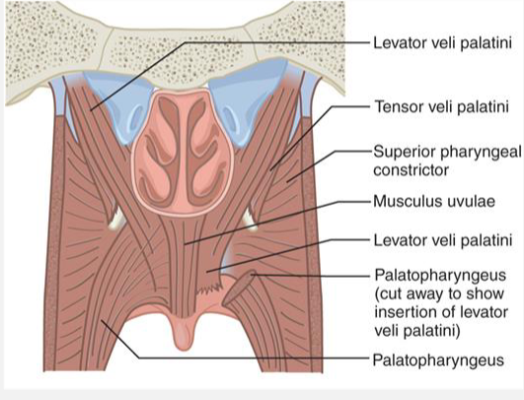
Superior Constrictor (Pharyngeal Ring)
Muscle of the pharyngeal wall that constricts (narrows) the pharynx.
Helps move the lateral pharyngeal walls inward to aid in velopharyngeal closure.

Palatopharyngeus (Posterior Faucial Pillar)
Runs in the posterior faucial pillars
Pulls the lateral pharyngeal walls inward and helps bring them against the velum during closure.

Palatoglossus (Anterior Faucial Pillar)
Found in the anterior faucial pillars
Responsible for lowering the velum quickly for nasal sounds and helping open the velopharyngeal port.

Musculus Uvulae
Midline muscle that creates a bulge (velar eminence) on the nasal surface of the velum
Adds bulk and stiffness for a tighter velopharyngeal seal.

Tensor Veli Palatini
Does not elevate the velum
Tenses the soft palate and opens the eustachian tube during swallowing for middle ear ventilation.
Coronal Velopharyngeal Closure
Velum moves posteriorly to contact the posterior pharyngeal wall (most common, ~70%).
Circular Velopharyngeal Closure
Velum, lateral pharyngeal walls, and posterior wall all contribute equally → forms a sphincter-like closure (~25%)
Sagittal Velopharyngeal Closure
Lateral pharyngeal walls move medially to meet in midline with minimal velar movement (least common, ~5%).
Pneumatic vs Non‑Pneumatic Activities
Non‑pneumatic activities (no airflow):
Examples: swallowing, gagging, vomiting
Velopharyngeal closure is very high and tight
Closure is firm and along entire pharynx
Purpose: Prevent material from entering the nasal cavity
Pneumatic activities (use airflow/pressure):
Examples: speech, blowing, singing, sucking
Closure is lower in the pharynx
Requires precise, rapid, coordinated movement
Purpose: Direct airflow and sound between oral and nasal cavities
Respiration for Speech Sounds
High-pressure sounds
• Plosives (p, b, t, d, k, g)
• Fricatives (f, v, s, z, ʃ, Θ, ð)
• Affricates (ʧ, ʤ)
Low-pressure sounds
• Liquids (l, r)
• Glides (w, j)
No-pressure sounds
• Nasals (n, m, ŋ)
Lip Anatomy
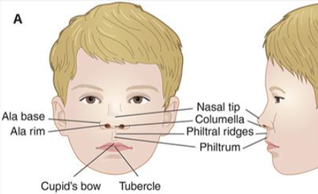
Philtrum: Midline vertical groove between nose and upper lip
Philtral ridges (columns): Raised borders of philtrum; fusion lines
Cupid’s bow: Double peak shape of upper lip border
White roll: Raised outline of the lip border
Vermilion: Red portion of the lips
Labial tubercle: Midline fullness of upper lip