AMD - Posterior Segment & Ocular Disease Spring 2026
1/105
There's no tags or description
Looks like no tags are added yet.
Name | Mastery | Learn | Test | Matching | Spaced | Call with Kai |
|---|
No analytics yet
Send a link to your students to track their progress
106 Terms
Leading cause of blindness in individuals >50yo?
AMD
Do more people have dry or wet AMD?
dry AMD
Do males or females have higher prevalence for AMD?
No
Risk for African Americans vs White/Hispanic for AMD
Risk is lower in African Americans
EXAM QUESTION: If you are unsure whether there are changes in the macula (from clinical exam, reduced acuity, or both) you must do what?
1) Run an OCT
2) Refer to Ophthalmology
3) Or BOTH
EXAM QUESTION: What is the primary problem in macular degeneration (AMD)?
poor vascular supply to RPE/photoreceptors
EXAM QUESTION: What is an additional problem besides poor vascular supply to RPE/photoreceptors in AMD?
Breakdown of cell membranes d/t oxidative stress
EXAM QUESTION: What are the major risk factors for AMD?
-age
-smoking
-race
-genetics
What is the biggest contributor to the risk factors for AMD?
Age
Are there many genes that are related to AMD?
Yes -- there are 103(ish)
What are the OTHER (besides the major ones) that are risk factors for AMD development?
1) Lower intake of dietary anti-oxidants and omega-3 fatty acids
2) High body mass index/waist circumference
3) CV disease
4) UV exposure
5) Baseline levels of serum cystatin C (kidney function)
6) Blue iris>green iris
7) Divorced > married
8) Hyperopia
EXAM QUESTION: If you see macular degeneration in younger patients, you should think of what?
Inflammation from infections, autoimmune disorders, white dot syndromes
Central serous
Inherited disorders (Stargardt disease, pattern dystrophy)
Trauma
EXAM QUESTION: Many practitioners will not use AMD diagnosis until when?
until drusen are apparent
What are the classic signs of dry AMD?
RPE dropout
Pigment clumping
Drusen
What is RPE dropout? What color?
RPE pigmentary change; dull yellow
What is pigment clumping? What color?
RPE pigmentary change & hyperplasia; black
What can drusen progress to long-term?
Geographic atrophy long-term
Severe loss of vision
What color are drusen?
Dull yellow
What characterizes wet AMD? (five things)
Choroidal neovasc (CNVM or MNV)
RPE hyperplasia
Old blood (yellow/red)
Hard exudates
Sub-RPE hemorrhage is green-black; subretinal heme is red
What are serous RPE detachment, pigment epithelial detachment (PED), localized retinal detachment all signs of?
Could be a sign of either wet or dry AMD
How could dry AMD lead to a serous RPE/retinal detachment?
fluid leaks from the choriocapillaris and detaches RPE and/or the retina from Bruchs membrane
How could wet AMD lead to a serous RPE/retinal detachment?
fluid leaks from a choroidal neovascular membrane
If you have a serous retinal detachment, the fluid will be assumed to be from what origin?
choriodal neovasc
What color is a serous RPE/retinal detachment d/t dry AMD?
yellow/orange
What color is a serous RPE/retinal detachment d/t wet AMD?
yellow, orange, red

Can non-exudative macular neovascularization (nonexudative MNV) leak?
yes -- it may ultimately leak
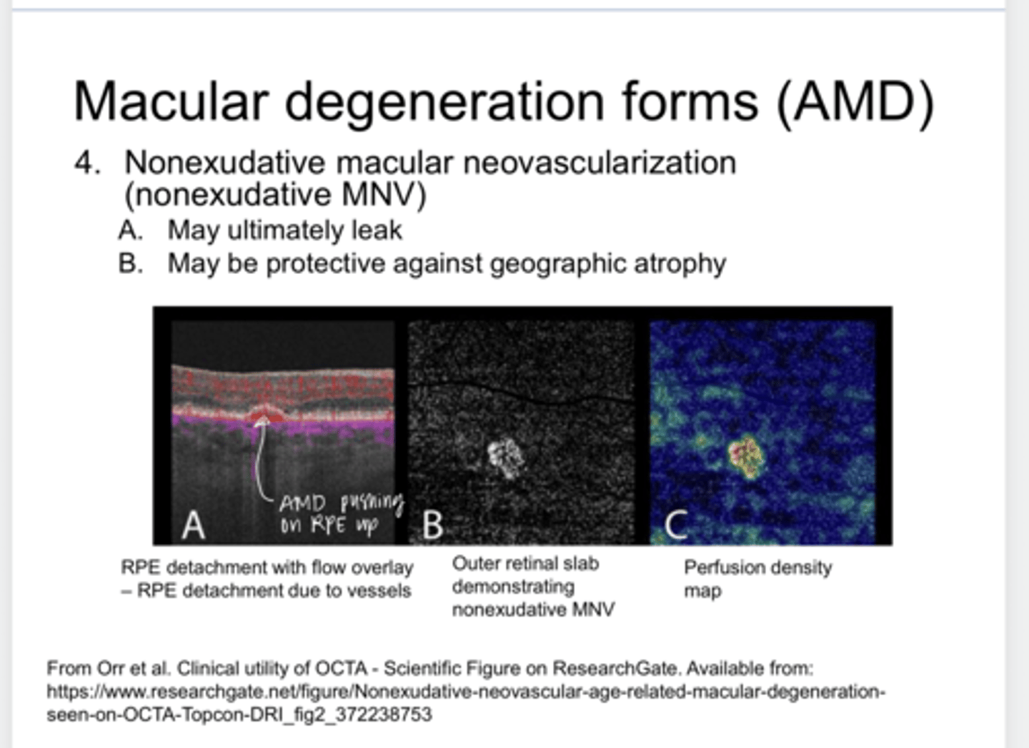
What can non-exudative macular neovascularization (nonexudative MNV) be protective against?
geographic atrophy

Why is non-exudative macular neovascularization (nonexudative MNV) protective against geographic atrophy?
Alleviates some of the hypoxic stress if these vessels are not leaky and provide more profusion
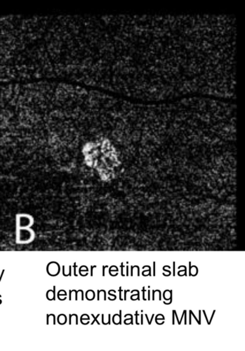
RPE Detachment w/ Flow Overlay d/t Vessels (Pic)
RPE Detachment w/ Flow Overlay d/t Vessels (Pic)

What happens when OCT-A image looks like this and you do a FA?
it WILL NOT leak

Will non-exudative MNV eventually leak?
Yes

Small drusen size
<63 microns
**A retinal vessel is about 125 microns in diameter so it’s roughly less than half a retinal vessel diameter
Small drusen name
Drupelet

Medium drusen size
64-125 microns
**A retinal vein is about 125 microns in diameter

Large drusen size
>125 microns
**A retinal vein is about 125 microns in diameter

Dry AMD (Pic)
Dry AMD (Pic)

Are small drusen usually a problem?
usually not

Are large drusen a problem?
Yes -- this is worrisome

Why are you concerned about large drusen?
Concerned about possibility of choroidal neovascularization

What are drusen composed of?
protein/fat

If drusen start to coalesce with poorly defined borders, what is this referred to as?
soft drusen
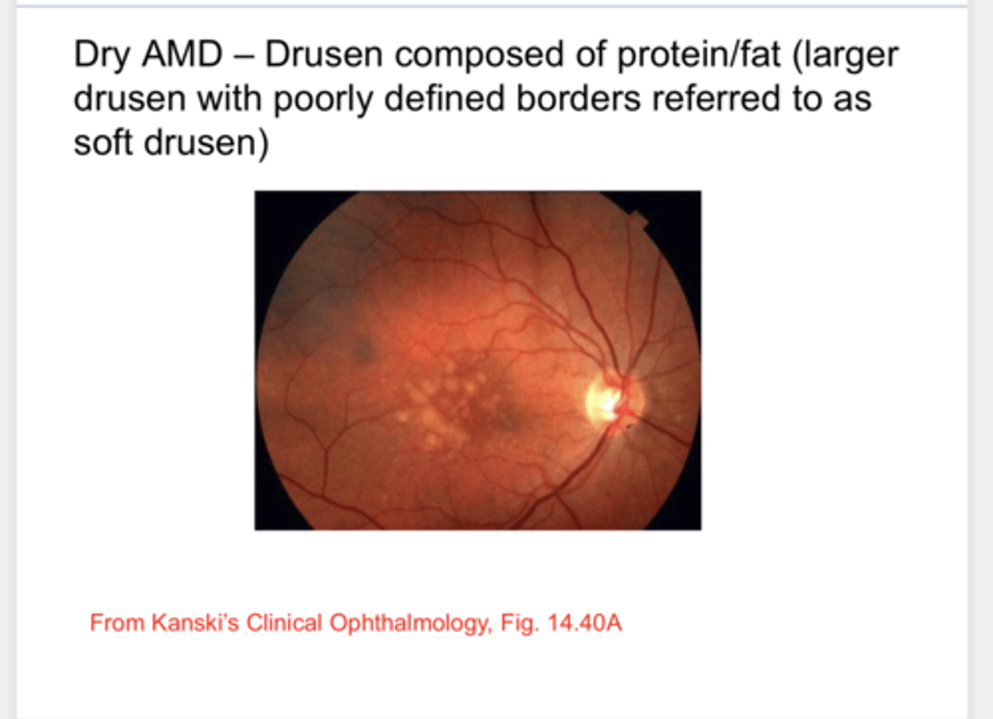
What should you do on this patient?
OCT -- to look for neovasc

Calcific drusen (Pic)
Calcific drusen (Pic)

What do calcific drusen look like?
hard exudate
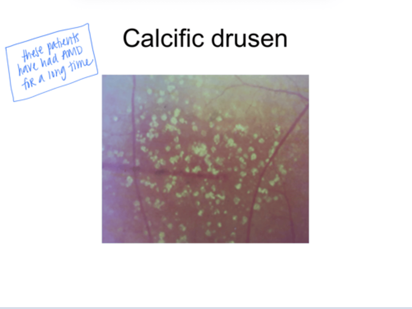
What color are calcific drusen?
-yellow, not very shiny
-well defined

What does calcific drusen indicate?
These patients have had AMD for a long time

What does this patient have?
a lot of soft drusen (coalesced drusen)
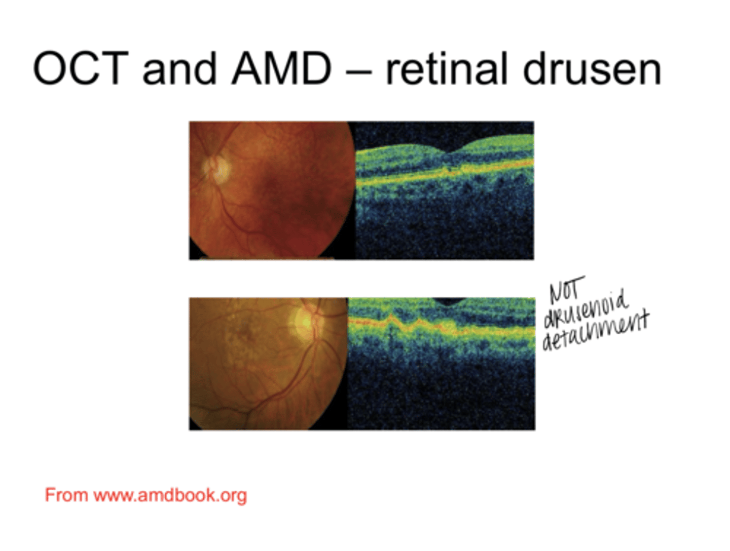
Drusen on an OCT (Pic)
Drusen on an OCT (Pic)
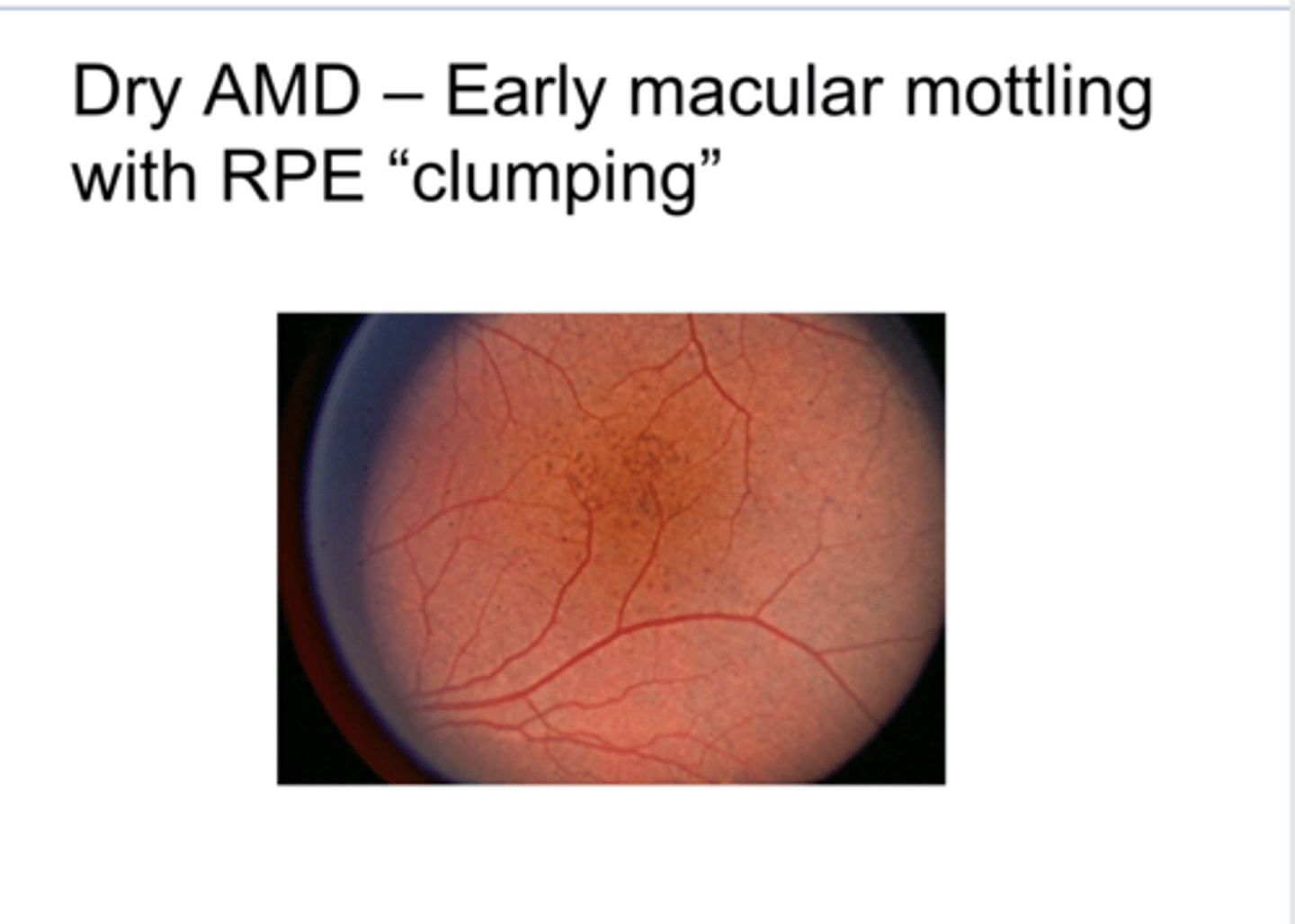
Dry AMD -- Early Macular Mottling with RPE Clumping (Pic)
Dry AMD -- Early Macular Mottling with RPE Clumping (Pic)

Can you get these pigmentary changes from other diseases besides AMD?
Yes
Does drusen ONLY happen in AMD?
Yes

Dry AMD -- Areas of RPE Dropout/RPE Window Defects (Pic)
Dry AMD -- Areas of RPE Dropout/RPE Window Defects (Pic)
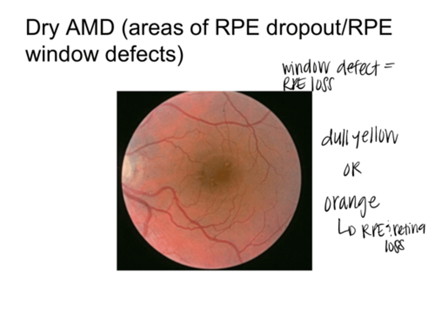
What color is RPE Window Defect & RPE loss?
dull yellow or orange

If RPE dropout is orange, why?
RPE and retina loss

Dry AMD -- RPE Clumping and RPE Dropout in Fundus Photo and FA (Pic)
Dry AMD -- RPE Clumping and RPE Dropout in Fundus Photo and FA (Pic)

RPE Clumping will be (hyper/hypo) fluorescent on FA
hypo -- RPE pigment clumps will block
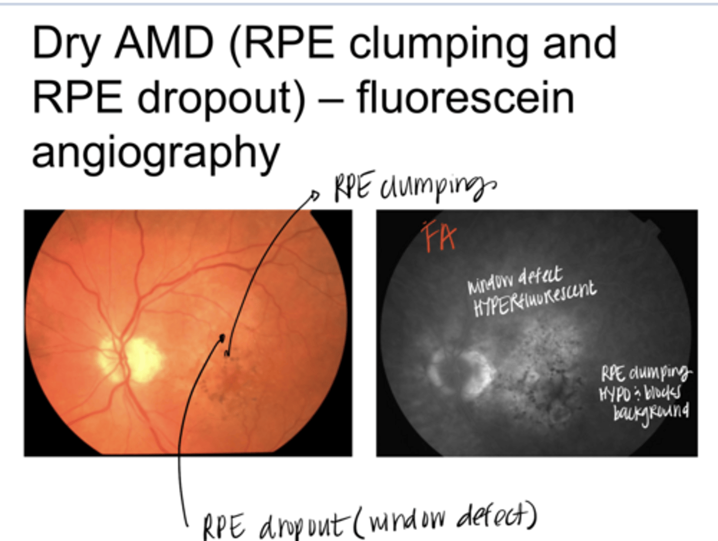
RPE Window defect will be (hyper/hypo) fluorescent on FA
hyperfluorescent -- no pigment to block

Dry AMD -- Drusen, RPE Clumping, Orange Areas are Window Defects with Effacement (Pic)
Dry AMD -- Drusen, RPE Clumping, Orange Areas are Window Defects with Effacement (Pic)
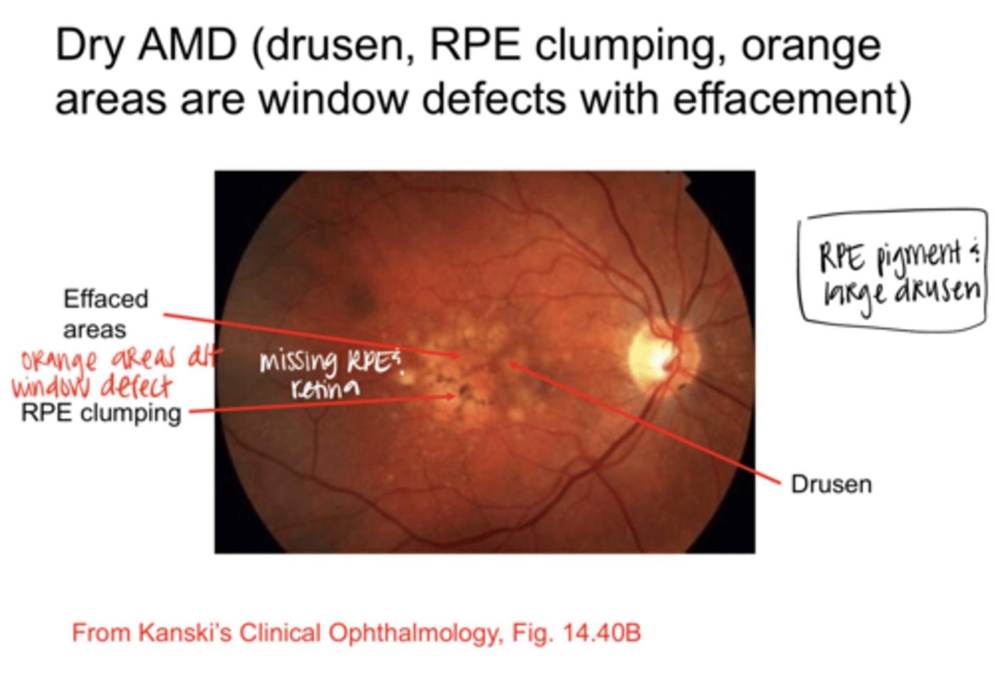
What size are the drusen in this pic? (see pic)
Large

What are effaced areas?
areas of missing RPE and retina (photoreceptors)

What color are effaced areas of AMD?
orange

What is the end stage of Dry AMD?
Geographic atrophy

What are characteristics of geographic atrophy?
choroidal BVs are showing through the RPE window defect & outer retina (possibly inner retina?)

Is vision good in this patient? (see pic)
No, the fovea is involved and VA will be terrible (20/400ish)
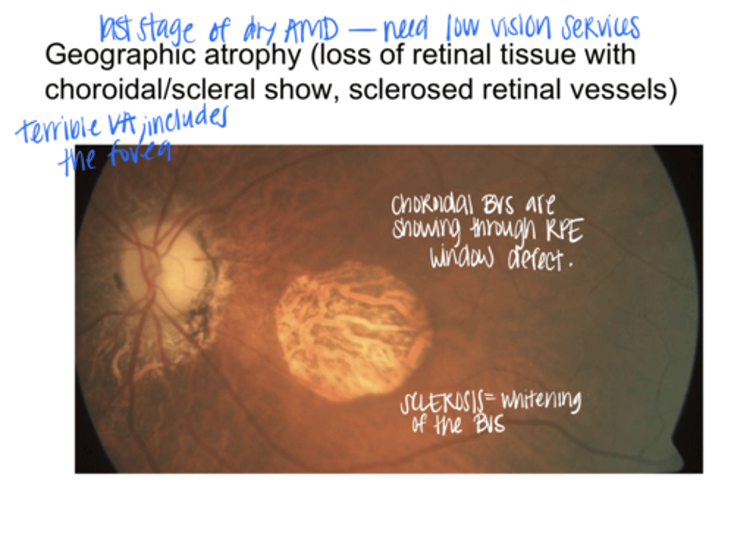
Whitening of BVs d/t what in geographic atrophy?
sclerosis
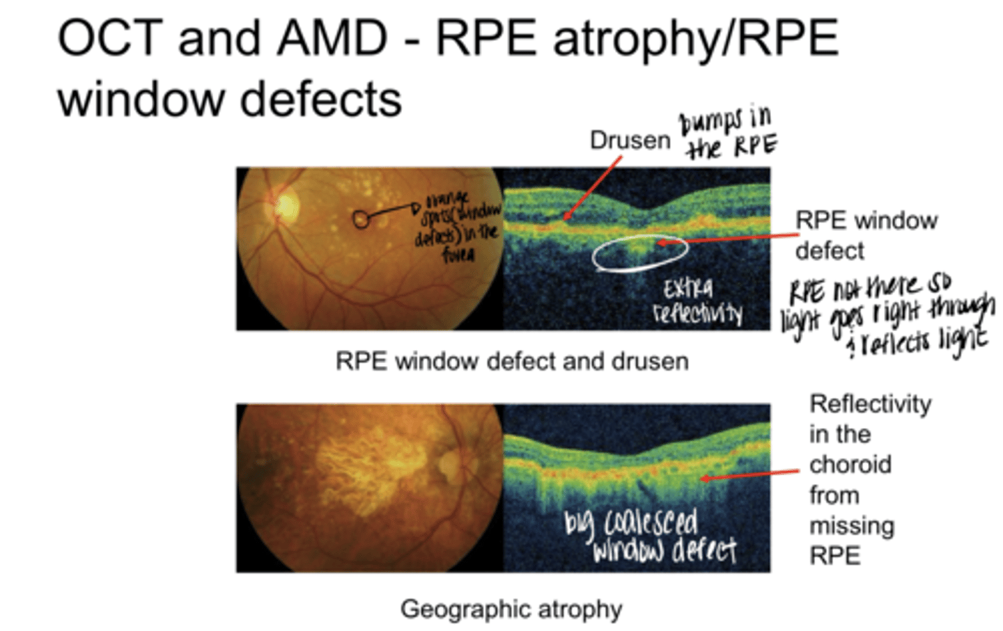
OCT and AMD -- RPE Atrophy and RPE Window Defects (Pic)

A RPE Window defect will be (hyper/hypo)reflective on OCT
hyper

Drusen will be (hyper/hypo)reflective on OCT
hyper

Is the RPE very thick in this patient? (see pic)
No -- reflectance off sclerosed vessels

What is present in this patient? (see pic)
Geographic atrophy -- coalesced window defects, shown by extra reflectivity in the choroid from missing RPE
EXAM QUESTION: Changes of dry macular degeneration summary?
RPE clumping
RPE Dropout/Window Defects
Drusen
Geographic atrophy
REVIEW: What color is RPE clumping?
dark pigment
REVIEW: What color is RPE dropout?
yellow or orange if effaced
REVIEW: What color is drusen?
dull yellow
REVIEW: Small, well defined drusen are (hard/soft) drusen
hard drusen
REVIEW: large, more poorly defined drusen are (hard/soft) drusen
soft

RPE/Retinal Detachment -- Serous Elevation from Choroidal Neovasc (Wet AMD) or Serous Fluid from Choriocapillaris (Dry AMD)
RPE/Retinal Detachment -- Serous Elevation from Choroidal Neovasc (Wet AMD) or Serous Fluid from Choriocapillaris (Dry AMD)
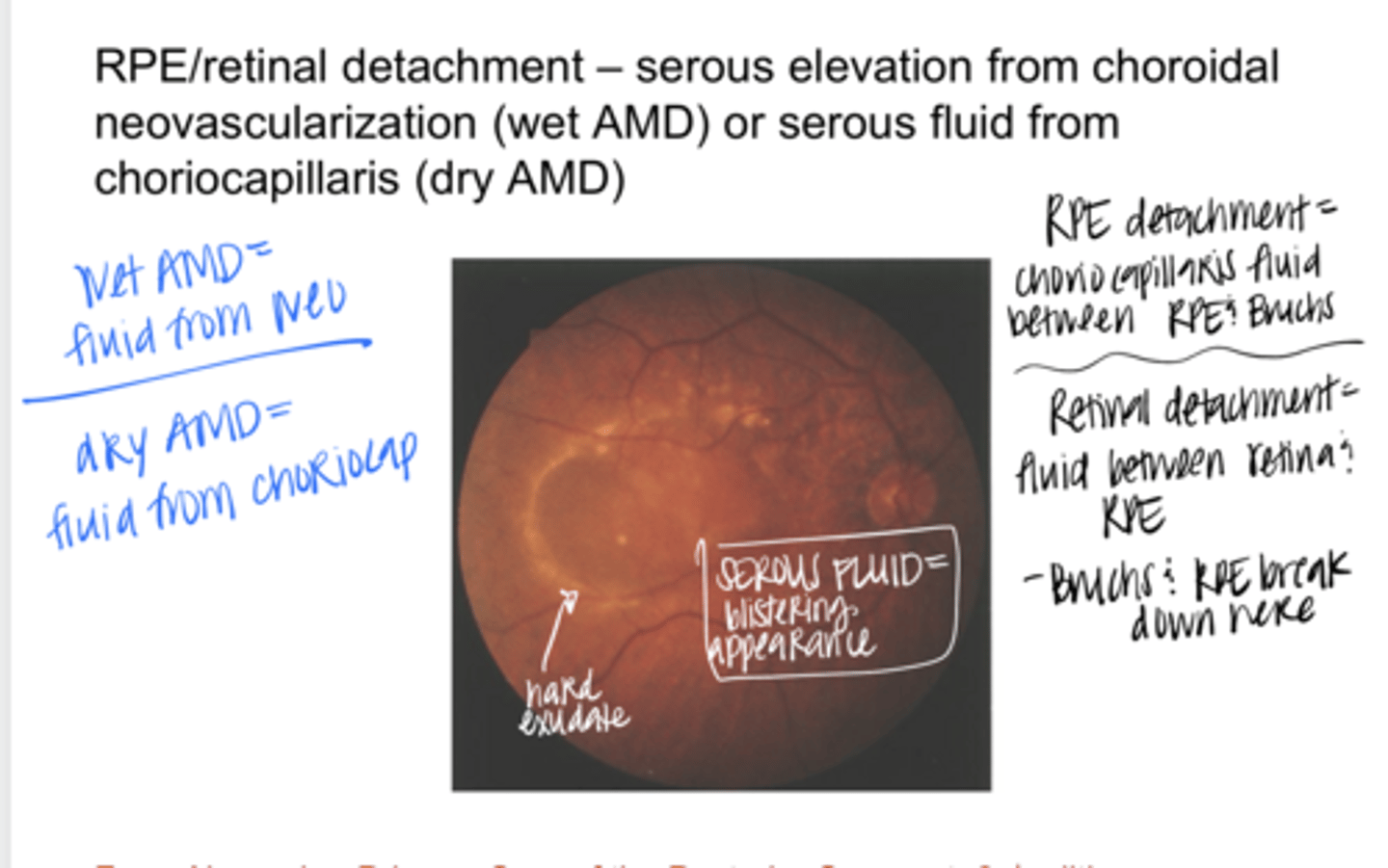
Is there fluid present in this retina? (see pic)
Yes -- blister like appearance

WET AMD Source of Serous Fluid
choroidal neovasc

DRY AMD Source of Serous Fluid
from choriocapillaris

Serous RPE Detachment (PED) with Small Serous Retinal Detachment (Pic)
Serous RPE Detachment (PED) with Small Serous Retinal Detachment (Pic)
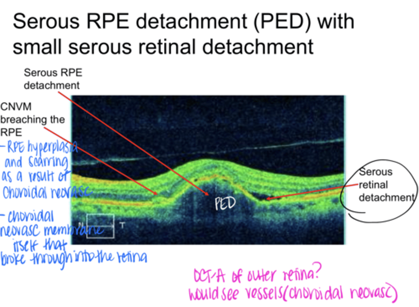
Where is the RPE Detachment in this pic?
See pic

Where is the serous retinal detachment in this pic?
See pic

What is the extra hyper-reflectivity in this pic near the RPE?
-RPE hyperplasia and scarring (result of choroidal neovasc)
-Choroidal neovasc membrane itself that has broken through the retina

If you did an OCT-A of this patient in the outer retina, what would you see?
Choroidal neovasc

Wet AMD w/ Subretinal Hemorrhage (Bright Red) & Sub-RPE Hemorrhage (Darker) (Pic)
Wet AMD w/ Subretinal Hemorrhage (Bright Red) & Sub-RPE Hemorrhage (Darker) (Pic)

How do we want to catch edema in the macula?
with OCT or Amsler grid
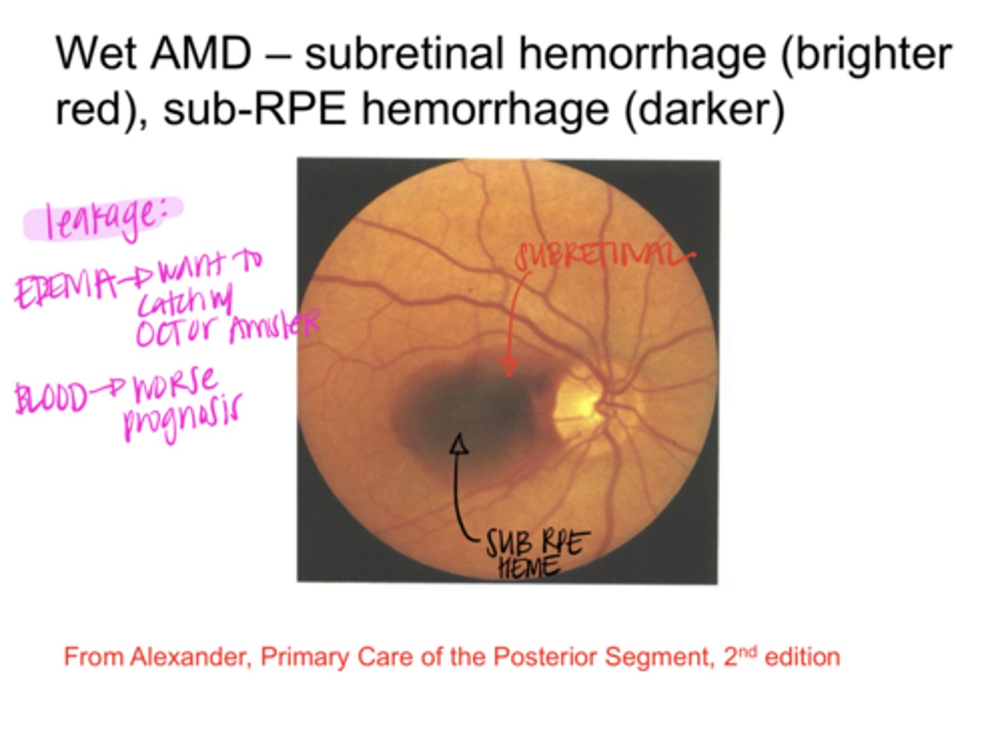
Blood in the retina leads to (better/worse) prognosis
worse

Darker blood in Wet AMD is indicative of a (subretinal heme/sub-RPE heme)
sub-RPE heme

Brighter blood in Wet AMD is indicative of a (subretinal heme/sub-RPE heme)
subretinal heme
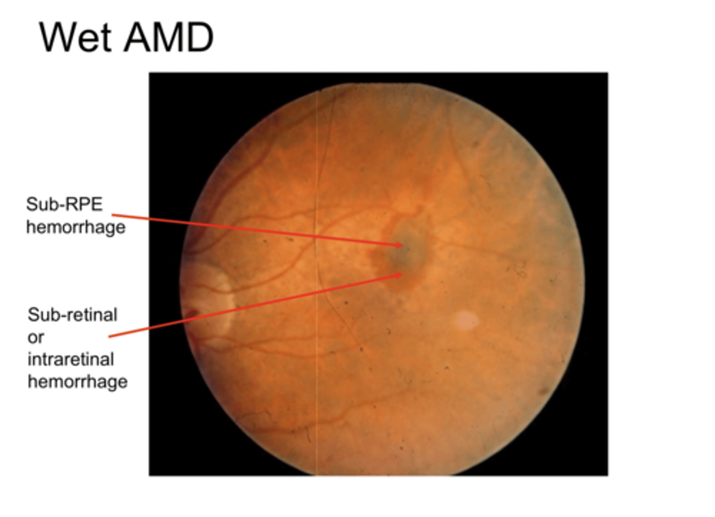
Wet AMD (Pic)
Wet AMD (Pic)
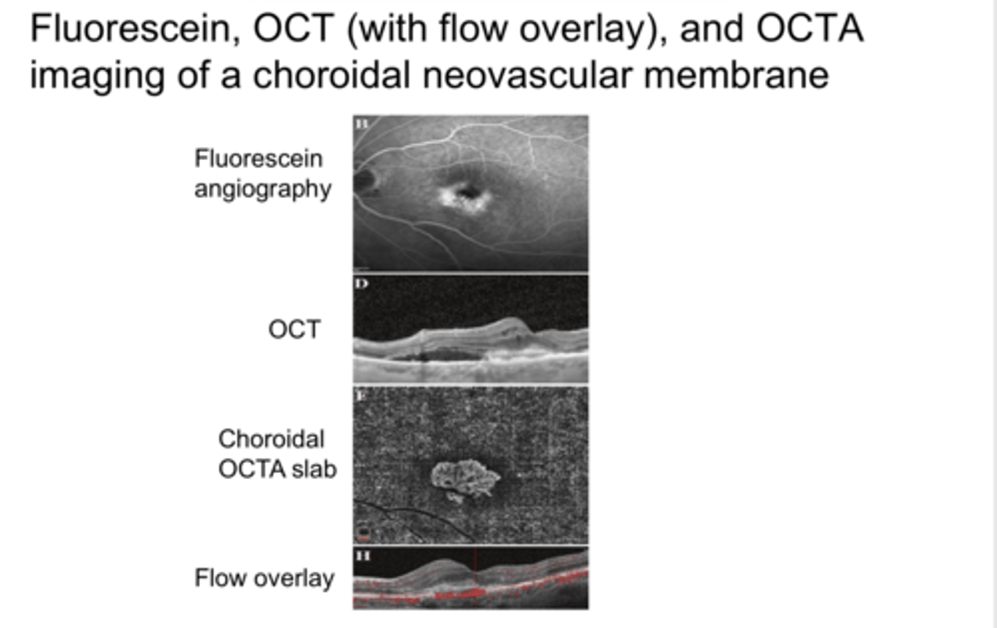
Choroidal Neovasc will be (hyper/hypo) fluorescent on FA
hyper

Choroidal Neovasc will be (hyper/hypo)reflective on OCT
hyper

Choroidal Neovasc presentation on OCT-A?
Neovasc vessels present in the choroidal OCT-A Slab
Classifications of Classic Choroidal Neovasc Membrane (CNVM) OR Type 1 Choroidal Neovascularization
-Well demarcated hyperfluorescence on FANG
-Subretinal Neovasc
Classifications of Occult Choroidal Neovasc Membrane (CNVM) OR Type 2 Choroidal Neovascularization
-Not well demarcated on FANG (diffuse fluorescence)
-Sub-RPE Neovasc
Classifications of Type 3 Choroidal Neovascularization
Starts in the retina and grows into the choroid

Classic vs Occult Choroidal Neovasc (Pic)
Classic vs Occult Choroidal Neovasc (Pic)
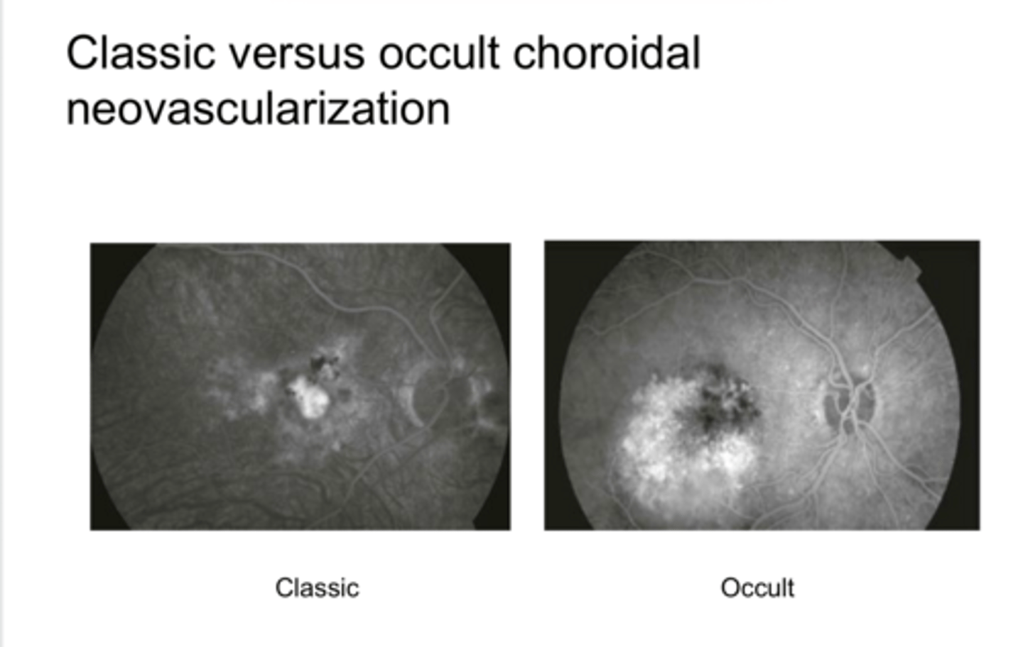
What does Classic (Type 1) Choroidal Neovasc Membrane look like on FA?
-hyperfluorescent
-well demarcated

What does Occult (Type 2) Choroidal Neovasc Membrane look like on FA?
-hyperfluorescent
-not well demarcated