Ultimate Exposures Review: Test 4 Geometric detail and distortion
1/87
There's no tags or description
Looks like no tags are added yet.
Name | Mastery | Learn | Test | Matching | Spaced | Call with Kai |
|---|
No analytics yet
Send a link to your students to track their progress
88 Terms
When a patient moves under his/ her conscious will either because he/ she can’t or isn’t willing to cooperate, it’s called ____ motion.
Voluntary
When motion is present on a radiograph and the cause of the motion is physiological function of the body, it’s called ______ motion.
Involuntary
Which of the following sets of exposure factors would be least likely to result in motion on the radiograph?
200 mA, 1/1000 sec., 100 kVp (least amount of exposure time)
To avoid shape distortion, what items must be parallel to one another?
1- CR
2- long axis of the part of interest
3- IR
4- tube face/ window
2,3,4
When shape distortion occurs and the mage appears smaller in one axis than the actual object, ______ is present. If the image appears larger in one axis than the actual object, _____ is present.
Foreshortening, elongation
Image sharpness or unsharpness can be determined by the number of ____ per mm.
Line pairs
What is the single most detrimental factor contributing to a lack of image sharpness?
Motion
The ____ unsharpness in a radiograph is the unsharpness caused by the image receptor system.
Material
Which of the following factors creates penumbra?
1- OID
2- SID
3- FS
4- intensifying screen
5- grid
3
A radiographic procedure performed by placing the object half way between the FS and the film in order to see structures that are too small to see by normal radiographic measures is called ______.
More than one, but not all of the above
Fractional focal spots are normally ______ mm in size.
None of the above
Which of the following contribute to magnification in the image?
1- long OID
2- short SID
3- large FSS
4- improper tube, part, film alignment
1,2,4
The two types of motion are _____ and ______.
Voluntary
Involuntary
If you could only pick one way to reduce either type of motion, what would it be?
It would be to reduce exposure time
When phototiming or fluoroing, the mA station should be ______ as much as possible when motion is a concern.
Increased
Give three types of immobilization devices:
sandbags
Velcro straps
Pediatric immobilizers (pig-o-stat)
What does “FSS” stand for?
Focal spot size
As FSS increases, unsharpness in the image _______.
Increases
The area on the anode bombarded by electrons is called the _____ FS.
Actual
The ______ FS is the FS as seen by the film which determines image sharpness.
Effective
As SID is increased, image sharpness ______
Increases
As OID is decreased, image unsharpness ______.
Decreases
Another term for size distortion is _____.
Magnification
Give the two types of shape distortion
elongation
Foreshortening
As distortion in the image increases, what happens to image sharpness? It _______
Decreases
A line of density and a space is referred to as a _____.
Line pair
What’s the only way to eliminate involuntary motion?
Short exposure time
Give two ways to eliminate voluntary motion
clear instructions
Immobilization
The blotchy, grainy, mottled pattern in a radiograph caused by an insufficient quantity of photons used to provide a given density is called __________.
Quantum mottle
Quantum mottle is usually attributable to the use of ______.
Low mAs
What three factors affect geometric unsharpness? ( List in order from least to most critical)
SID
FSS
OID
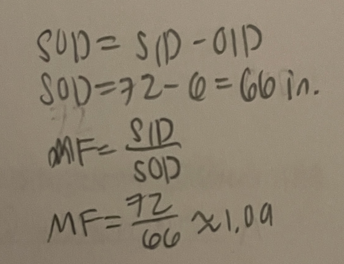
Give the formula for finding SOD.
SOD=SID-OID
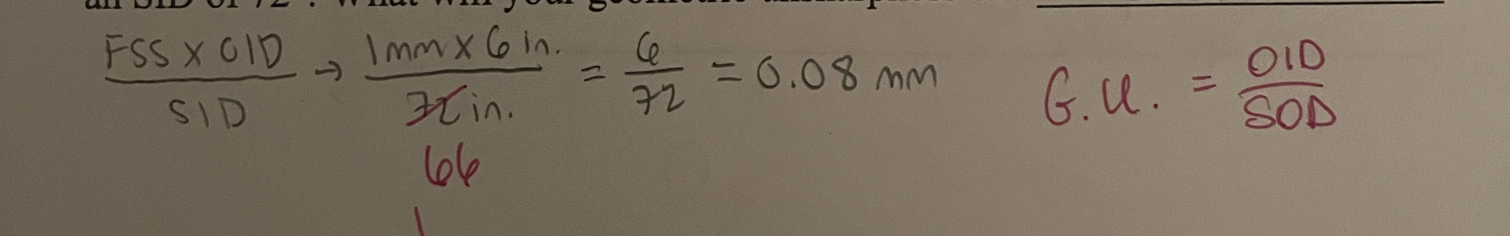
You’re doing a portable lateral C-Spine. You’re using a FSS of 1 mm, and OID of 6”, and an SID of 72”. What will your geometric unsharpness be?
0.1
You’re doing a portable lateral C-Spine. You’re using a FSS of 1 mm, and OID of 6”, and an SID of 72”. What will the magnification factor be?
1.09
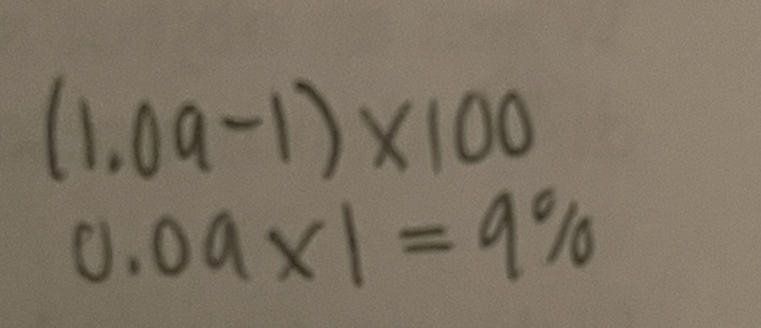
You’re doing a portable lateral C-Spine. You’re using a FSS of 1 mm, and OID of 6”, and an SID of 72”. What will the magnification % be?
9%
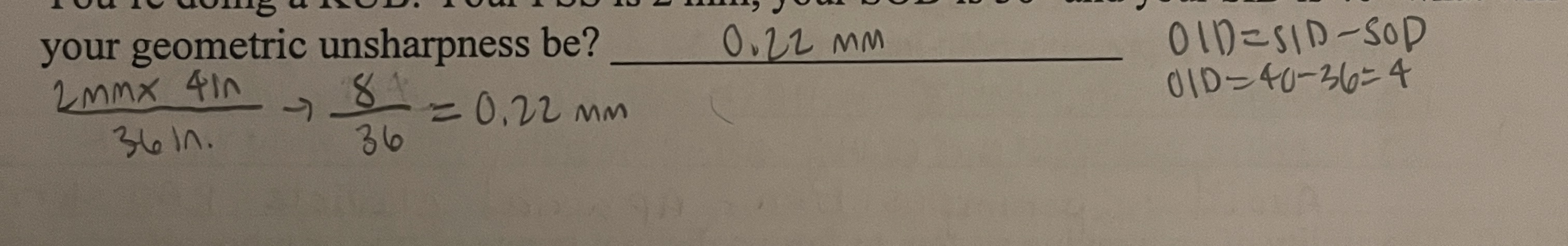
You’re doing a KUB. Your FSS is 2 mm, your SOD is 36” and your SID is 40” what will your geometric unsharpness be?
0.22 mm
The unsharp area around the structures in the image is termed _____.
Penumbra
Double focus tubes enable the RT to choose a _______.
Larger or smaller focal spot size
As OID increases, image unsharpness _____.
Increases
As SID increases, image sharpness _______.
Increases
If an object in the radiograph has increased in length and width by the same proportion, _____ distortion has occurred.
Size
Shape distortion is also called ______ distortion.
True
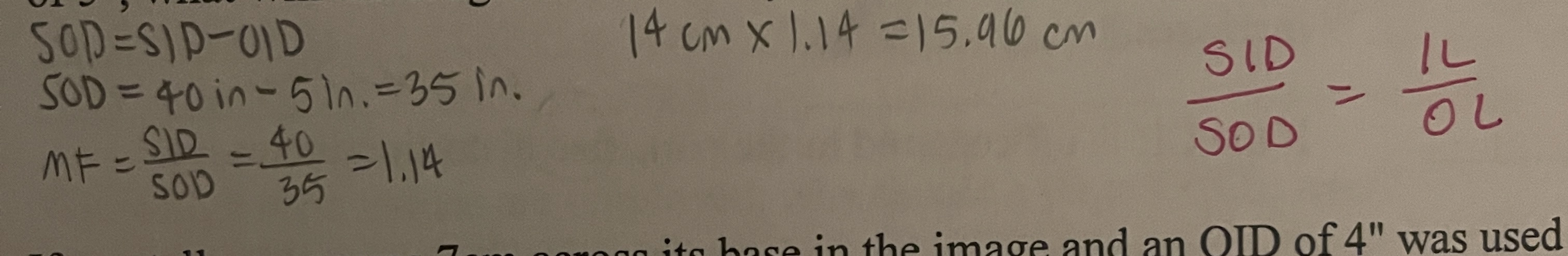
If a kidney measures 14 cm in length and it is radiographed at an SID of 40” and an OID of 5”, what will the length of the kidney in the image be?
15.96 cm
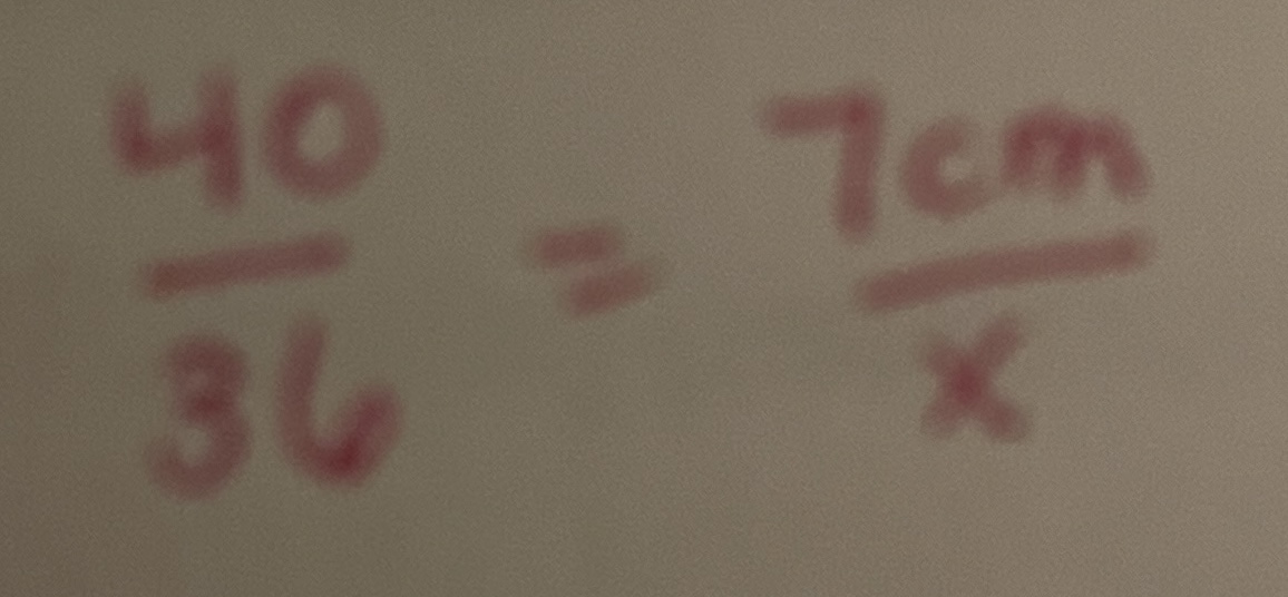
If a patella measures 7 cm across its base in the image and an OID of 4” was used and an SID of 40” was used, what is the true width of the base of the patella?
6.3 cm
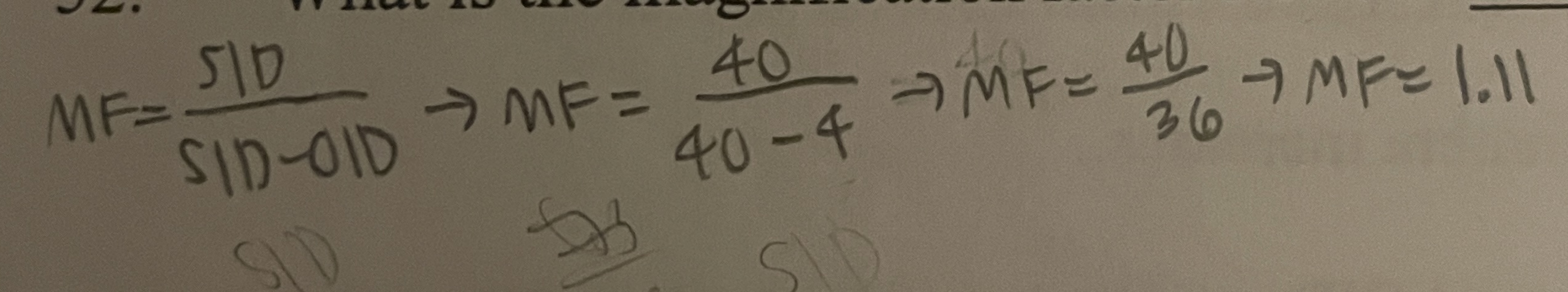
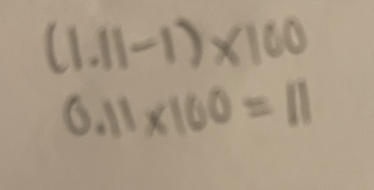
If a patella measures 7 cm across its base in the image and an OID of 4” was used and an SID of 40” was used, what is the magnification factor?
1.11
If a patella measures 7 cm across its base in the image and an OID of 4” was used and an SID of 40” was used, what is the magnification %?
11%
What must be used when macroradiography is utilized to restore sharpness to the image?
A fractional focal spot
When an objects shape has been misrepresented in the image, ______ distortion is present.
Shape
If the part can’t be placed parallel to the film, what must be done with the CR?
The CR must be angled perpendicular to the part
Give two instances where shape distortion can be used advantageously and give a projection that represents both instance.
To visualize joint spaces more clearly- AP/ AP oblique projection of the knee
Avoid superimposition - AP axial clavicle, RAO sternum
The ability of an imaging system to form a distant image of two or more objects located close together is termed ________, and is measured in _______ (units).
spatial resolution
lp/mm
True or false:
The CR should be centered to the film, regardless of where the part is on the film in order to maintain shape distortion.
False
True or false:
SID has a greater effect on size distortion than OID
False
True or false:
As SID decreases, size distortion increases and sharpness of detail decreases.
True
True or false:
As OID decreases, size distortion increases and sharpness of detail increases
False
True or false:
If the CR is off centered to the part, size distortion increases
False
True or false:
The penumbra in an image is more noticeable about the periphery of the image than in the center of the image.
True
True or false:
As SID increases, penumbra decreases
True
True or false:
As OID decreases, penumbra decreases
True
True or false:
As FSS increases, penumbra increases
True
True or false:
It is best to use the shortest OID and the longest practical SID when concerned with magnification
True
True or false:
The more lp/mm that can be clearly resolved when using a resolution test template pattern, the sharper the image.
True
True or false:
As FSS increases, size distortion increases
False
True or false:
Motion decreases unsharpness
False
Why should changes in SID or OID be noted on the radiograph and requisition?
To ensure accurate measurement and reproducibility of the radiograph, as they affect magnification and image quality
Why is explaining the procedure to the patient important?
Helps reduce anxiety, ensures cooperation, and decreases voluntary motion
When should you immobilize and how much immobilization is necessary?
When the patient is unable to remain still and the amount should be just enough to prevent movement without causing discomfort or injury
Give one advantage and disadvantage to decreasing SID.
Advantage: decreases patient exposure
Disadvantage: increases size distortion and decreases image sharpness
What would be a disadvantage to increasing kVp to alter your exposure time?
It can reduce image contrast, leading to a more flat image
When high mAs values are employed, why should a large FSS be used?
To prevent heat buildup and ensure safe operation of the xray tube
Explain why it is important to keep the CR in the center of the area of interest.
It ensures optimal image clarity and minimizes shape distortion
Modulation transfer function
Measures the inevitable loss of information and sharpness of detail between the beam that leaves the body and the beam that records the image. Measures the limitations of imaging systems as they relate to material unsharpness
Resolution
The ability of an imaging system to record, as separate images, a specific number of lines within a limited space
Line spread function
Evaluates the imaging systems ability to record an extremely narrow beam of X-rays passing through a slit 10 microns wide
Aliasing (Moire pattern)
Occurs when the spatial frequency exceeds the Nyquist frequency and incoming data is sampled less than two times per cycle.
Photographic properties
Visibility of detail
Geometric properties
Sharpness of detail
Contrast
Visibility of detail
Density
Visibility of detail
Definition of detail
Sharpness of detail
Distortion
Sharpness of detail
Which of the following setups will result in the best possible sharpness of detail?
No grid, direct exposure
Which FS is the smallest?
Actual
Which would provide the best sharpness of detail for a lumbar spine - AP or PA?
AP because of decreased OID
Decreasing the exposure time and increasing mA to maintain density is obviously the easiest way to reduce motion on a film and is also the best way as it has negligible effects on other image qualities. But, what if you use the highest mA possible and exposure time still needs to be reduced? Give two other ways you could decrease exposure time to maintain density.
increase kVp
Decrease SID
Give an exam in which macroradiography would be utilized.
Angiography
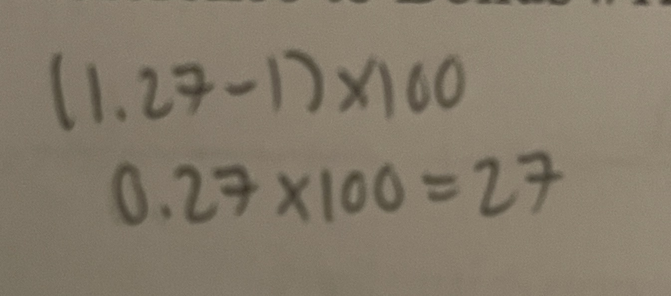
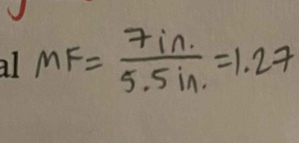
If an object imaged at 4” OID measures 7” long in the image and it is 5.5” in actual length, what is its magnification factor?
1.27
If an object imaged at 4” OID measures 7” long in the image and it is 5.5” in actual length, what is The magnification %?
27%