Thalamus
1/27
There's no tags or description
Looks like no tags are added yet.
Name | Mastery | Learn | Test | Matching | Spaced | Call with Kai | Chat |
|---|
No analytics yet
Send a link to your students to track their progress
28 Terms
Describe the Thalamus
Comprised of?
Function?
Comprised of:
dorsal thalamus + thalamic reticular nucleus (ReT)
Functions:
Conveys Info (highway)
sensory pathways → cerebral cortex
(except for olfaction)
Lower centers → cerebral cortext
Lower centers ex: basal ganglia, cerebellum, hypothalamus
To areas involved in attention and executive functions.
Gate Info transfer → cerebral cortex
Coordinates cortical arousal
Describe the connections between the thalamus and the cortex projections
Thalamus Nuclei (except ReT) and Cerebral Cortext = reciprocally connected
Via Excitatory projection neurons
Corticothalamic fibers → individual thalamic nuclei
modulatory inputs (info processing)
Corticothalamic > thalamocortical projections
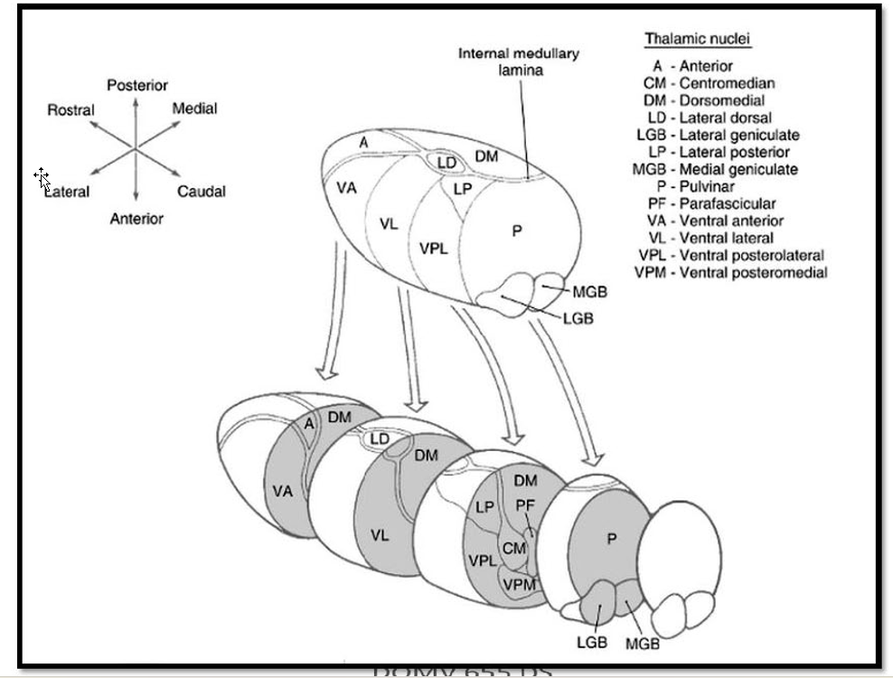
Draw out the thalamic nuclei
Describe the Differences between specific and non-specific:
Function
Electrical stimulation produces?
Specific Vs Non
Specific
send efferent (thalamocortical fibers) → specific functional cerebral cortical areas → relay information to these areas.
Electrical stimulation → rapid, localized responses in ipsilateral cortex.
Non-specific
send efferent → widespread areas of cortex → more generalized cortical activation.
Electrical stimulation: → widespread activity in both hemispheres w/ longer time delay.
What are Relay Nuclei?
A/E?
Location
Consists of?
Relay Nuclei:
type of specific nuclei
A/E:
A: subcortical inputs
(sensory pathways, cerebellum, basal nuclei, mammillary bodies)
E: primary cortical areas (e.g., Sl, VI, MI)
represent final segment of pathways conveying sensory info to cortex
Location:
Anterior/Lat. Thalamus
Consist of:
Ventral Posterior nucleus: Somatosensory
Lateral Geniculate Nucleus: Vision
Medial Geniculate Nucleus: Audition
Describe the Ventral Posterior Complex (VP):
VPL vs VPM’s site of termination
Projects to? location?
VP:
VPL (ventralpostolateral):
site of termination of the somatosensory pathways of the body
VPM (ventralposteromedial):
site of termination for trigeminothalamic pathways (face/oral cavity)
Projects to:
primary somatosensory cortex (Sl)
in postcentral gyrus and posterior paracentral gryus of the parietal lobe.
Describe the VPI
Location
Function? What also have similar function?
Projections
ventral posterior inferior nucleus (VPI)
Location: small area in between VPL and VPM
Function: receive vestibular Info
***NOTE: (oral part of VPL also receives this)***
Projections:
to parieto-insular vestibular cortex (PIVC) @ posterior end of insula
Vestibular Cortex of parietal lobe
@ depths of central sulcus (3a)
@ rostral tip of intraparietal sulcus (2v).
Describe the VPMpc
AKA?
Afferent
Efferents?
medial parvicellular portion of the ventral posteromedial nucleus (VPMpc)-
AKA: ventromedial basal (VMb) nucleus-
Afferents:
solitary nucleus
Taste (directly)
GVA (via parabrachial nucleus)
NOTE: parabrachial nucleus surrounds the SCP in dorsal lat. Pons
Efferents:
Taste:
gustatory cortex of inner frontal operculum and insula
GVA:
Insula
GI tract, cardiovascular input, and respiratory input represented sequentially caudal to the taste representation.
Describe the VMpo
Location:
A/E
ventromedial posterior thalamic nucleus (VMpo)
Location: More caudally in ventral medial thalamus
Afferent:
Pain/Temp from Anterolateral Tract
Efferent:
dorsal posterior insula
caudal to viscerosensory representation in the insula
What is the interoceptive Cortext?
The dorsal insula receiving input from VMb and VMpo can be considered "interoceptive cortex" representing the physiological condition of the body.
What constitutes the metathalamus?
The lateral and medial geniculate bodies constitute the metathalamus and may be considered the caudal continuation of the ventral nuclear mass of the lateral thalamus.
Describe the LGN:
Location
A/E
Describe the MGN:
A/E
Lateral Geniculate Nucleus (LGN):
Location:
small, rostrolaterally directed projection from the posterior thalamus
A/E:
A: retina of both eyes
E: primary visual cortex (VI)
Found on banks of the calcarine sulcus on the medial surface of the occipital lobe
Medial Geniculate Nucleus (MGN):
A/E:
A: inferior colliculus via its brachium
E: primary auditory cortex (A1)
associated w/ transverse temporal gyri of Heschl
List the nuclei associated w/ the motor relay nuclei of thalamus
Ventral Anterior nucleus (VA)
Ventral Lateral nucleus (VL)
Describe the VA
A/E
Function?
ventral anterior nucleus (VA)
Afferents:
basal nuclei
internal segment of the globus pallidus
Targets parvocellular parts of VA
pars reticulata
Targets medial magnocellular part
Efferent:
frontal eye fields (BA 8) + prefrontal cortex
Function:
Contributes to loop circuits of basal nuclei system (oculomotor, motor, associative)
Involved in motor planning and behavior.
Describe the VL
Afferent?
VLO vs VLp:
A/E
Function
ventral lateral nucleus (VL)
Afferent:
basal nuclei + cerebellum.
VLO (VLa)
Rostral part of VL
A/E:
A: GPi
E: premotor cortices, including SMA.
Function:
Contributes to motor loop circuit of basal nuclei,
involved in motor planning.
VLp:
Caudal Portion
A/E:
A: deep nuclei of cerebellum
E: M1
Function:
Contributes to cerebellar circuits
modulating motor activities, e.g., limb movements.
Describe the Limbic Nuclei
Function
Consists of?
Limbic Nuclei:
Function:
contribute to memory circuitry of the limbic system.
(Circuit of Papez)
Consists Of:
Anterior nuclear group
Lateral dorsal nucleus (LD)
***NOTE: LD nucleus can be considered a dorsal extension of the anterior nucleus and has similar limbic connections.***
Describe the Anterior Nuclear Group
Comprised of?
Location
A/E
anterior nuclear group
Comprised of:
three nuclei enclosed by the split internal medullary lamina
NOTE: refered collectively as anterior nucleus
Location:
caudal lateral to interventricular foramen
A/E:
A: hippocampal formation
Via fornix/Mammillary bodies (mammilothalamic tract)
E: cingulate cortex of the limbic lobe
Describe Association Nuclei
Function
Location
Properties
Comprised of?
Association nuclei (higher- order relay)
Function:
send projections to association cortices (e.g., parieto-occipital, prefrontal)
relay information from primary cortical + subcortical inputs.
Location:
medial thalamus and posterior part of dorsal thalamus.
Properties:
Largest Nuclei of Thalamus
Strong reciprocal connections
Comprised of:
Mediodorsal nucleus (MD)
Pulivinar
Lateral Posterior nucleus
Describe MD
Function
A/E
Damage =?
mediodorsal nucleus
Function:
Involved in: attention, decision making, behavioral planning
Via connections w/ amygdala interfaces with emotion networks of the limbic system
A/E:
A:
amygdala,
olfactory cortex,
entorhinal cortex,
substantia nigra,
anterolateral system.
E: prefrontal cortext (reciprocal)
Including FEF and Anterior cingulate cortext
Damage =:
executive functions (e.g., judgement, decision making)
affective behaviors.
Describe Pulvinar and LP
Location
A/E
Function
Pulvinar and Lateral posterior nucleus (LP):
Location/Properties:
back of thalamus dorsal and lateral to the midbrain.
merges w/ pulvinar and its caudal borders are difficult to distinguish.
commonly considered with the pulvinar.
Consists of 4 subnuclei
A/E:
A:
superior colliculus
visual cortex of occipital lobe (vision related)
pretectum
unimodal sensory and association cortices of the parietal and temporal lobe.
E:
multimodal association cortices of the posterior parietal lobe and lateral temporal lobe.
Function: spatial attention
Describe the Gating Function of the Thalamus:
Describe the Two Gates
What happens in non-REM sleep vs Wakefullness
Gating Function:
Two Gates:
Tonic Mode:
During Wakefulness/REM sleep:
thalamocortical relay neurons = slightly depolarized via modulatory inputs → tonic discharge → transfer into to cortex
Burst Mode:
relay neurons = hyperpolarized via inhibitory input from the ReT or withdrawal of excitatory inputs from brainstem → Bursting discharge → functional disconnection between thalamic inputs and thalamocortical projections
Non-REM sleep vs Wakefullness:
Non-REM: rhythmic burst discharge,
Wakefullness: many relay neurons = Arrhythmic burst mode;
→ increase signal-to-noise ratio → Novel Stimuli detection
Presumably, attention and analysis of stimulus = switch to tonic mode.
Describe Non-specific Nuclei:
Described as?
Comprised of?
Non-specific Nuclei:
traditionally described as having diffuse projections to cerebral cortex Nuclei
Comprised of:
Midline nuclei (ex: paratenial nucleus and nucleus reuniens)
Intralaminar nuclei
Describe the midline nuclei
Properties
A/E
Reciprocally connected to?
Function
midline nuclei
Properties:
small and difficult to distinguish in humans and relatively poorly understood.
A/E:
A:
hypothalamus,
basal forebrain,
brainstem reticular formation.
E:
ventral striatum
amydala.
Reciprocally connected to:
Limbic cortex (anterior cingulate cortex, entorhinal cortex)
hippocampus
Function:
connections of the midline nuclei suggest limbic functions.
Describe the IL:
Location
Caudal Group
Consists of?
Afferent?
Function
Rostral Group
Afferent
Function
Efferent
Intralaminar nuclei:
Location:
surrounded by internal medullary lamina
Caudal Group:
Consists of centromedian nucleus (CM) + parafascicular nucleus (Pf)
Afferent:
GPi (largely collaterals of projections to VLO).
Function:
participates in basal nuclei circuitry;
Increases Cortical/Striatal neuron excitability → cortico-basal nuclear-thalamocortical loop circuits
Rostral Group:
Afferent:
ascending reticular activing system
anterolateral system
cerebellum
Function:
Regulates arousal + cognitive alertness
IE: an arousal and alerting function.
Efferent:
striatum and widespread areas of cerebral cortex
Describe ReT:
Properties
Afferent
Key Difference
Function
Reticular nucleus (ReT):
Properties:
Derivative of ventral thalamus
Contain GABAergic neurons that project to all thalamic nuclei
Afferent:
collaterals from thalamocortical + corticothalamic projection neurons
Key Difference:
Unlike nuclei of the dorsal thalamus, does NOT project to the cerebral cortex.
Function:
Saliency or focusing on a sensory or motor modality.
By gating activity within thalamic nuclei
synchronization of cortical activity during Non-REM sleep
contributing to sleep spindle + delta wave activity.
Describe the thalamic radiations
What is it?
Types
Projects through?
Describe the four thalamic radiations:
Consists of?
Travels Through?
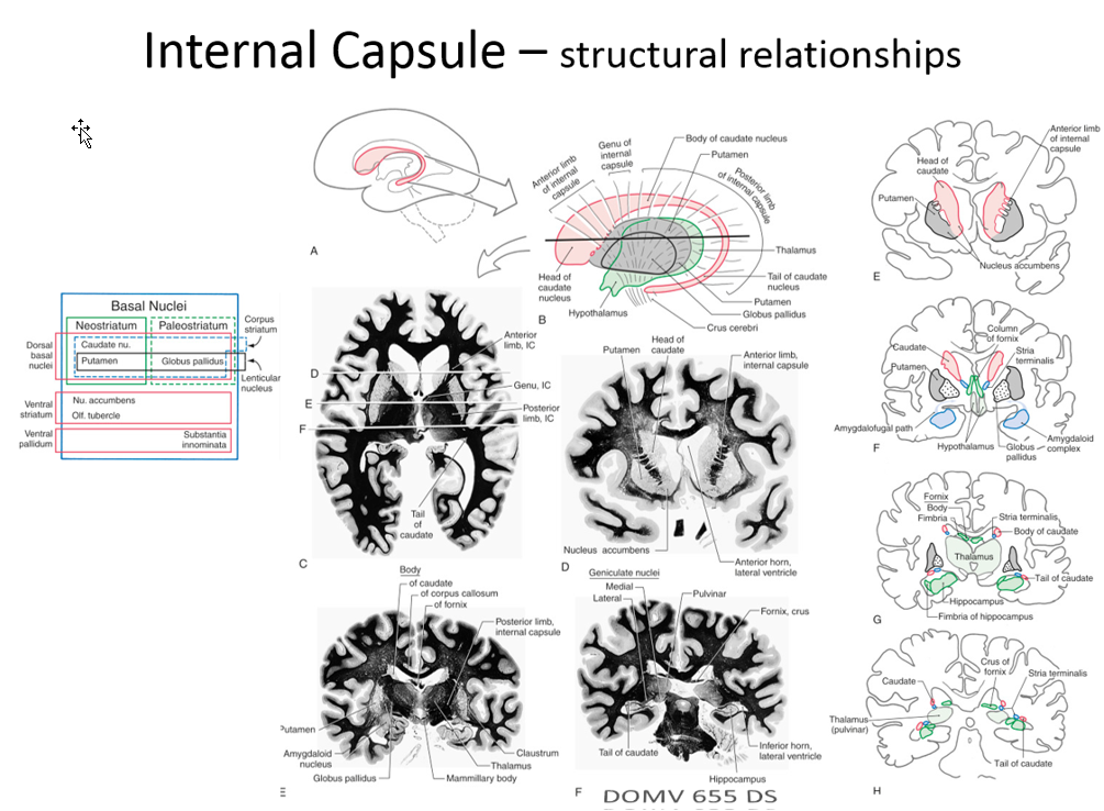
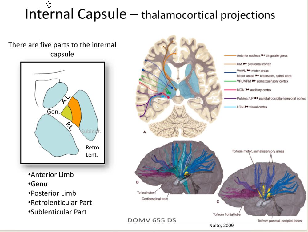
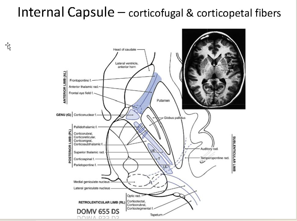
Thalamic Radiations:
What is it?:
fibers connecting the thalamus with the cerebral cortex
Types:
Anterior thalamic radiation
Superior thalamic radiation
Posterior thalamic radiation
Inferior thalamic radiation
Projects through Internal Capsule
Anterior Radiation:
Consists of:
fibers connecting MD + anterior nucleus → frontal lobe and cingulate cortex.
Travels Through:
anterior limb of internal capsule
Superior Radiation:
Consists of
fibers connecting VP → parietal lobe ; VA-VL → frontal lobe.
Travels through
posterior limb of internal capsule.
NOTE: May also be referred to as the central thalamic radiation.
Posterior Radiation: •
Consist of:
fibers connecting LGN → occipital lobe (geniculocalcarine tract- optic radiations)
Travels Through:
retrolenticular part of the internal capsule
Note: Also, contains pulvinar projections to the occipital lobe and posterior parietal lobe.
Inferior Radiation:
Consists of
fibers connecting MGN → temporal lobe (auditory radiations).
Travels through
sublenticular part of internal capsule.
Note: Also, contains pulvinar projections to the temporal lobe.