Clinical Skills Lecture - Exam 2 (derm)
1/128
There's no tags or description
Looks like no tags are added yet.
Name | Mastery | Learn | Test | Matching | Spaced | Call with Kai |
|---|
No analytics yet
Send a link to your students to track their progress
129 Terms
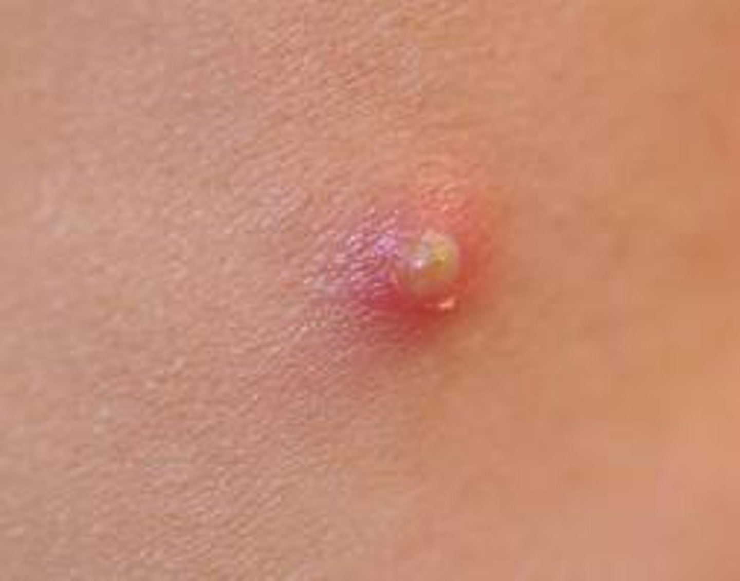
sebaceous (epidermoid) cyst
-closed sac lined w/ epidermis
-filled w/ fluid or semi-solid
-ruptures easy so don't squeeze!
Furuncle (boil)
infected sebaceous cyst
staph
carbuncle
many sebaceous cysts w/ multiple pockets
urticaria (hives)
-pruritic papules, plaques, and wheals
-if severe is angioedema which can be fatal
-IgE-mediated to allergen or idiopathic
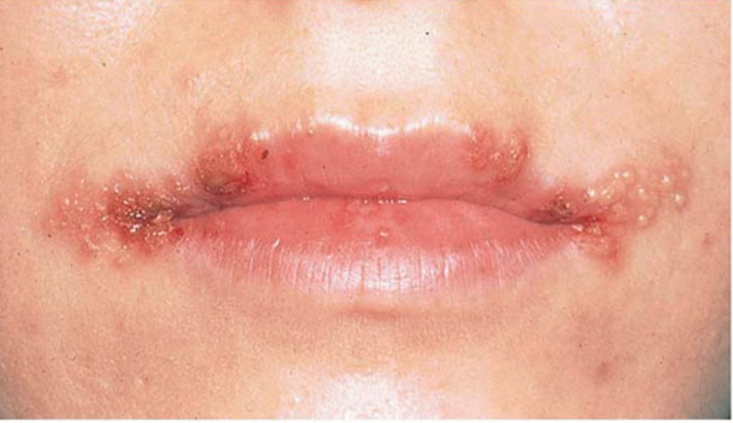
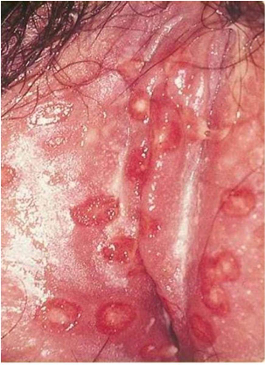
HSV-1
oral
HSV-2
genital
Herpes zoster (shingles)
reactivation of varicella in immunocompromised
-unilateral b/c along dermatomes
-pain before rash occurs
scabies
female mites burrow under skin and lay eggs
-pruritus secondary to sensitization to eggs, mites, and their feces
-worse after hot shower or at night
-extremely contagious
main areas to check for scabies
hands
flexor surfaces
axillae
waist
inner thighs
feet
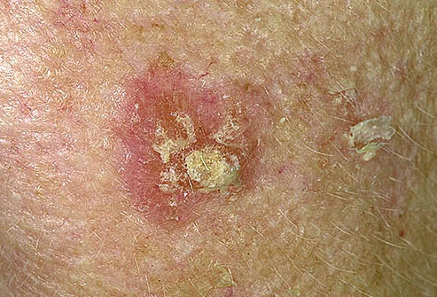
actinic keratosis
rough scaly papule secondary to sun exposure
pre-SCC
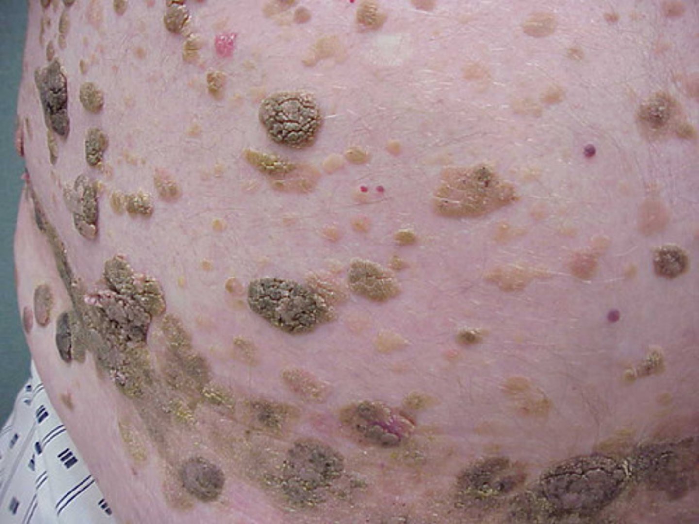
seborrheic keratosis
stuck on papules
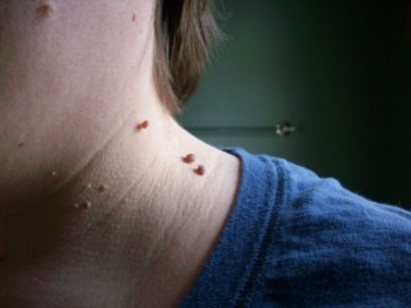
acrochordon (skin tag)
pedunculated papules
lipoma
fatty tumor
benign, soft, rubbery, subq
verruca vulgaris
firm, hyper-keratotic pap
-dots are thrombosed capillaries
-spontaneous resolution
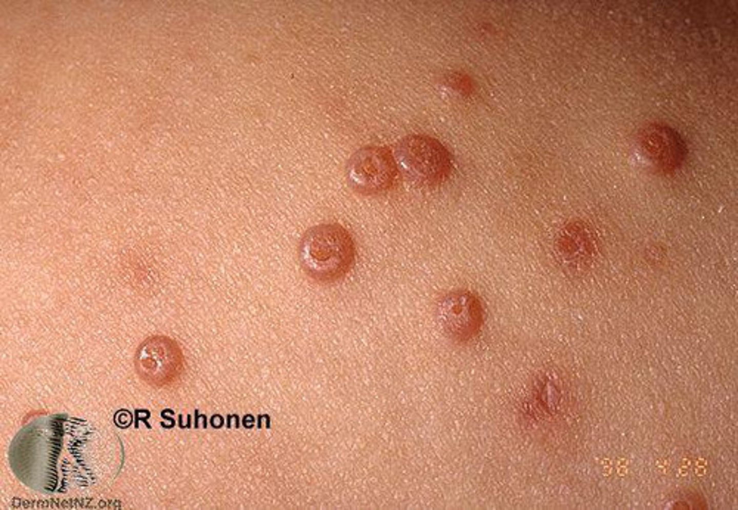
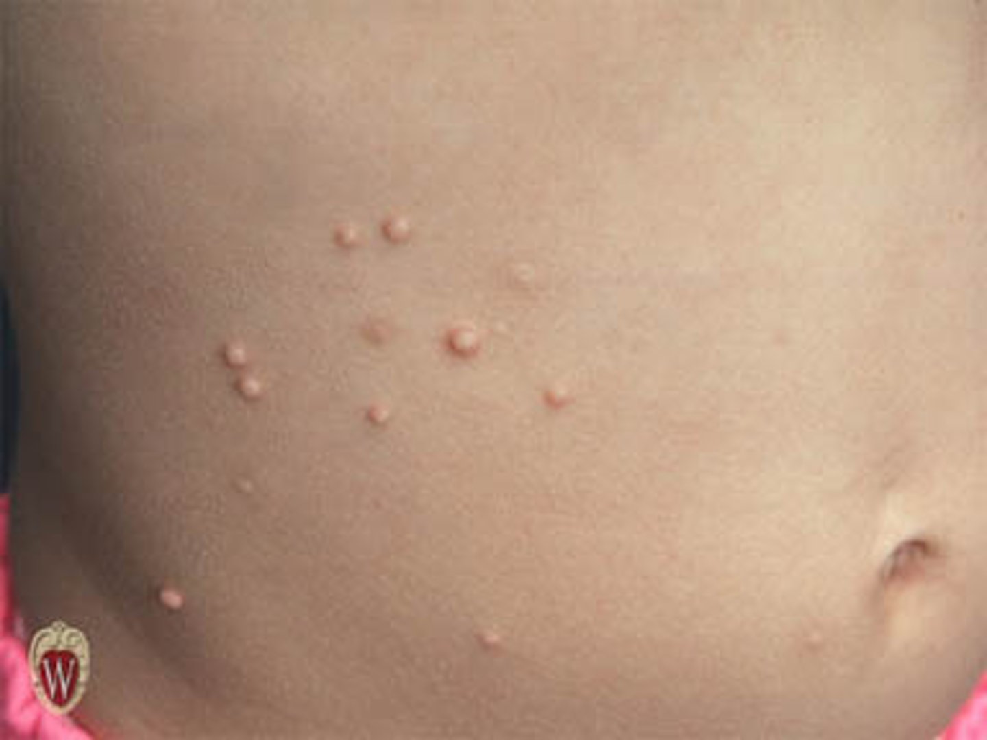
molluscum contagiosum
-epidermal viral inf
-looks like wart but has umbilicated center or white core
-spread via direct contact
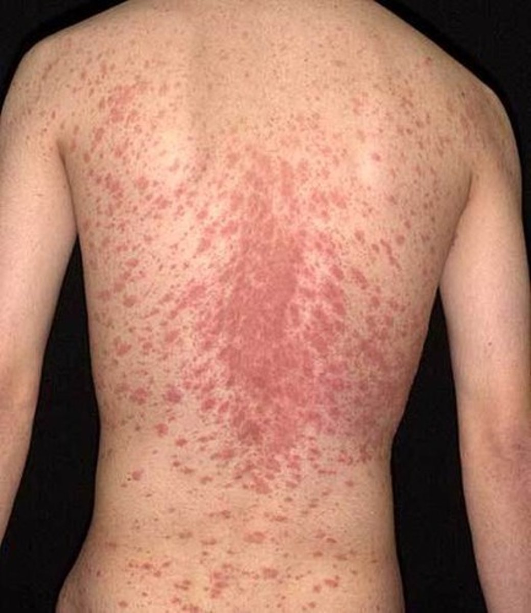
pityriasis rosea
viral infection
-"herald patch" which is salmon colored oval on trunk/arms
-2 weeks later = generalized eruption of papules w/ centripetal scaling
"Christmas tree pattern"
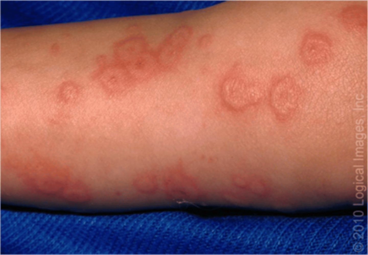
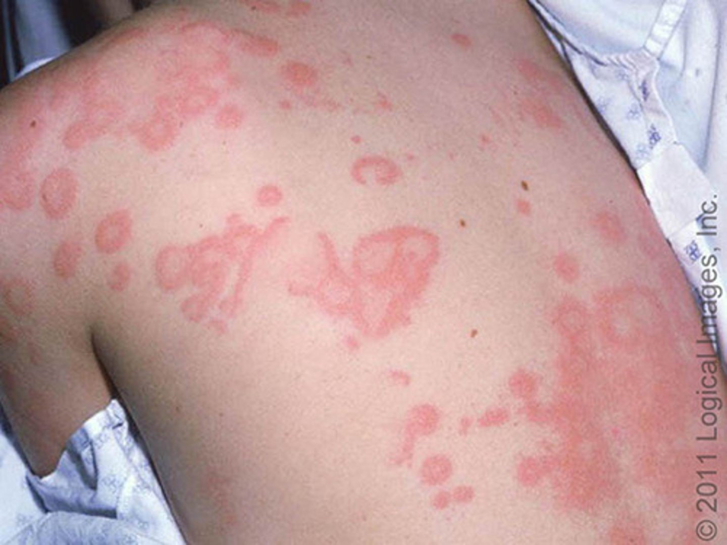
erythema multiforme
reaction of blood vessels in the epidermis and mucous membranes
-drug reaction, infection, idiopathic
-"target lesions" palms, soles, mucous membranes
-resolve in 2-4 weeks
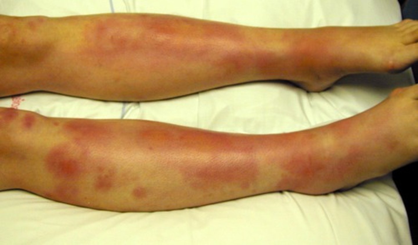
erythema nodosum
large, painful subq nodules w/ overlying erythema
-more in females 15-30
-strep, drug reactions, pregnancy, TB, sarcoidosis, IBS
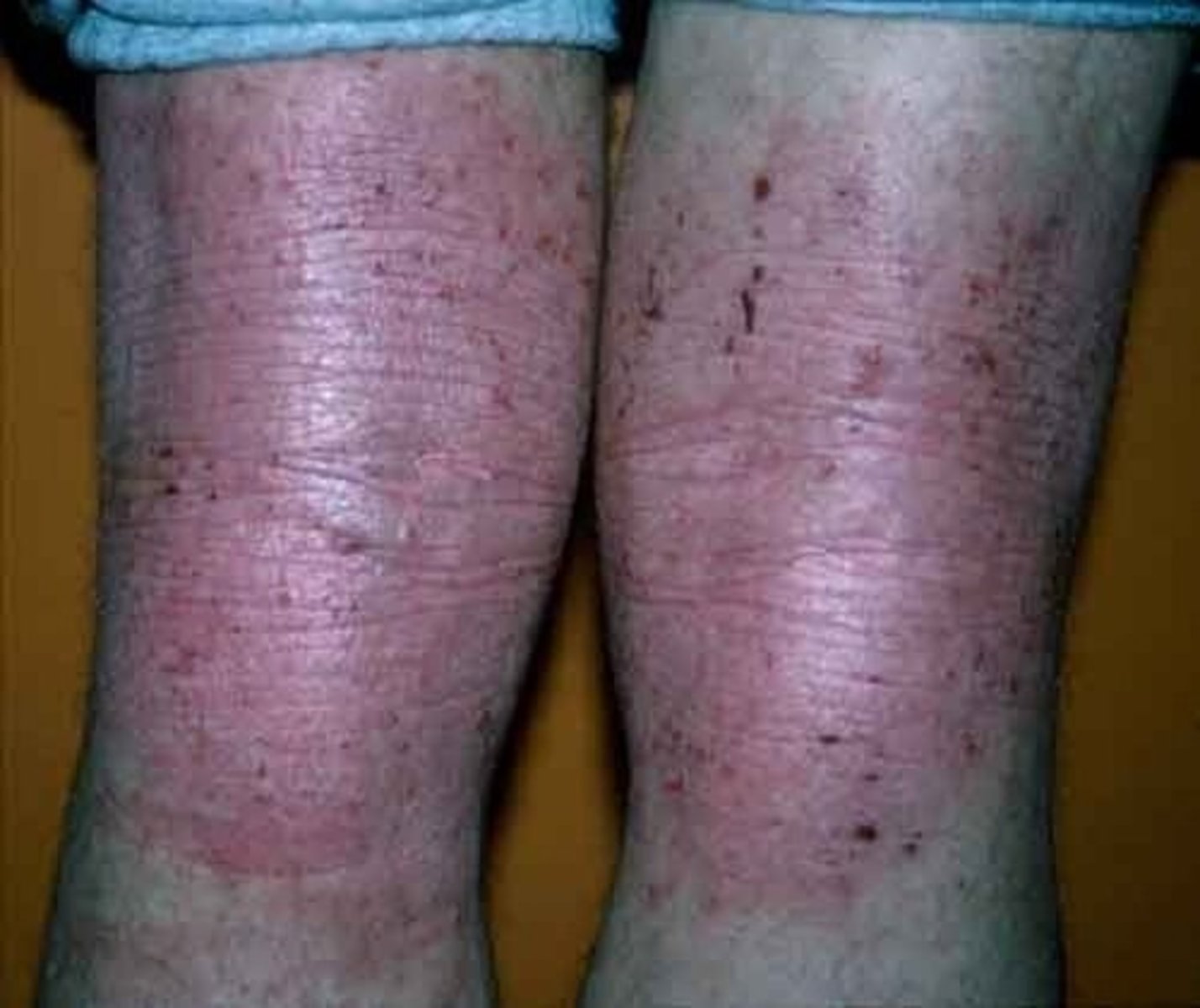
contact dermatitis
pruritic vesicles due to allergic rxn
-plants, nickel, dyes
-immunologic rxn
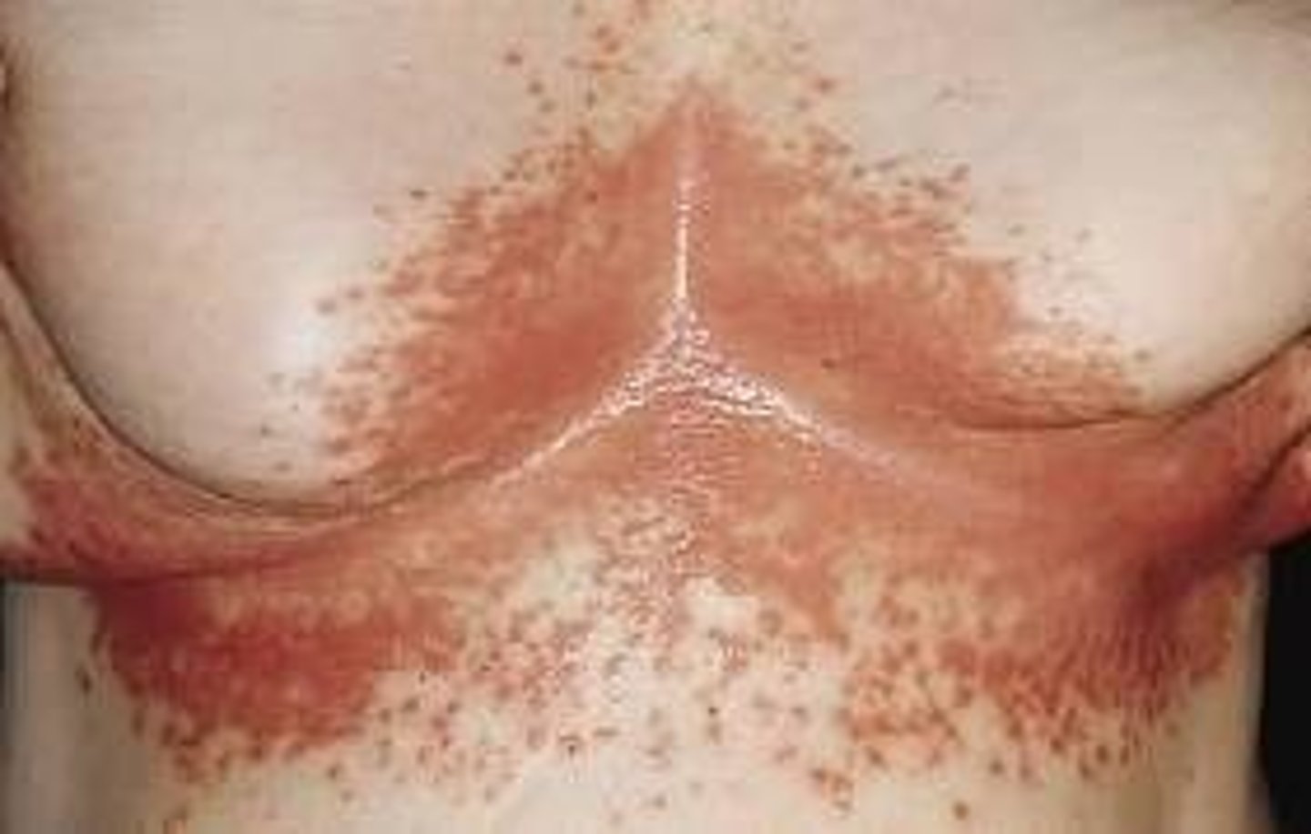
cutaneous candidiasis
moist areas
-more in diabetes and immunocompromised
-pustule progresses to red eroded patch w/ satellite lesions
impetigo
staph or strep
-colonizes in minor breaks in skin
-honey colored crusts
-very contagious
vitiligo
-white macs and patches
-loss of pigment due to lack of melanin
-autoimmune
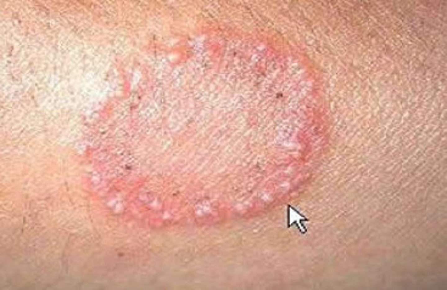
tinea
sharply marginated, peripheral enlargement with central clearing
pruritic
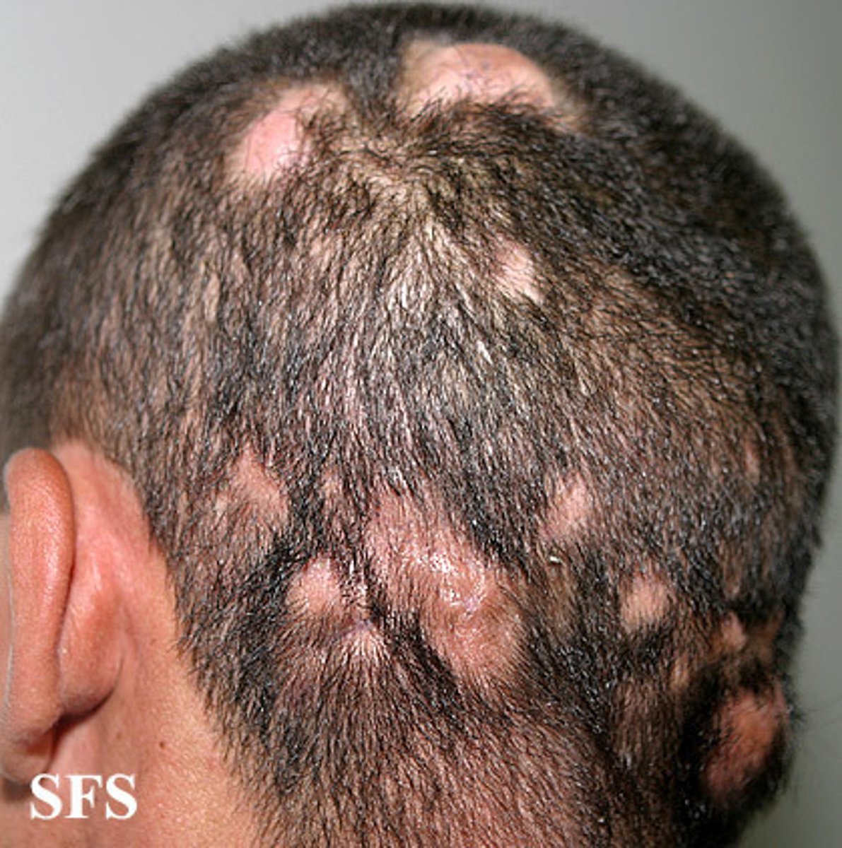
tinea capitis
fungal infection of scalp
-patches of alopecia
tinea corporis
scaling annular rash
"ringworm"
tinea cruris
jock itch
tinea pedis
athletes foot
-dermatophyte infection
-webs of feet
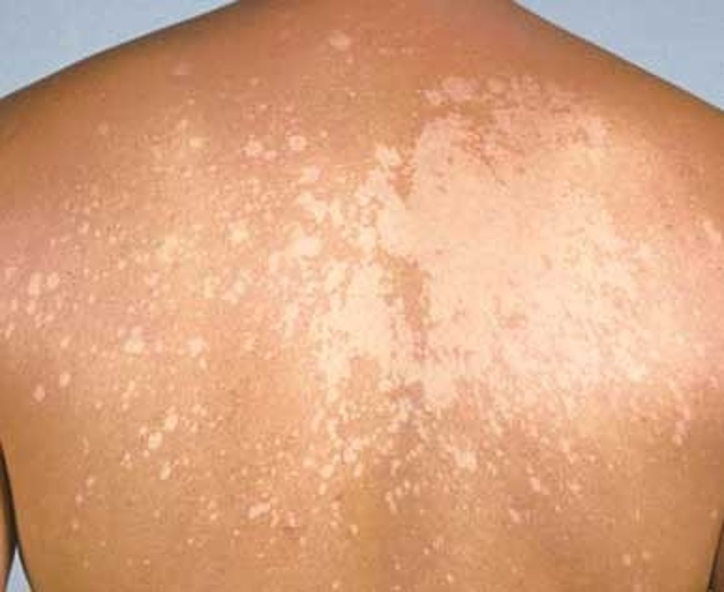
tinea versicolor
pityriasis versicolor
-chronic, asymptomatic yeast infection w/ scaling oval patches
-affected skin will lighten
-oily areas
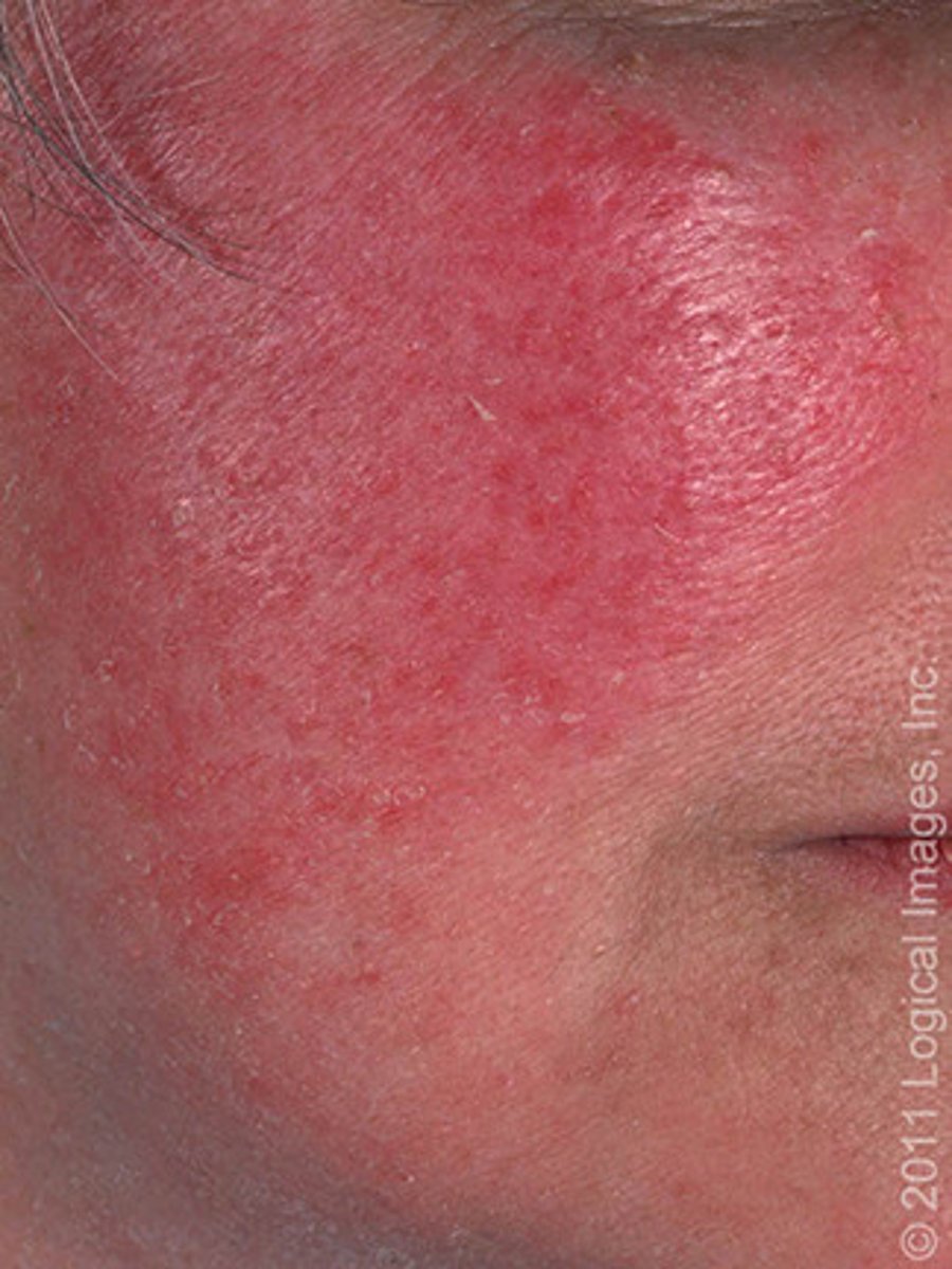
rosacea
flushing, papules, pustules
-can be confused w/ acne
-thickened disfigured skin on nose (rhinophyma)
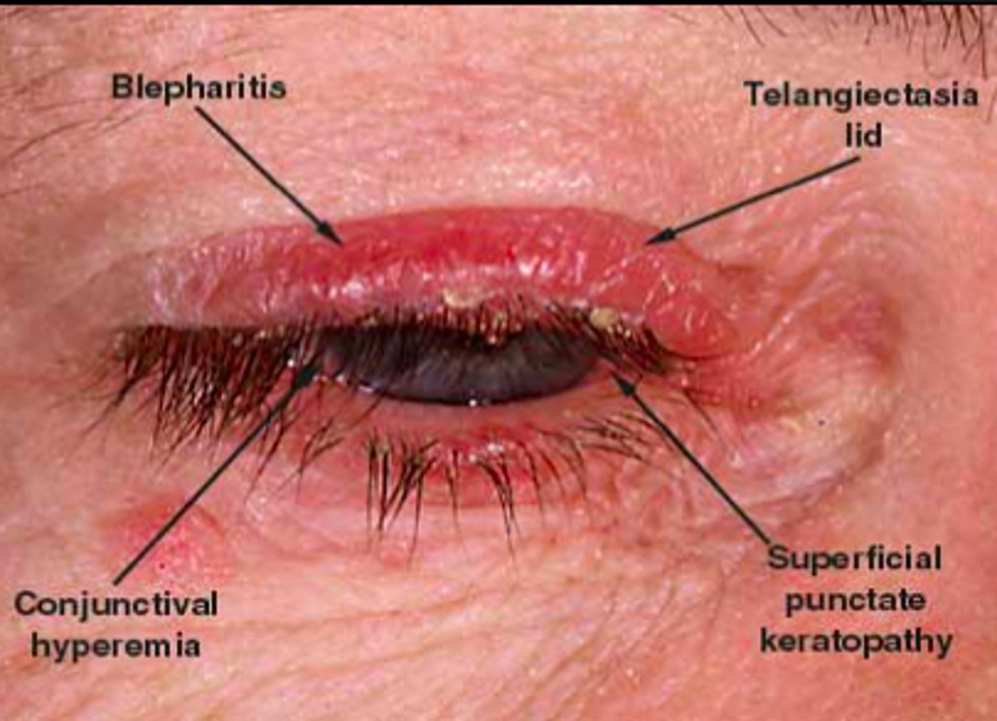
ocular rosacea
eye irritation
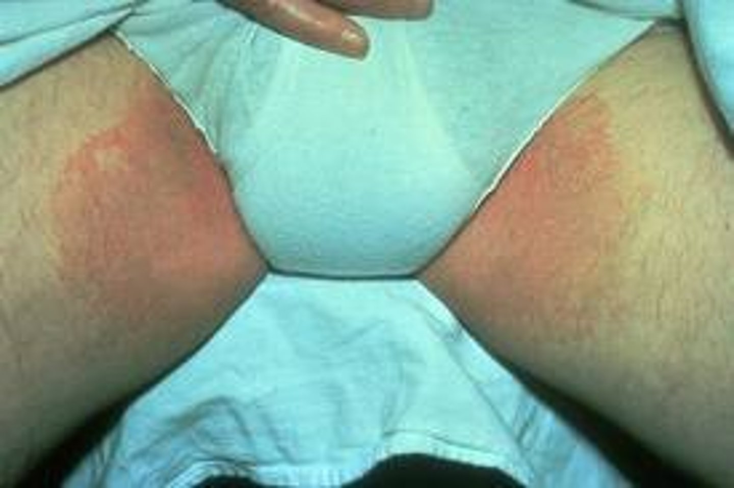
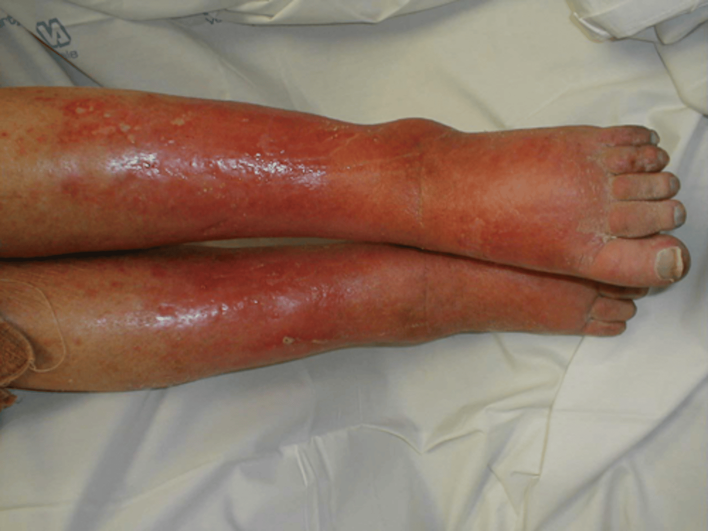
erysipelas
-dermis and epidermis w/ defined borders
-superficial edema and palpable
-strep
is a less severe cellulitis
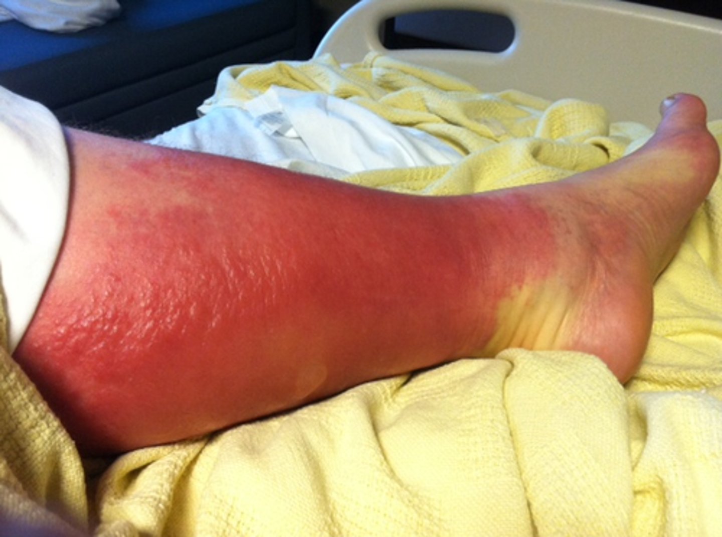
cellulitis
bacterial infection of dermis and subcutaneous tissue
-red, hot, tender
-staph, strep, H. flu enters through break in skin
marked w/ pen to trace growth
decubitis ulcer
bedsore
-areas of high pressure @ bony prominences
-tissue becomes ischemic then necrotic
acne vulgaris
most common skin condition
-androgen stimulation, skin microbiome, innate cell responses, genetics, diet
open comedones
black heads
closed comedones
white heads
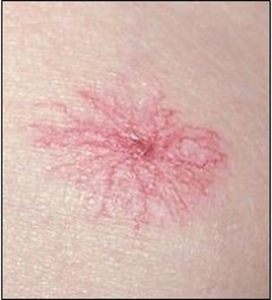
telangiectasia (spider angioma)
red body with "legs"
dilated small vessels that are red or blue
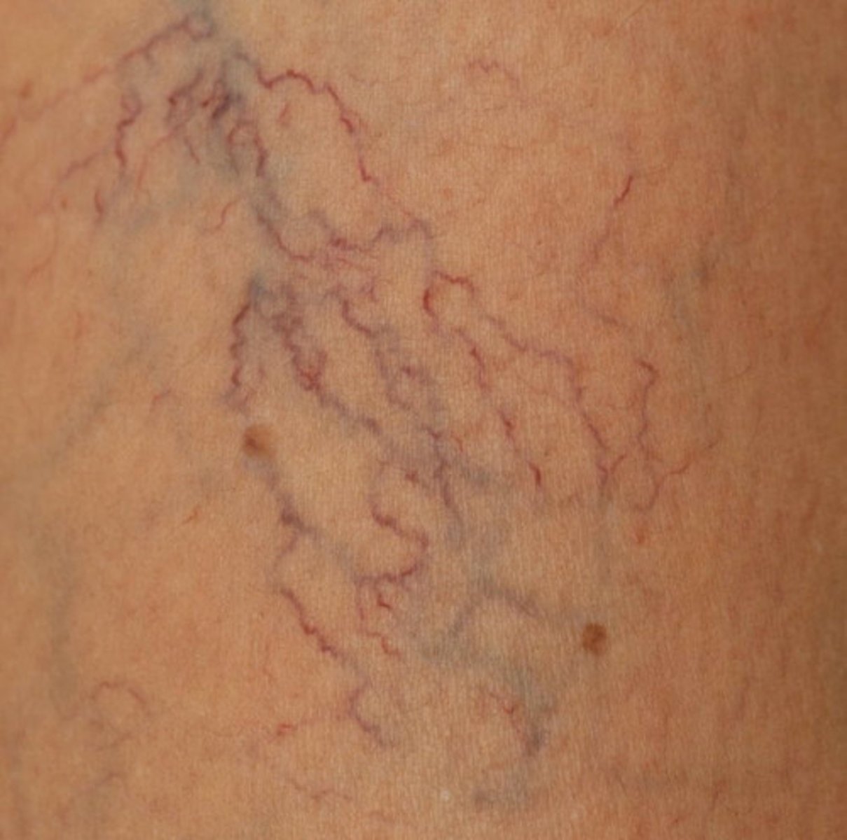
spider veins
blue superficial varicose veins with legs
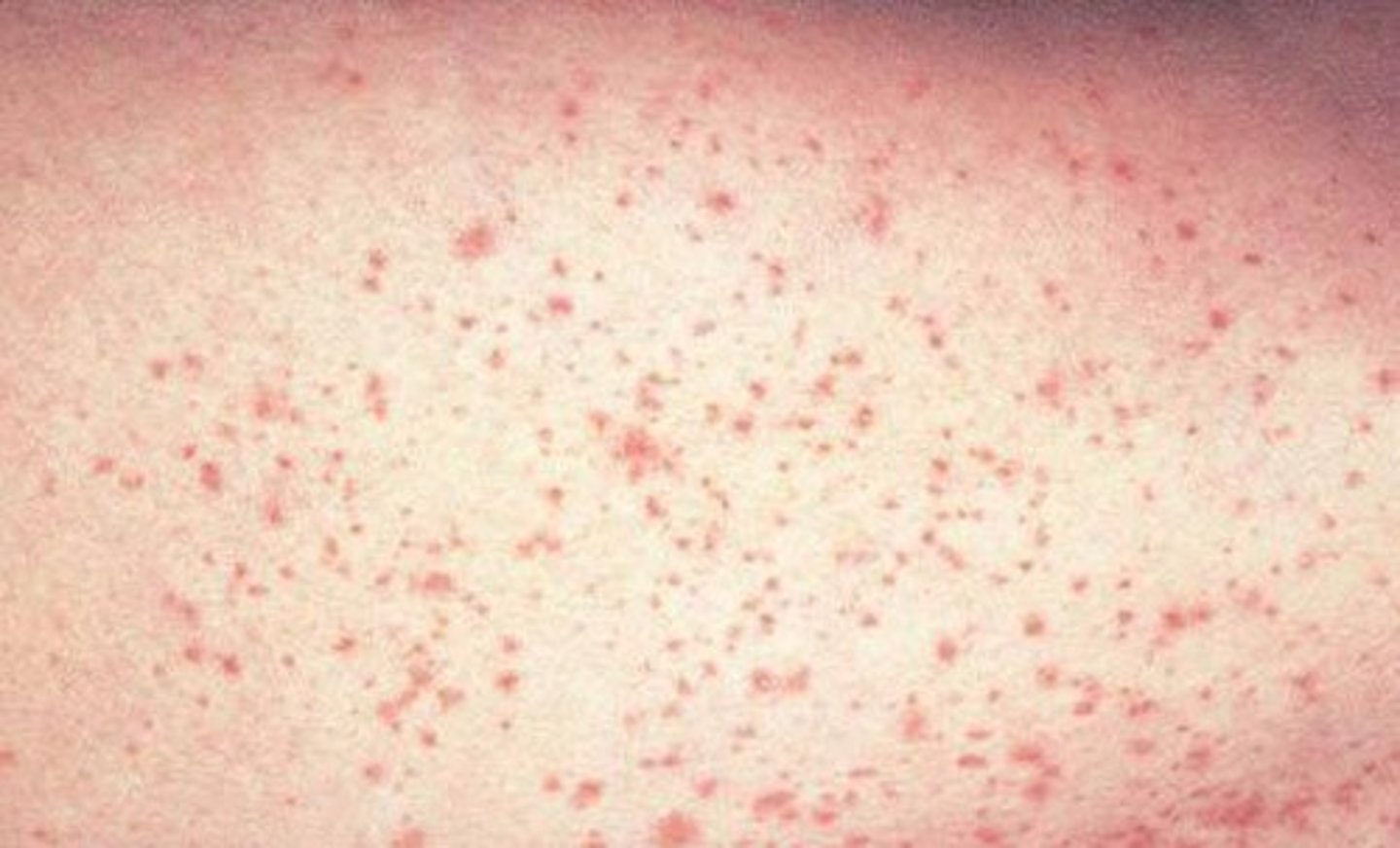
petechia
1-3 mm when blood leaks out of vessels
transient
purpura
larger than petechiae
-blood leaks out of vessels here too
ecchymosis
bruising
-greater than 3 mm
-if associated w/ edema = contusion
hematoma
pool of blood under skin resulting in swelling
swollen bruise
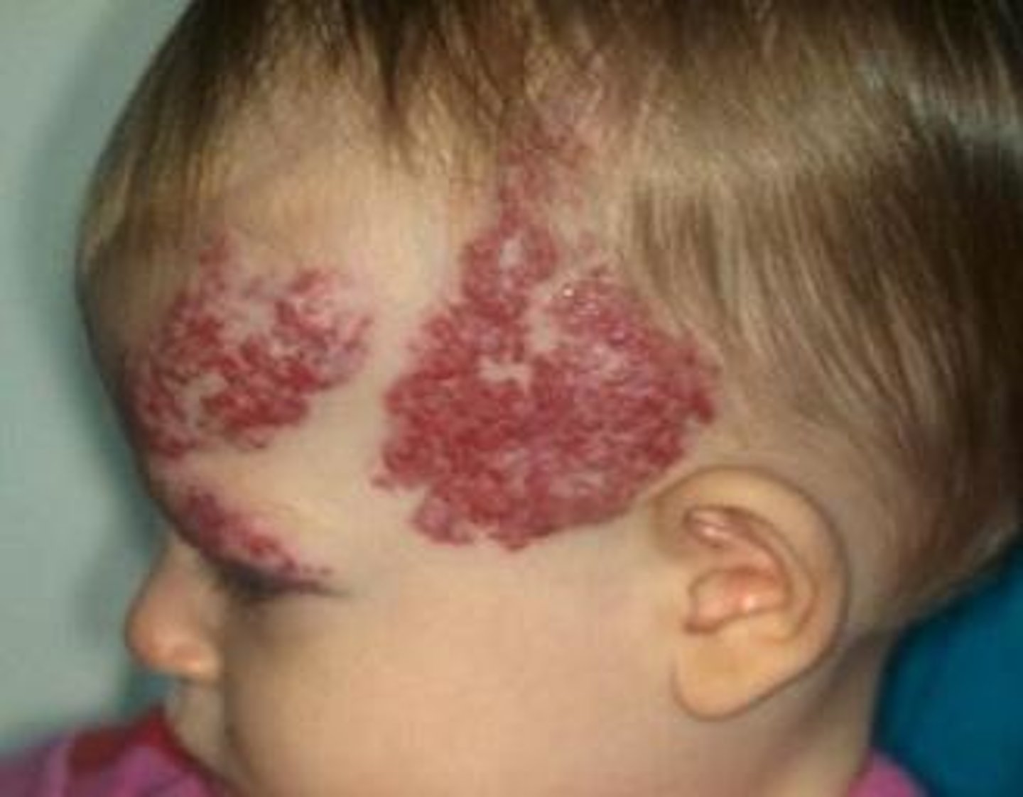
cavernous hemangioma
-congenital, soft and spongy
-can contain alot of blood
-first few weeks of life, gone by age 9
head, neck, viscera, liver, pancreas
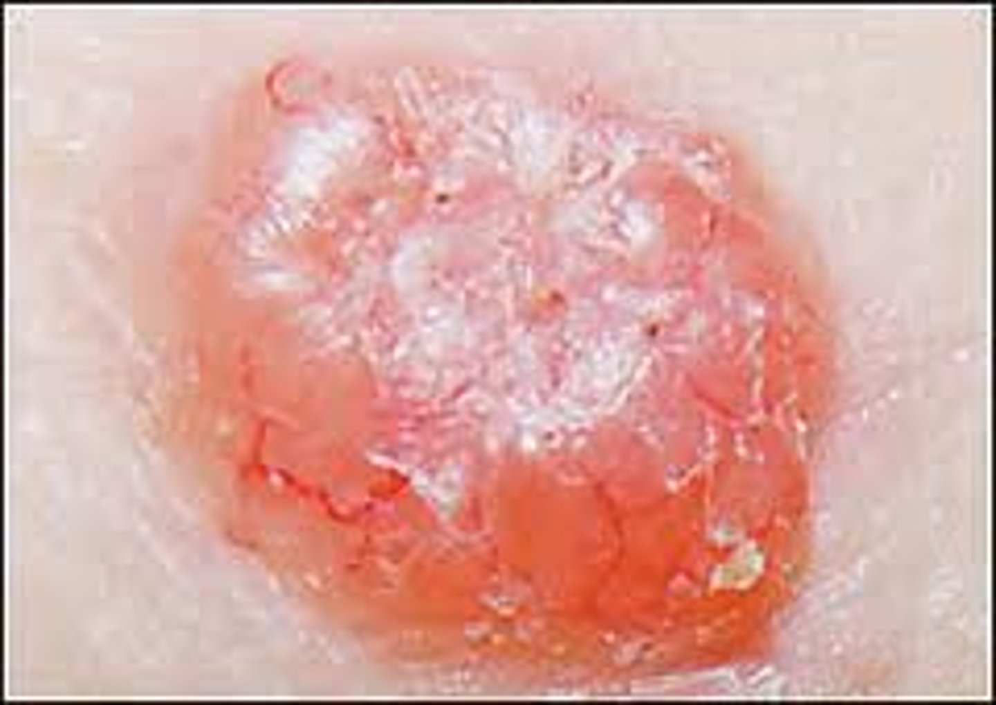
BCC
pink papule/plaque with pearly appearance and surrounding telangiectasias
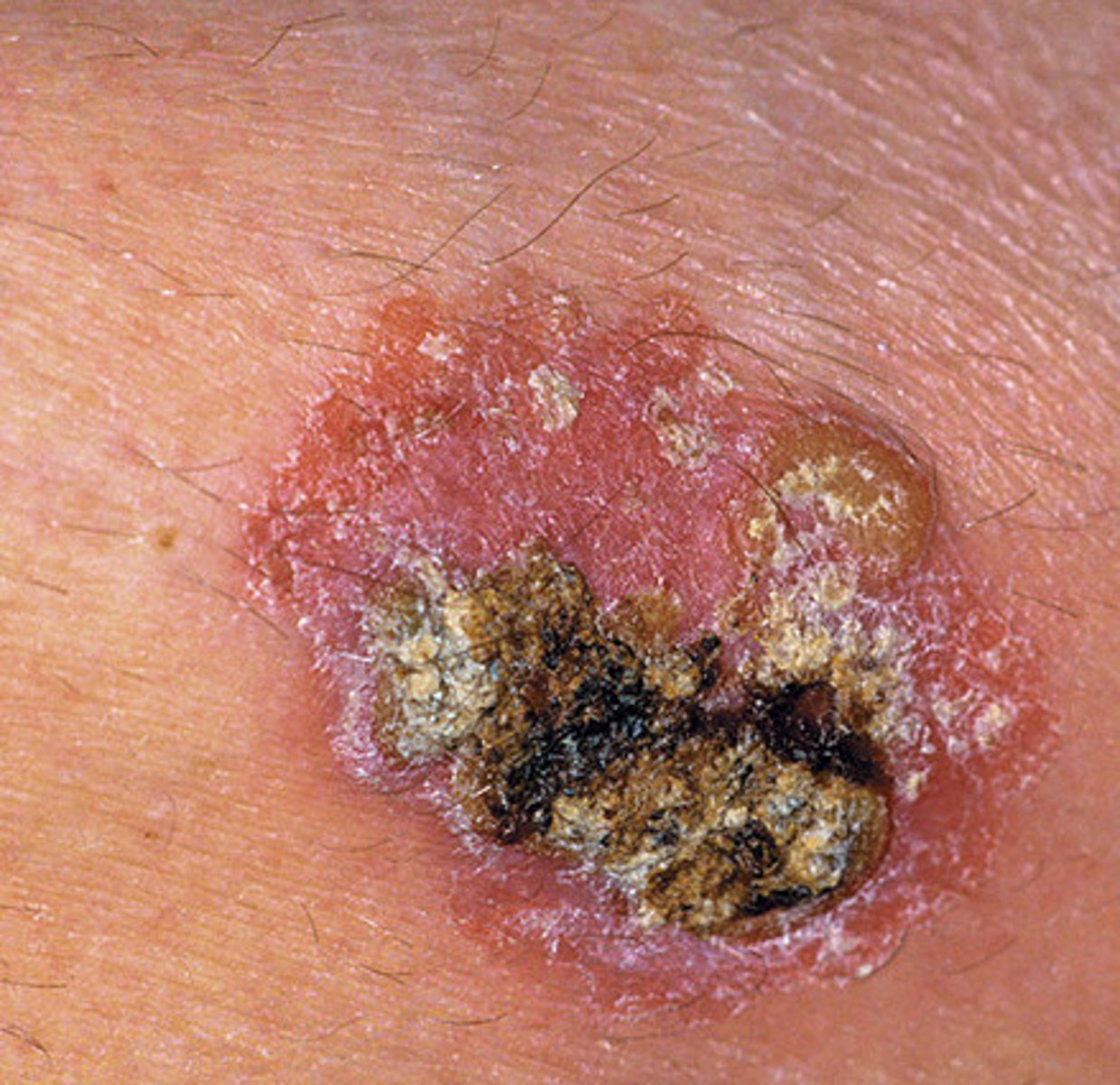
SCC
firmer edges than BCC
-keratotic
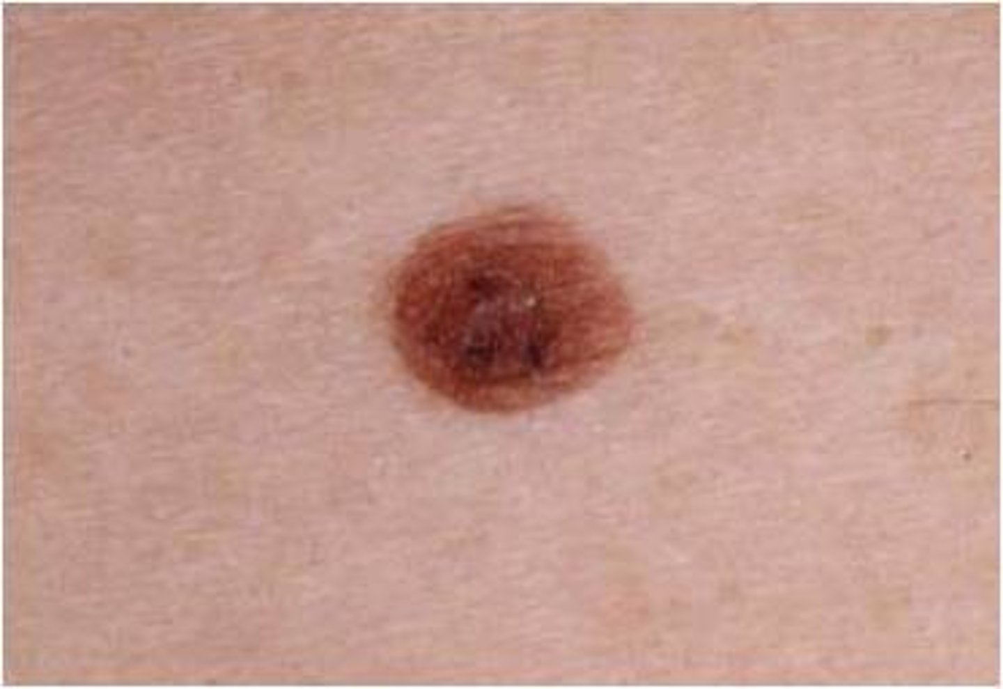
malignant melanoma
30% pre-existing nevi
-increasing prevalence
what to inspect for on scalp
scaliness
masses
lesions
what to inspect for on skull
size
contour
deformities
depressions
masses
what to inspect for on face
facial expression
contours
symmetry
involuntary movements
masses
edema
examination for skin on face
color
texture
thickness
hair distribution
lesions
rashes
acne
4 functions of skin
1. retain body fluid
2. protect underlying tissue
3. synthesize vitamin D
4. help control body temperature
appendages of skin
hair
nails
glands
melanin
genetically determined
levels increase w/ sun exposure
oxyhemoglobin
reddening or erythema of skin
deoxyhemoglobin
pallor of skin
carotene
yellow pigment of skin due to vitamin A
eating carrots in kids
bilirubin
yellow-brown pigment from breakdown of RBC
jaundice
vellus hair
short, thin, light-colored barely noticeable hair
face hair
terminal hair
thick, long, and dark
during puberty, replaces vellus hair
beard and pubic hair
how long does it take the nail to grow from base to digital edge
3-4 months
skin ROS
rash
lumps
sores
pruritus
dryness
new lesions or changes to existing ones
hair and nail changes
skin color changes in skin ROS
pallor
erythema
cyanosis
jaundice
increased/decreased pigment
heliotrope
what is heliotrope
purple rash around the eyelids
lupus
inspection of hair
distribution
quantity - can be increased, sparse, or expected
palpation of hair
texture - silky, brittle, or coarse
-hair pull and hair tug test
hair pull test
-examines shedding from roots
-gently grab 50-60 hairs and pull away firmly from scalp
Abnormal = 6+ easily pull out
hair tug test
-hair fragility testing
-hold a group of hair in one hand and pull shafts with opposite hand
androgenetic alopecia
male pattern baldness
alopecia areata
sharply outlines hair loss that may regrow
alopecia totalis
total loss of hair
pediculosis
lice infestation
-direct contact
-inspect for nits, hair shaft and above ears and nape of neck
step 1 of skin exam - inspection - positioning patient
seated, dressed in a gown
step 1 of skin exam - inspection - examination
-head and neck including face and ears
-upper back
-shoulders, arms, hands
-chest and abdomen
-anterior thighs and legs
-feet and toes
step 1 of skin exam - inspection - final step
position patient in standing position
-lower back
-posterior thighs and legs
-genitalia
step 1 of skin exam - inspection - what to note
-color
-moisture (dry, diaphoretic, oily)
-edema
-excoriations
-scars
what are excoriations
scratches
what to note if rashes/lesions are present
number
size in cm
color
texture
distribution/location
configuration
ABCDEs of melanoma
Asymmetry
Border
Color
Diameter
Evolving (can be more HPI)
Nevus
congenital or acquired in highly pigmented area
-flat or raised
step 2 of skin exam - palpation
-temperature (compare backs of hands)
-texture (rough or smooth)
-mobility and turgor
-tenderness
-lesions for consistency
mobility and turgor of skin
increased = dehydrated
decreased = hydrated
rash descriptions
number
size
color
shape
texture
location
configuration
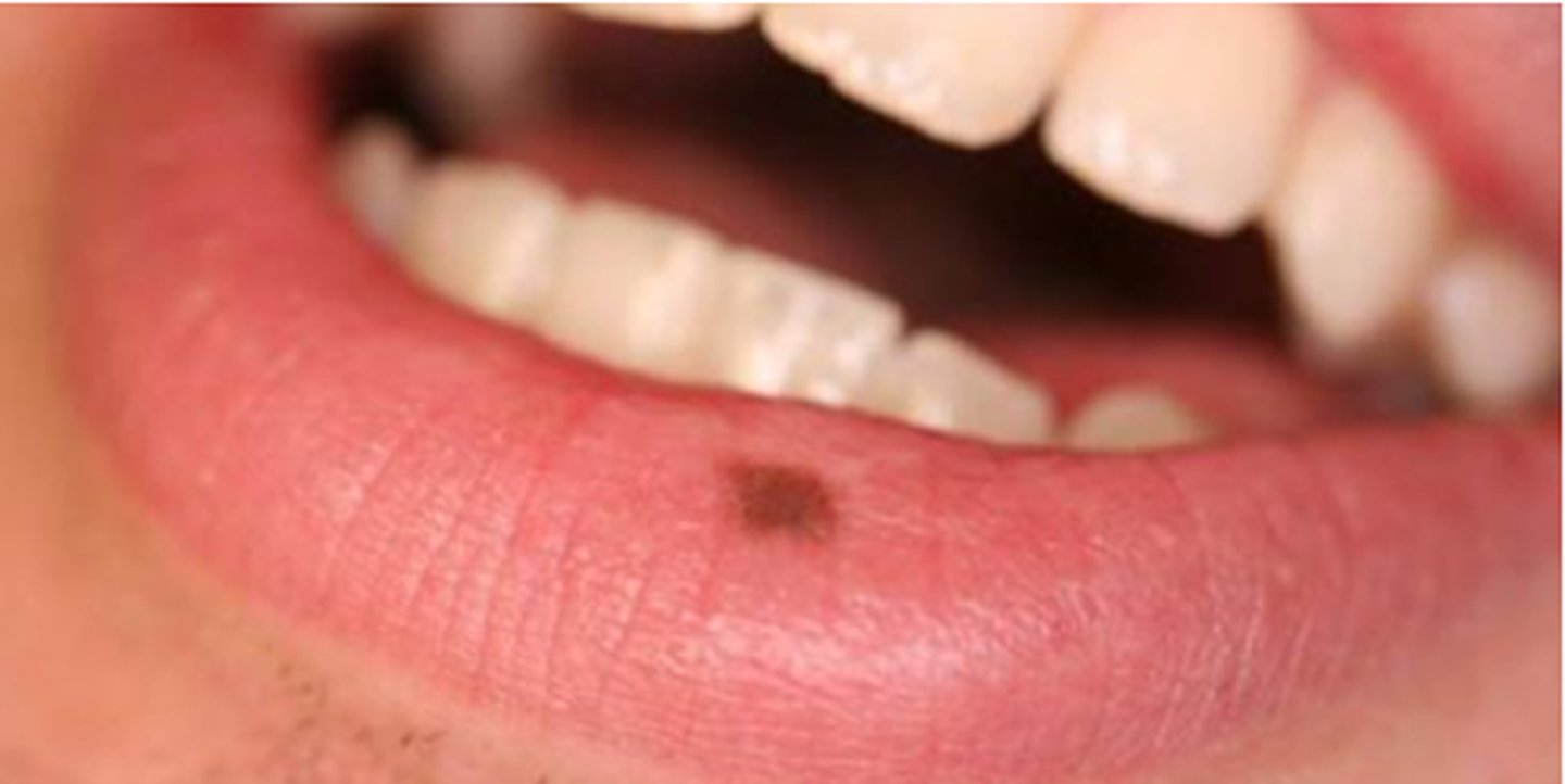
macule
flat, distinct colored area
less than 1cm
ex: freckle
patch
flat, distinct discolored area
over 1cm
ex: vitiligo
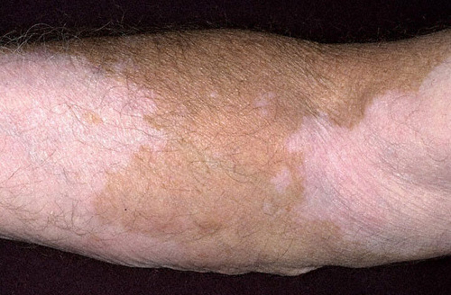
vitiligo description
say paler than surrounding skin
papule
elevated lesion
smaller than 1 cm
plaque
elevated lesion
larger than 1 cm
ex: psoriasis
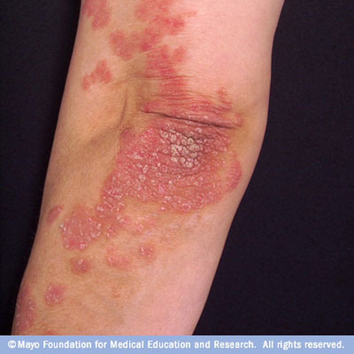
psoriasis description
pink silvery-white scale
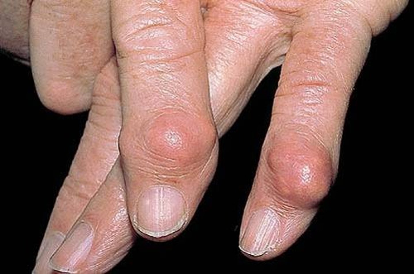
nodule
mass bigger than 0.5cm
deeper than papule
ex: rheumatoid nodule
wheal
irregular, transient, localized edema
ex: hives, mosquito bite
vesicle
lesion filled w/ serous fluid smaller than 1cm
ex: herpes zoster
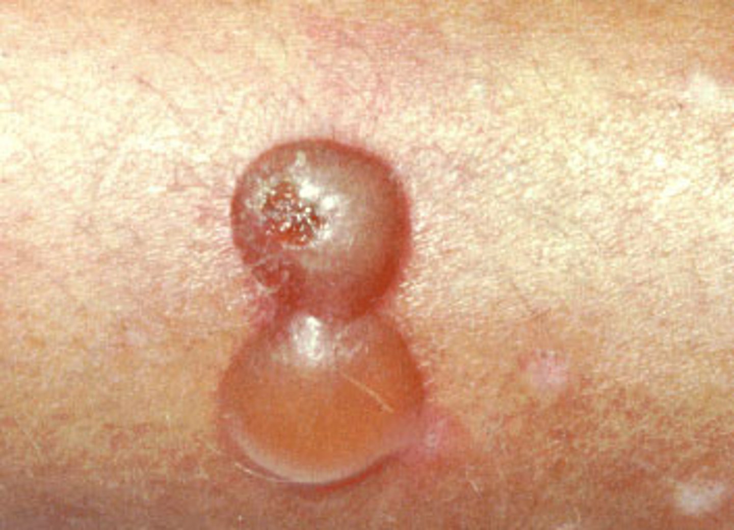
bulla
fluid filled lesion bigger than 1 cm
ex: burn
pustule
pus-filled lesion
ex: acne
erosion
loss of epidermis
moist w/o bleeding
ex: ruptured blister
ulcer
deep loss of skin surface
can bleed and scar
ex: diabetic foot ulcer
fissure
linear crack in skin
crust
dried residue of serum, blood, or pus
IS A SCAB
scale
flakes of exfoliated epidermis
ex: cradle cap, sunburn, dandruff
lichenification
thick, rough skin
chronic scratching
increased visibility of normal skin furrows
measuring lesions
measure long axis then perpendicular to that axis