Chp 12 (Nervous Tissue)
1/44
There's no tags or description
Looks like no tags are added yet.
Name | Mastery | Learn | Test | Matching | Spaced | Call with Kai |
|---|
No analytics yet
Send a link to your students to track their progress
45 Terms
Overall Functions of Nervous System
Coordinates all systems functions, maintain homeostasis
Responsible for all perceptions, behaviours, memories, and voluntary movements
3 Different Functions of Nervous System
Sensory: detect changes in environment (stimulus), relay info to CNS
Integrative: analyze info and make decisions (interneurons)
Motor: respond to stimuli by initiating action
Organization of Nervous System (The 2 Systems)
Central Nervous System (CNS): brain and spinal cord
Peripheral Nervous System (PNS): all nervous tissue outside of CNS, including nerves and sensory receptors
Neurons
-cells that transmit signals
-dont divide
-electrically excitable
Neuroglia
-supportive cells that protect n nourish neurons
-can divide
-not electrically excitable
-4 kinds in CNS, 2 in PNS
Cell Body
-contains nucleus
-nissl bodies (Rough ER)
-neurofibrils give shape and support
-microtubules move material inside/along axon
Cell Processes (Nerve Fibers / Neurites)
-dendrites: conduct signals TO cell body (receiving)
-highly branched
-unmyelinated
-axons: conduct signals AWAY from cell body → other body cells, starting at trigger zone
-ends in thin axon terminals (with synaptic end bulbs at tips)
filled with neurotransmitters
Explain the process of signal communication btw Dendrites, Cell Body, and Axons
-Dendrites receive the signal
-send it towards the cell body, where its processed
-sent down long axon.
At the end bulbs, Ach is released into a synapse, thus triggering a muscle/nerve activation. Ach-ase breaks down the Ach to be recycled
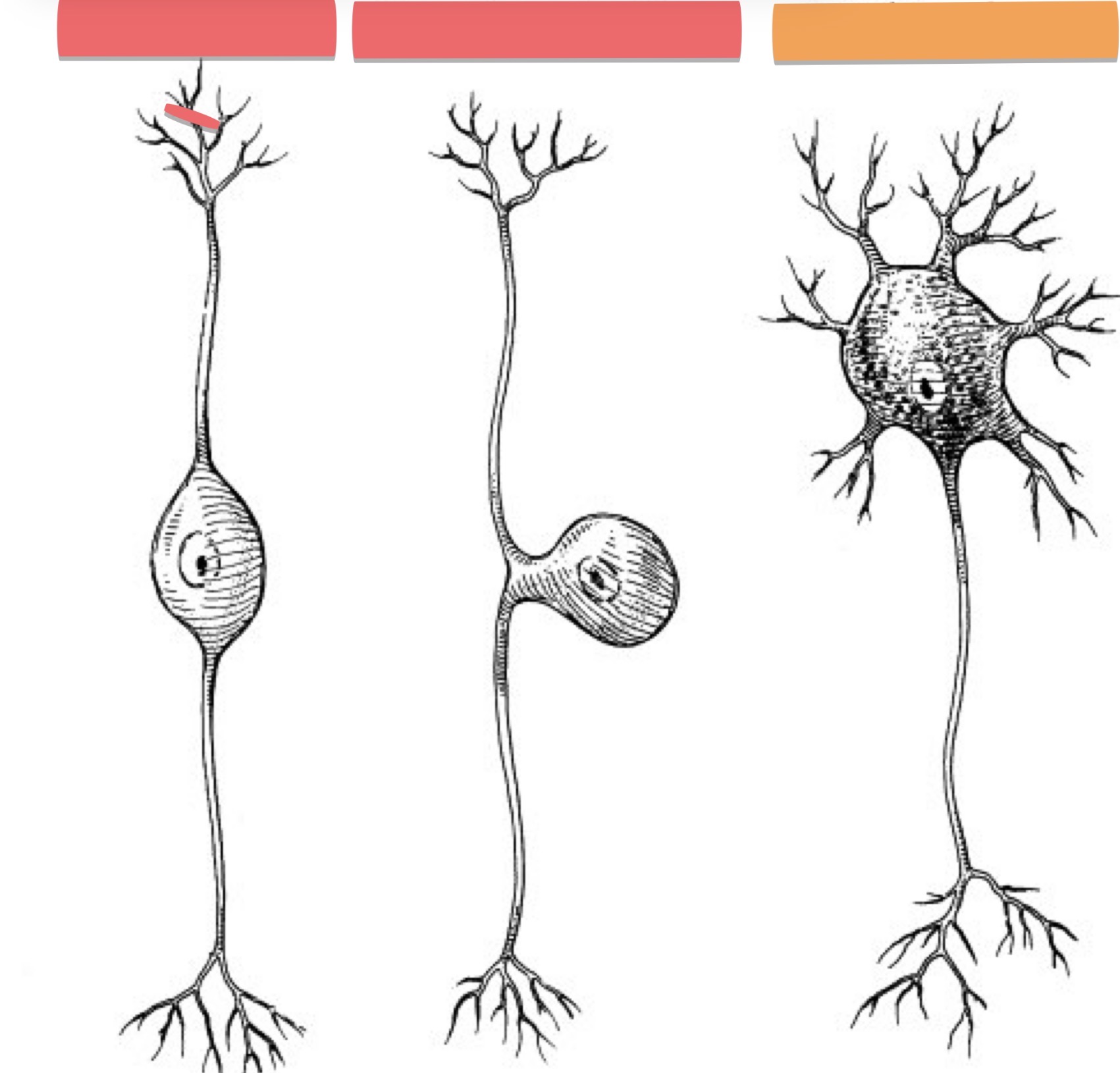
What Neuron is this?
Multipolar
several dendrites, one long axon
Majority of neurons in CNS (interneurons), all motor neurons
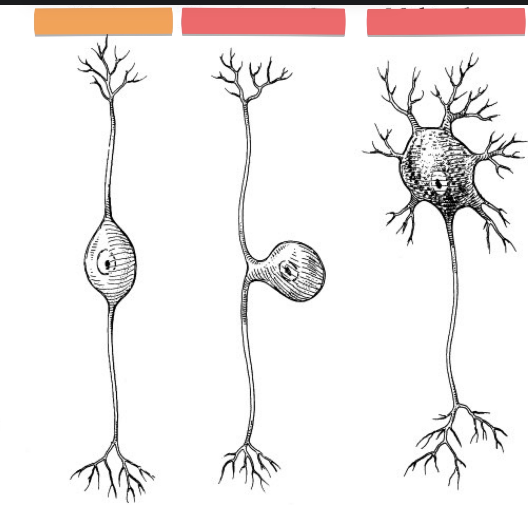
What Neuron is this?
Bipolar
one dendrite and axon
Eye retina, inner ear, olfactory area of brain
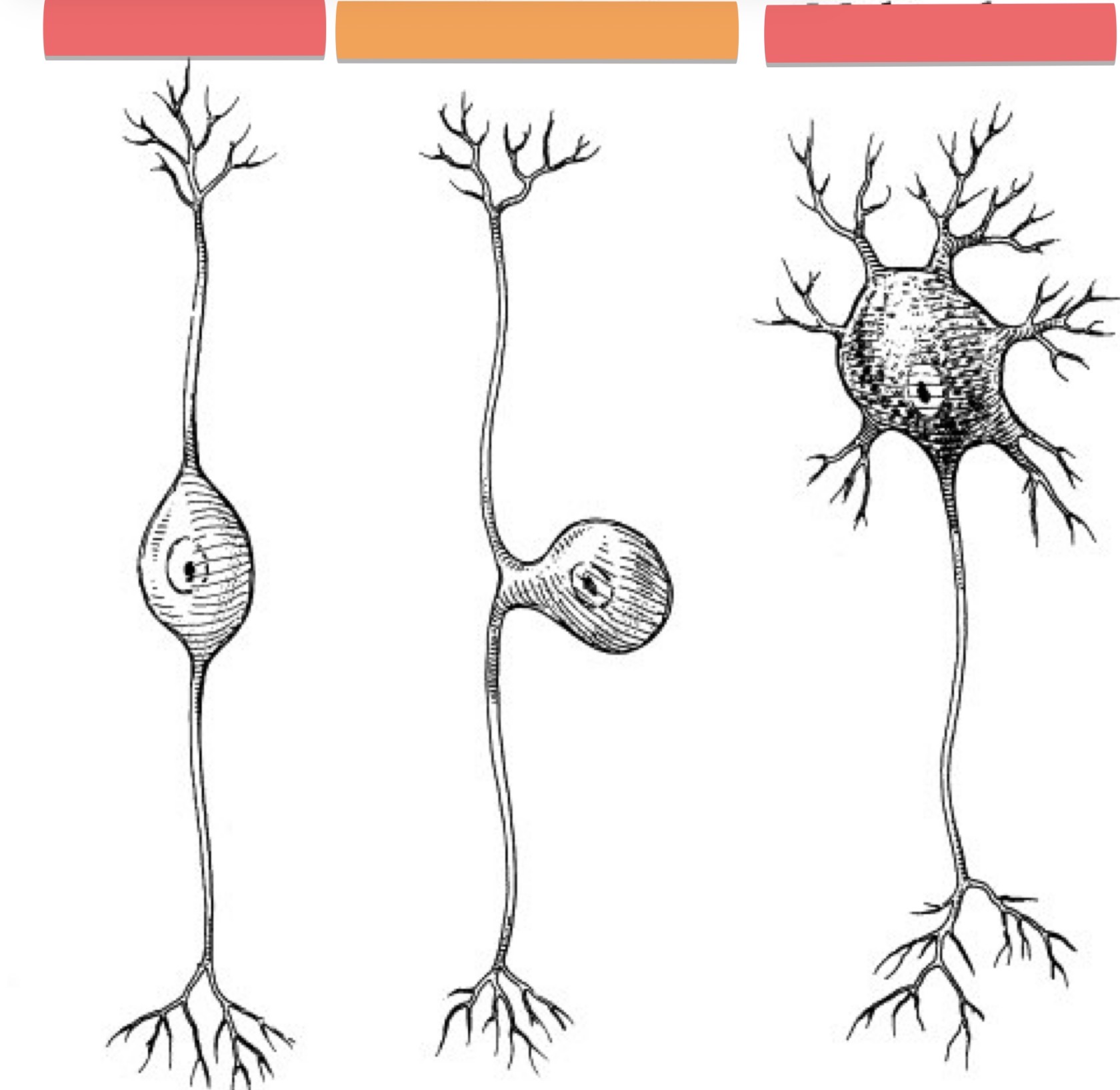
What Neuron is this?
Pseudounipolar
Single fused axon and dendrite (which acts as sensory receptors)
Sensory neurons
Functional Neuron Classifications + How do they all work tgt in carrying out a function?
(Classified whether they transmit AP’s towards or away from CNS)
sensory / afferent (away)
Relay sensory info TO CNS through cranial or spinal nerves
Motor / efferent
Send commands FROM CNS to muscles and glands (effectors) through cranial or spinal nerves
Interneurons / association
Connect sensory to motor neurons in CNS, most numerous
Sensory neurons receive info from inside n outside the body n sends it to the CNS, Interneurons connect sensory and motor neurons tgt for the signal to flow through, the motor neurons initiate the command to the gland, muscle etc.
Eg; i pick up a hot coal (sensory tells CNS its hot), signal flows through (interneurons) my body, then i let go (motor)
Astrocytes (4 Neuroglial Cell Types in CNS)
-star shape
-cover blood cap’s, forms blood-brain barrier
-regulate proper ion + neurotransmitter concentrations in inter. Fluid
-provide structural support for neurons
-regulate growth in embryos
-influence synapses formation
-similar to satellite cells
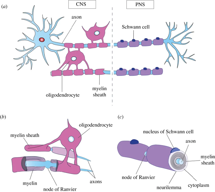
Oligodendrocytes (4 Neuroglial Cell Types in CNS)
-form n maintain myelin sheath (lipid n protein covering) around axons in CNS
myelin insulates axons and speeds up signals btw neurons
-similar to Schwann cells
Microglial Cells (4 Neuroglial Cell Types in CNS)
-phagocytic
clear microbes n dead cells
Ependymal Cells (4 Neuroglial Cell Types in CNS)
-form epithelial membrane lining ventricles of brain and central canal
-make n circulates cerebrospinal fluid (CSF)
forms blood-CSF barrier
Satellite Cells (2 Neuroglial Cell Types in PNS)
-surround cell body of PNS neurons
-structurally support neurons
-regulate exchange of mtaerial btw cell body of neurons and inter. Fluid
-similar to astrocytes
Schwann Cells (2 Neuroglial Cell Types in PNS)
-encircle + maintain axons in PNS, ensures neuronal survival
-produce myelin sheaths that surrounds axon in PNS
-similar to oligo’s
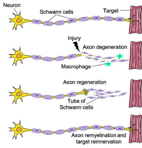
PNS Myelination
-myelin is made from Schwann cells
-outer plasma membrane of a schwann forms nuerolemma
-PNS can regenerate
IF cell body is intact, schwann’s are functional, n scar tissue formation isnt too rapid
Distal axon degenerates, regen. Tube is formed by Schwann’s
CNS Myelination
-myelin is made from Oligo’s
-cell processes wrap around CNS axons, but cell bodies dont surround axons, so no neurolemma (= little regen.)
Clusters of Cell Body + Bundles of Axons in PNS n CNS
PNS
cell body clusters: ganglion
Axon bundles: nerves
CNS
celll body clusters: nucelus
Axon bundles: tracts
White v Gray Matter
White: myelinated axons
Gray: cell bodies, dendrites, axon terminals, unmyelinated axons, neuroglia
-gray is typicall found in the middle or around the edges of the brain n spinal cord
Electrical Signals in Neurons
-excitable cells communicate via AP’s n GP’s
-AP’s communicate through short/long distances, but GP’s are for short only
Production of an AP/GP depends on a resting membrane potential (RMP) and certain ion channels
Channels are needed so ions can diffuse through cell membrane
Leak Channels (Types of Channels in Neurons)
alt btw open/close randomly, K channels more numerous than Na channels
dendrites, cell bodies, axons of all neurons
Ligand-gated Channels (Types of Channels in Neurons)
need specific chemical binding to open/close (eg; neurotransmitter, hormone)
Dendrites of SOME sensory neurons, dendrites + cell bodies of inter + motor neurons
Mechanically Gated Channels (Types of Channels in Neurons)
open/close in response to mechanical stimuli (eg; vibration, pressure)
Dendrites of SOME sensory neurons (eg; touch, pressure like skin, and auditory like ear)
Voltage-Gated Chanels (Types of Channels in Neurons)
open/close in response to a membrane potential change, generates/conducts AP’s
In axons of all neurons
Graded Potentials / GP’s (local membrane changes)
-in response to a stimulus (eg; change in MP) can be tiny/large and in any direction (inc. or dec. = graded)
-mainly in dendrites and cell body, uses mechanically + ligand channels
-if sufficient (reach threshold), triggers an AP (can travel long distances)
GP’s can vary in size (depending on stimulus) but all AP’s are the same
Resting Membrane Potential (RMP)
-nerve cells are excitable because
RMP is polarized (conc. Of ions n net charge is diff from inside to outside cell membrane)
Specific ion channels
-more - ions inside and + ions outside
-voltage across membrane is -70mV for a neuron
-Na and Cl are high in ECF
-K, proteins, PO4- are high in cytosol
What transporter is responsible for the conc. Gradient of Na+ and K+ ions?
Na/K ATPase
OR
Na/K Pump
Flow of Ions
-ions must flow in/out of cell for signals (changes in potential) to take place
-flow of ions occurs through channels in membrane
ions flow from high to low conc., and towards opposite charge
Hyperpolarization and Depolarization
Hyper: membrane becomes more - than MRP (-70 → -80)
taking in anion (-) or losing cation (+)
Further from threshold, less likely to trigger AP
Depolarization: membrane becomes more + than MRP (-70 → -60)
taking in cation
Closer to threshold, more likely to trigger AP
Action Potentials (AP’s)
-rapid depolarization of MP followed by restoration to MRP (-70 mV)
-strength of an AP is ALWAYS the same
-involved voltage channels (Na n K)
-travels whole axon, releases neurotransmitter at synapse
Phases of AP
-chemical or mechanical stimulus causes GP to reach at least -55mV (threshold), then 2 phases
depolarizing: voltage Na channels open, Na rushes in, MP becomes +30mV
Repolarizing: Na channels inactive (Na inflow stops), K channels open to let K out, MP back to -70mV
Refractory Period (period where a neuron CANT generate another AP), 2 types
absolute: inactivated Na channels must return to resting state before reopened
Relative: suprathreshold stimulus can start an AP, K channels still open, but Na channels closed, hyperpolarization
Stimulus Strength of AP’s
Subthreshold stimulus: GP insufficient in reaching threshold = no AP
Threshold stimulus: sufficient depolarzation = 1 AP
Suprathreshold stimulus: multiple rounds of depolarization = many AP’s
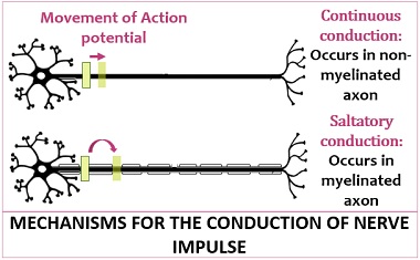
Propagation of AP’s
-communication through ur body only happens when AP travels from trigger zone → axon terminals
travel = propagation, 2 kinds
Continous Conduction (Unmyelinated Axons):
-travels down ENTIRE axon, overall slower
Saltatory Conduction (Myelinated Axons):
-myelin sheaths pass Na down quickly along axon, since they dont interact with them (so just skips past all myelin parts), overall faster
lipid portion of myelin is hydrophobic (no like Na)
-so, myelin = faster signal conduction
Factors Affecting Propagation Speed
Axon diameter (larger = faster)
Myelination Amount (more myelin = faster)
temperature (hotter = faster)
Clinical Manipulation of AP’s
-anesthetics (eg; procaine, lidocaine) act by blocking the opening of voltage Na channels
no gen. Of AP’s = pain sensations arent transmitted to CNS
Signal Transmission In Synapses
-synapse: junction btw neurons and another cell (2 kinds)
Electrical: ionic current spreads to next cell through gap junctions, faster, can synchronize groups of cells/neurons
Chemical: one way info transfer from a PRE-synaptic neuron → POST-synaptic cell
Chemical Synapses
AP reaches end bulb
Voltage channels open
Ca flows inward triggering release of neurotransmitter
Neuro. Crosses synaptic cleft (space btw neurons), binds to receptor on post-cell, causes ion channels to open
Ions rush in post-cell
Triggers depolarization or hyperpolarization (depends on what channels open)
Post-synaptic potential is generated
-postsynaptic potential = temporary graded change in MP of the post-synaptic cell
Excitatory and Inhibitory Potentials
-a neurotransmitter can be either inhibitory or excitatory
excitatory:
when NT Causes opening of ligand channels, depolarization happens (Post-cell more likely to reach threshold = start AP)
Depolarizing postsynaptic potential = EPSP (Excitatory Postsynaptic Potential)
Inhibitory:
when NT Causes opening of ligand Cl or K channels, hyperpolarization happens (post-cell less likely to reach threshold = no AP)
Hyperpolarizing postsynaptic potential = IPSP (Inhibitory Postsynaptic Potential)
-EPSP and IPSP are both GP’s and present in PNS and CNS
-one NT can be excitatory and inhibitory in diff locations (depends on receptors and channels)
Summation
Spatial: NT’s released from several end bulbs onto one neuron
Temporal: NT’s released from 2 or more firings of same end bulbs onto in rapid succession
Summation of all postsynaptic potentials (EPSP+IPSP) from all pre-synaptic NT’s will determine whether an AP is triggered + how frequent
Small Molecule NT’s
Ach: released by many PNS + some CNS neurons, excitatory on NMJ but inhibitory at other synapses
Amino Acids:
Glutamate: released by most excitatory neurons in brain, inactivated by reuptake by neurons and by neuroglia
GABA: inhibitory NT for 1/3 of brain’s synapses
Biogenic Amines (modified amino acids)
norepinephrine, seratonin (happiness), dopamine (mood)
CO1 + Nitric Oxide: Vasodilation
Neuropeptides
-3-40 amino acids, excitatory/inhibitory effects in CNS and PNS
Endorphins: natural painkiller (blocks release of substance P
Substance P: enhances pain perception
Removal of NT’s
-once NT are released into cleft, removed by
Diffusion: NT’s move away from cleft (down gradient)
Enzymatic Degradation: enzymes degrade NT (eg; Ach-esterase)
Uptake by Glial Cells / Reuptake by Neurons: NT get into neurons or glial cells by transporters