Chp 14 (Brain and Cranial Nerves)
1/31
There's no tags or description
Looks like no tags are added yet.
Name | Mastery | Learn | Test | Matching | Spaced | Call with Kai |
|---|
No analytics yet
Send a link to your students to track their progress
32 Terms
Brains Contribution to Homeostasis
-receiving sensory input
-integrating new and stores info
-making decisions and responses through motor activities
What protects the brain?
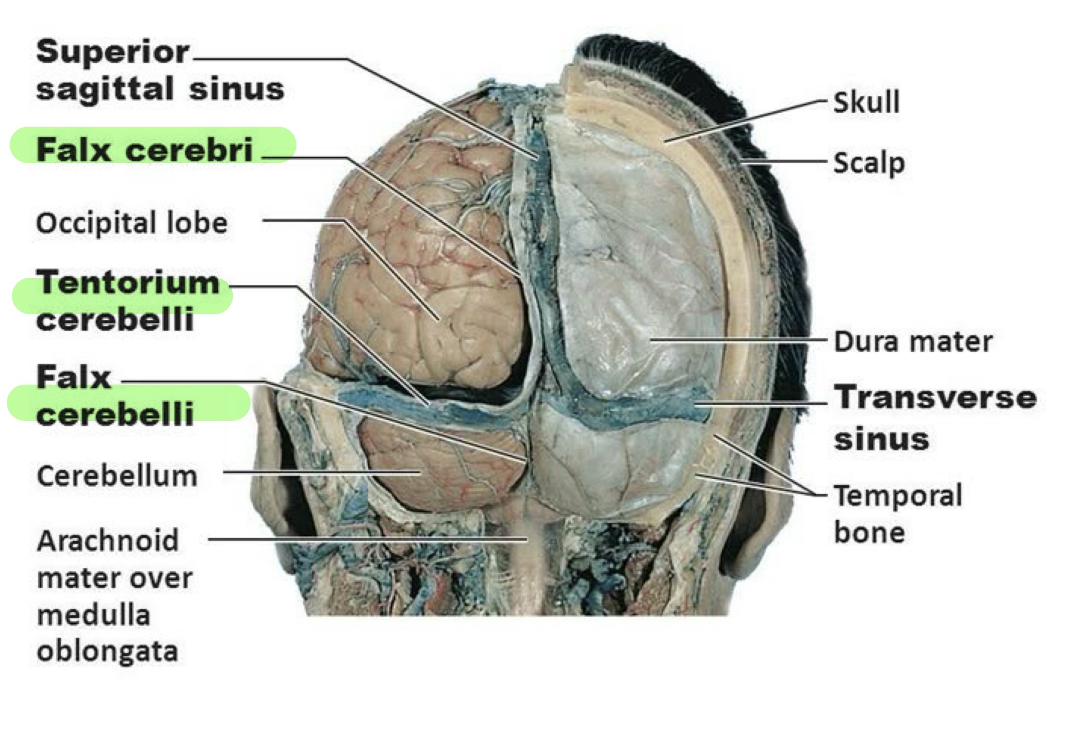
-Cranial Meninges (same as spinal) And Bones
dura has 2 layers: periosteal (external) meningeal (internal) and 3 extensions:
Falx Cerebri: splits cerebrum hemispheres
Falx Cerebelli: splits cerebellum hemispheres
Tentarium Cerebelli: splits cerebrum and cerebellum
venous sinus drains blood and CSF
Brain Circulation
Blood flows TO brain: internal carotid → vertebral arteries
Blood flow away from brain TO heart: dural venous → int. Jugular veins
-interruption of oxygen can lead to weakening/damage of braiN
-measured by fMRI
Blood-Brain Barrier
-prevents harmful substances entering, formed by
Tight junctions: seal brain capillaries’ endothelial cells
Basement membrane: thick, surrounds endothelial cells
Astrocytes: press against cap’s, secretes substance to allow passage btw blood n neurons
Cerebrospinal Fluid (CSF) Functions
-colourless, mainly water, bathes CNS
mechanical protection: absorbs shock n protects the delicate tissues of the CNS from jolts, allows brain to float
Chemical Protection: optimal chemical environment for neuronal signlaing
Circulation: medium for nutrient, waste, exchange btw blood and nervous tissue
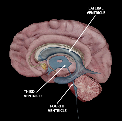
4 CSF Filled Ventricles
2 Lateral: for each hemisphere in cerebrum
seperated by septum pellucidum
3rd: along midline, above hypothalamus, btw R/L of thalamus
4th: btw brainstem and cerebellum
Where is CSF produced?
-choroid plexuses (Cap’s covered by ependymal cells found in ventricle walls)
cells secerete CSF via blood plasma filtration, and joined by tight junctions to make blood-CSF barrier
Circulation of CSF
Venous blood: Lat ventricles → 3rd → 4th → subarachnoid space → arachnoid villi of dural venous sinuses → heart n lungs
THEN
Arterial blood: 4th’s choroid plexuses → 3rd’s choroid plexuses → lat’s choroid plexuses
Reabsorption of CSF
-reabsorbed through arachnoid villi (looks like grapes)
Hydrocephalus
-build of CSF pressure due to drainage blockage of CSF (eg; tumour, inflammation)
allowed in babies (for fontanels to expand)
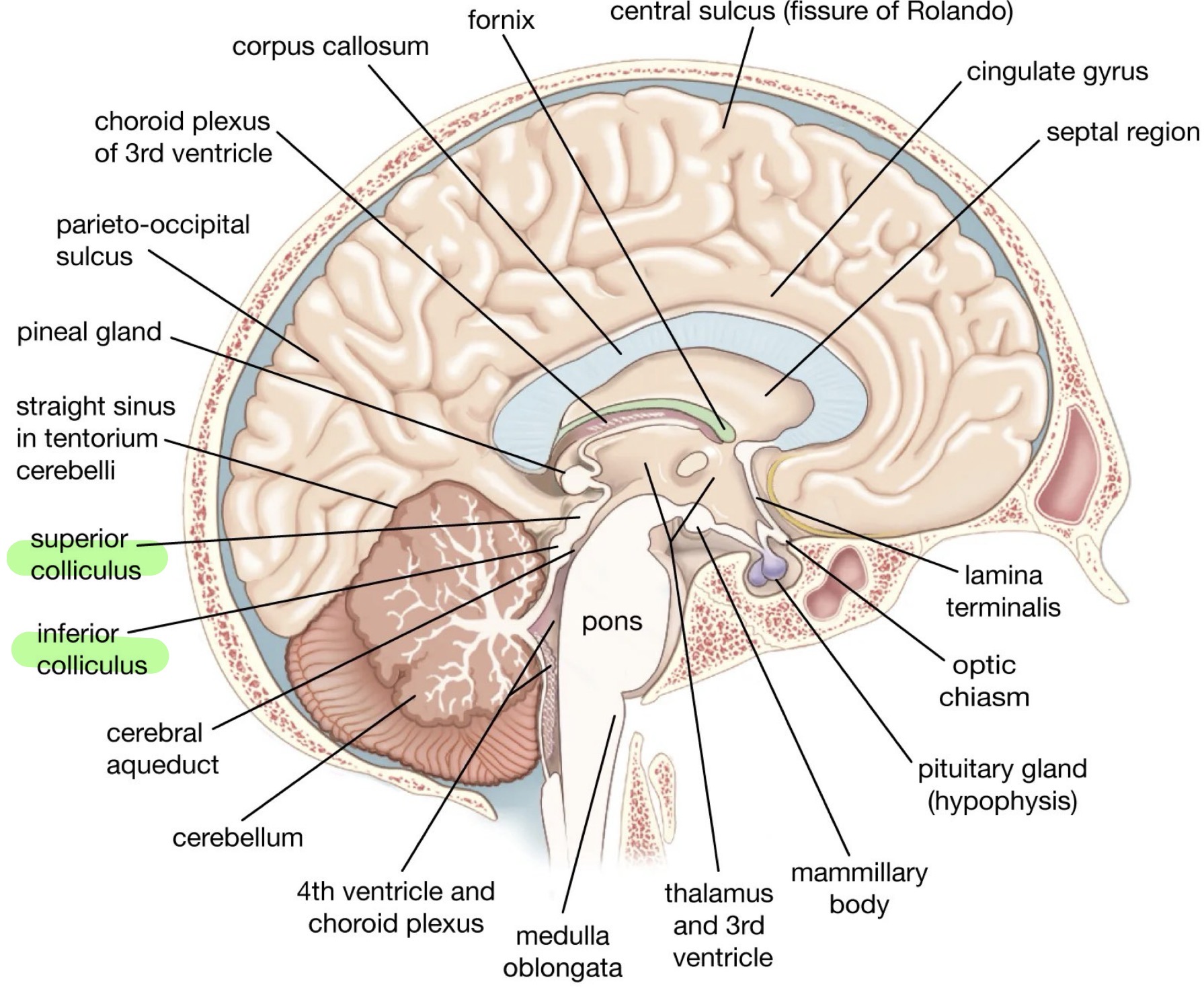
Brainstem Parts
medulla oblangata
Pons
Midbrain
Reticular foramen covers brain stem
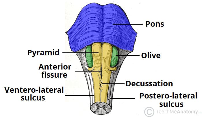
Medulla Oblangata
-Has ascending sensory + descending motor tracts, nuclei
-pyramids = ventral surface bulge
motor tracts that pass from cerebrum to spinal cord
Control voluntary movement in limbs n trunk
-decussation (crossing) of pyramids, crosses over
L side of brain controls R side body functions (and vice versa)
Body Functions Medulla Controls
Cardiovascular Centre: force n rate of heartbeat, BV diameter
Respiratory Center: rhythm of breathing
Reflex Center: coughing, sneezing, swallowing
-gustatory, cochlear, and vestibular nuclei, and nuclei for cranial nerves (8-12)
somatic sensation nuclei (eg; touch, proprioception)
Pons
-has nuclei and tracts
-connects brain parts via tracts
-helps in breathing
-has nuclei from cranial nerves (5-8)
Midbrain
-has cerebral aqueduct, connects 3rd n 4th ventricle
-has sensory + motor tracts, nuclei (visual and auditory info)
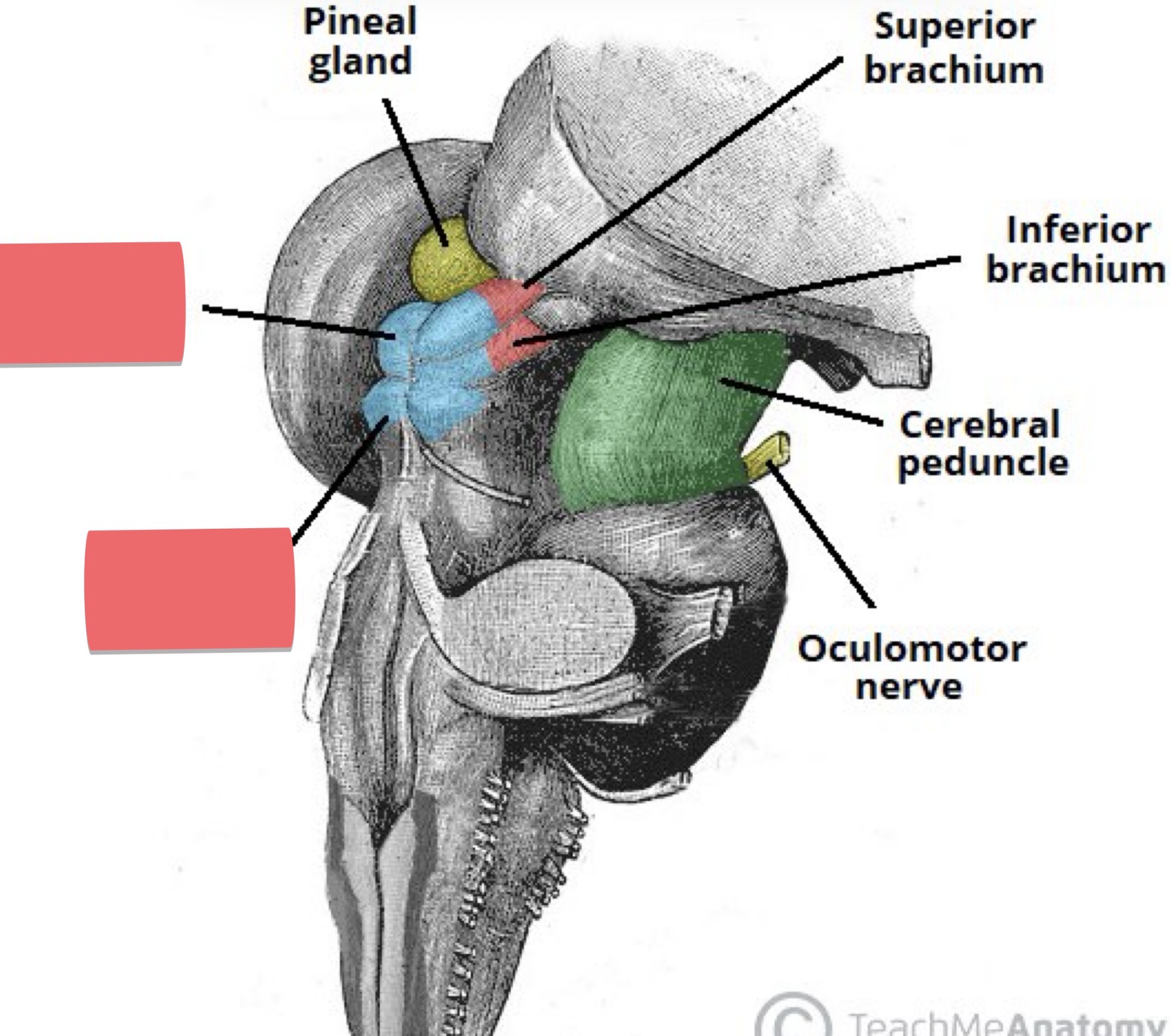
Anterior and Posterior Midbrain
Ant. (descending): cerebral peduncles = paired axons
Post. (Ascending): tectum = 4 rounded elevations
2 superior elevation (superior colliculi)
Reflex center for visual (eg; object tracking)
2 inferior elevation (inferior colliculi)
Auditory pathway, relays from ear to thalamus, reflex center for startle reflex
Substantia nigra: release dopamine (death of these cells = Parkinsons)
Nuceli from cranial nerves (3-4)
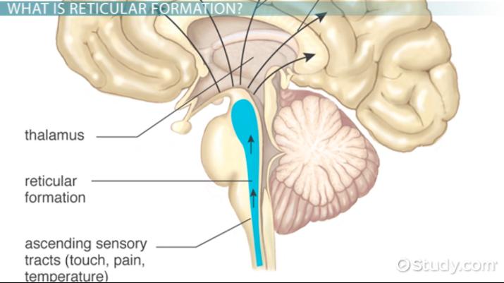
Reticular Formation
-area in brainstem where white + grey matter have netlike arrangement
-has reticular activating system (RAS)
maintains consciousness with stimuli
Alerts cerebral cortex to sensory signals to wake up
Cerebellum
-evaluates movements initiated by motor areas in cerebrum being carried out
-adjusts posture
-programs and fine tunes voluntary and involuntary movements
-store memories of learned movements (eg; typing)
-dmg can cause atoxia (inability to coordinate movements)
no proprioception, act drunk
Thalamus
-2 halves held tgt by interthalamic adhesion, has 7 major nuclei groups
-relay station for sensory and motor functions
sensory
Small piece of received Sensory info from spinal cord n brainstem goes through thalamus → cerebral cortex (like focusing on one sound in a noisy room, like a filter)
Pain, temp, pressure, hearing
Motor
Send info from cerebellum n basal nuclei → primary motor area of cortex
Relay station btw cerebrum parts
-consciousness, learning, memory, emotions
Hypothalamus
-has 12 nuclei in 4 regions
-controls many body activities, a major regulator of homeostasis
-connects to pituitary gland, ECF → blood (where endocrine n nervous meet)
Hypothalamus Functions
Control of ANS: regulator of visceral activities (eg; regulation of HR, food movement in GI tract)
Hormone Production: releasing or inhibiting hormones released into capillary network (stimulate/inhibit secretion of anterior pituitary hormones), make ADH
Regulation of Emotional n Behavioural Patterns: w/ limbic system, expression of rage, pain, pleasure
Regulation of eating and drinking: thirst, feeding, and satiety centers
Control of Body Temp: when blood flowing through hypo is too high/low, hypo directs ANS accordingly
Regulation of Circadian Rhythms n Consciousness: linked to input from retina to establish 24hr cycle
Epithalamus
-above behind thalamus, contains pineal gland
endocrine gland, secretes melatonin during darkness, promotes sleepiness
Circumventricular Organs
-lie in 3rd ventricle’s wall, no BBB
-monitors chemical changes in blood
-coordinate homeostatic activities of endocrine and nervous
-regulates BP, hunger, thirst
Cerebrum
-gives us high thinking
-outer cerebral cortex, inner cerebral white matter, deep gray matter nuclei, gyri (bumps)
-fissures (deep grooves) and sulcus (shallower grooves) in btw folds
Cerebral Hemispheres n Lobes
5 lobes: Frontal, Temporal, Occipital, Parietal, insula (deep)
All split by longitudinal fissure
Pre-Central Gyrus: primary MOTOR area
Post-Central Gyrus: primary SOMATO SENSORY area
Each hemisphere receives sensory input n generates motor outputs to OPPOSITE side of body
Cerebral White Matters
Under cortex, myelinated axons running in 3 directions
Association tracts: connect/transmit NI’s btw gyri in SAME hemisphere
Commisural Tracts: from gyri of 1 hemisphere to other
Projection Tracts: form descending/ascending tracts that transmit impulses from cerebrum to other parts of brain and spinal cord (eg; diencephalon)
Limbic System
-establishment of emotional states
pain, pleasure, anger, memory, etc
3 Types of Areas in Cerebral Cortex
Sensory Areas: perception of sensations
Motor Areas: initiate movements
Association Areas: memory, emotions, reasoning, personality
Sensory Areas
-reception and interpretation of sensory impulses
-primary visual, auditory, gustatory, olfactory, somatosensory areas
Primary Somatosensory Area
-postcentral gyrus
-input from touch, proprioception, pain, tickle, thermal sensations
-mainly for localization of origin and intensity of sensation
Primary Visual Area
-tip of occipital bone
-receives impulses that convey info (eg; shape, color, movement) for vision
Primary Auditory Area
-superior part of temporal lobe