L24- Brain Ageing

0.0(0)
Studied by 0 people
call kaiCall Kai
learnLearn
examPractice Test
spaced repetitionSpaced Repetition
heart puzzleMatch
flashcardsFlashcards
GameKnowt Play
Card Sorting

1/50

encourage image

There's no tags or description

Looks like no tags are added yet.

Last updated 6:14 PM on 5/24/26
Name
Mastery
Learn
Test
Matching
Spaced
Call with Kai

No analytics yet

Send a link to your students to track their progress

51 Terms

1
New cards

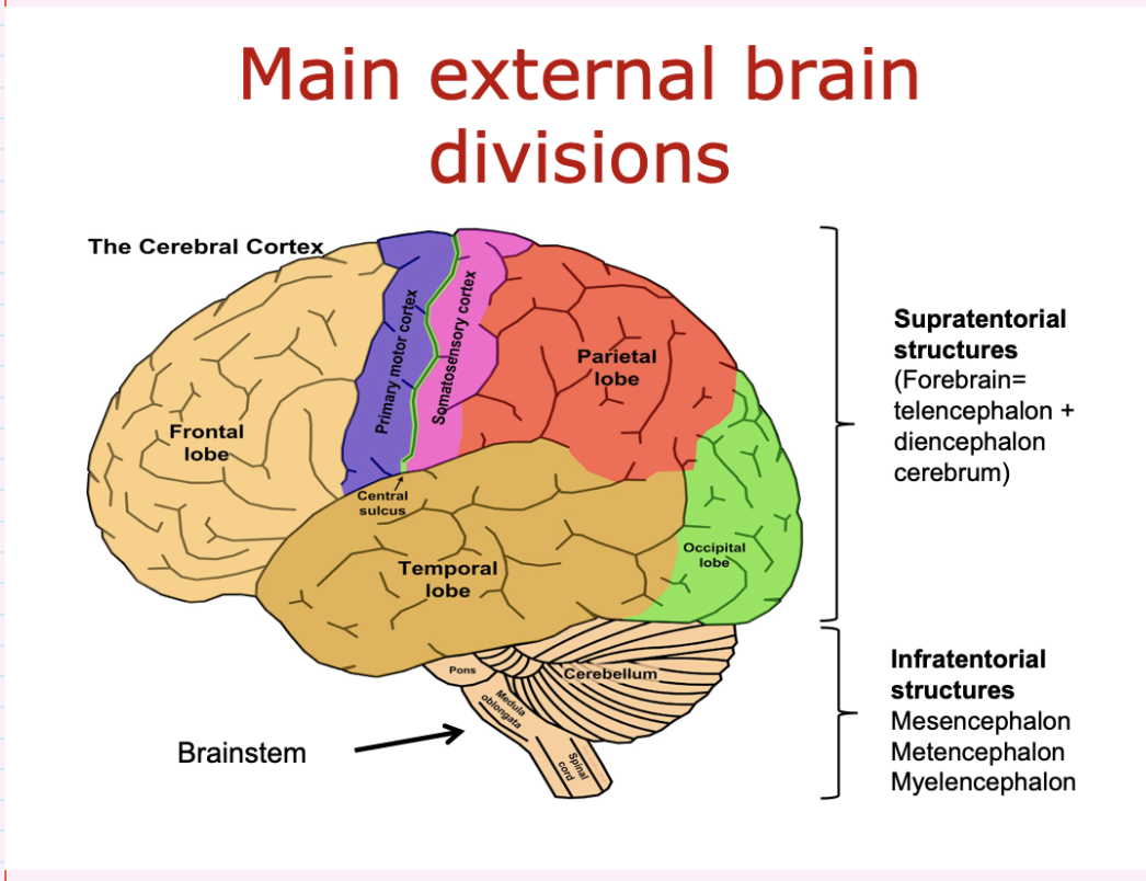
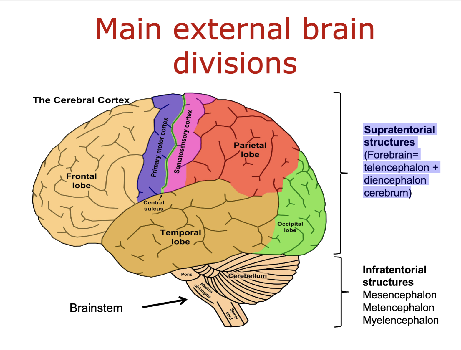
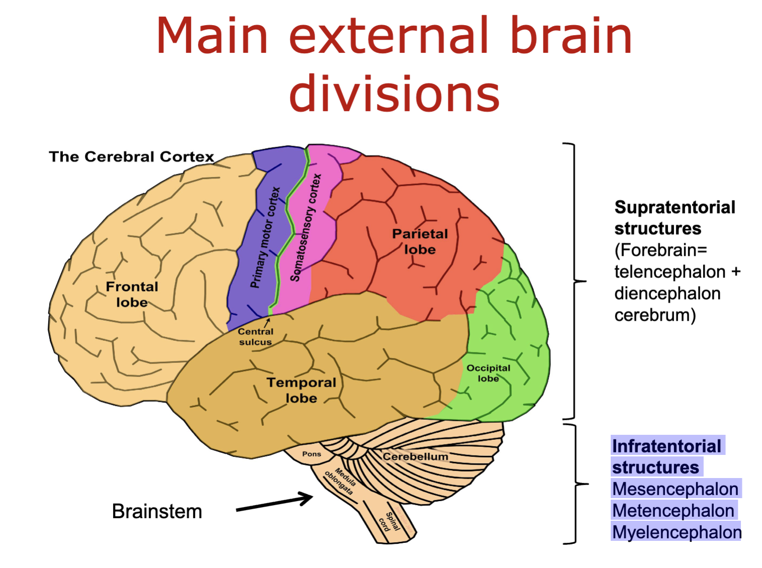
what are the main external brain divisions

  • frontal lobe

  • temporal lobe

  • occipital lobe

  • parietal lobe

  • brainstem

  • cerebellum

<ul><li><p>frontal lobe</p></li><li><p>temporal lobe</p></li><li><p>occipital lobe</p></li><li><p>parietal lobe </p></li><li><p>brainstem</p></li><li><p>cerebellum</p></li></ul><p></p>
2
New cards

what are the supratentorial structures of the brain

forebrain:

  • telencephalon

  • diencephalon cerebrum

<p>forebrain:</p><ul><li><p>telencephalon</p></li><li><p>diencephalon cerebrum</p></li></ul><p></p>
3
New cards

what are the infratentorial structures of the brain

  • Mesencephalon

  • Metencephalon

  • Myelencephalon

<ul><li><p>Mesencephalon</p></li></ul><ul><li><p>Metencephalon</p></li><li><p>Myelencephalon</p></li></ul><p></p>
4
New cards

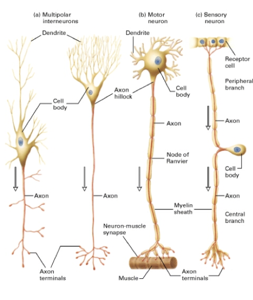
what are the different types of neurones in the brain

  1. multipolar neurones

  • interneurones

  • motorneurones

  1. sensory/bipolar neurones

<ol><li><p>multipolar neurones</p></li></ol><ul><li><p>interneurones</p></li></ul><ul><li><p>motorneurones</p></li></ul><ol start="2"><li><p>sensory/bipolar neurones </p></li></ol><p></p>
5
New cards

what do multipolar neurones do

  1. Interneurones- form all the neural wiring within the CNS.

  2. Motor Neurons- carry signals from the CNS to muscles and glands (efferent neurones).

Multipolar neurones have many processes originating from the cell body (e.g. spinal motor neurones, pyramidal neurones, Purkinje cells.)

6
New cards

what do sensory/bipolar neurons do

  • Carry messages from the body's sense receptors (eyes, ears, etc.) to the CNS (afferent neurones)

  • Account for 0.9% of all neurones

  • They have two axons (instead of an axon and a dendrite). One axon communicates with the sense organ; the other axon communicates with the CNS (e.g. dorsal root ganglion cells in the spinal cord).

7
New cards

what are the 2 forms of diagnostic brain imaging techniques

structure- CT and MRI/MRS

function- PECT/SPECT and EEG/MEG
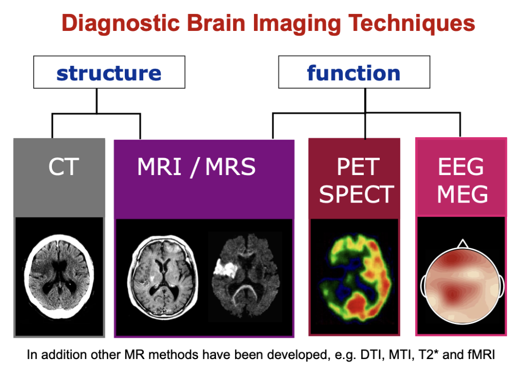
<p>structure- CT and MRI/MRS</p><p>function- PECT/SPECT and EEG/MEG</p>
8
New cards

how does the functional capacity of the brain change throughout life
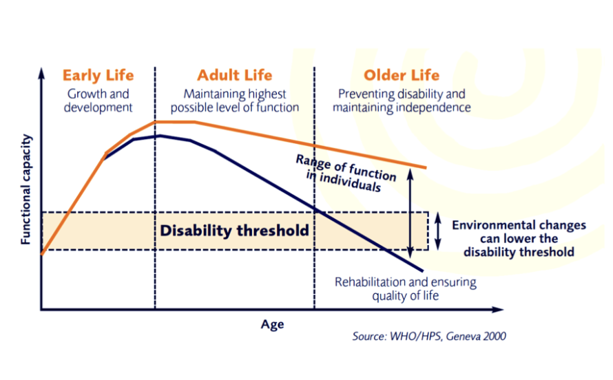
knowt flashcard image
9
New cards

what is meant by multifactorial susceptibility

a mix of genetic and acquired causes
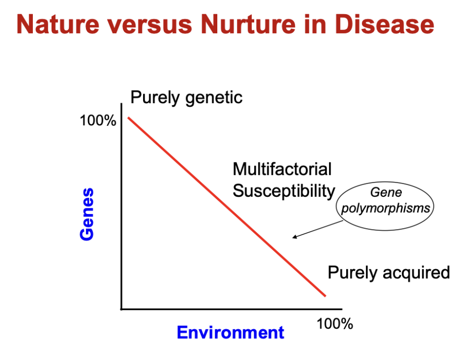
<p>a mix of genetic and acquired causes </p>
10
New cards

how does ageing and brain function relate
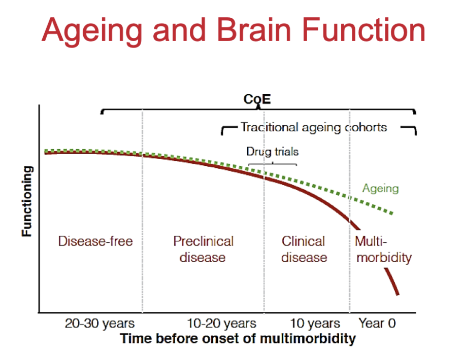
knowt flashcard image
11
New cards

what happens to the brain as we age

  • ventricles enlarge

  • cortex gets thinner- lose about 0.5% per year
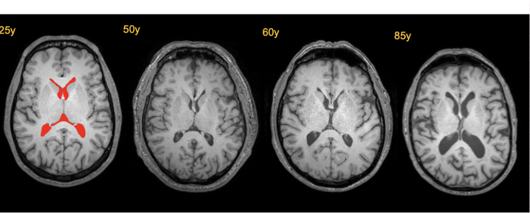
<ul><li><p>ventricles enlarge</p></li><li><p>cortex gets thinner- lose about 0.5% per year</p></li></ul><p></p>
12
New cards

what happens to brain atrophy during ageing

  • Reduced brain volume (atrophy)

  • Widened sulci (more space between gyri)

  • Enlarged ventricles

  • In dementia:

    • Changes are more pronounced

    • Greater loss of cortical and subcortical tissue
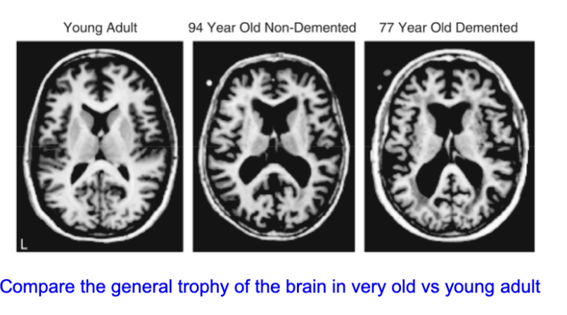
<ul><li><p><strong>Reduced brain volume (atrophy)</strong></p></li><li><p><strong>Widened sulci</strong> (more space between gyri)</p></li><li><p><strong>Enlarged ventricles</strong></p></li></ul><ul><li><p>In dementia:</p><ul><li><p>Changes are <strong>more pronounced</strong></p></li><li><p>Greater loss of <strong>cortical and subcortical tissue</strong></p></li></ul></li></ul><p></p>
13
New cards

what happens to the frontal lobe during ageing

frontal lobe volume declines during ageing
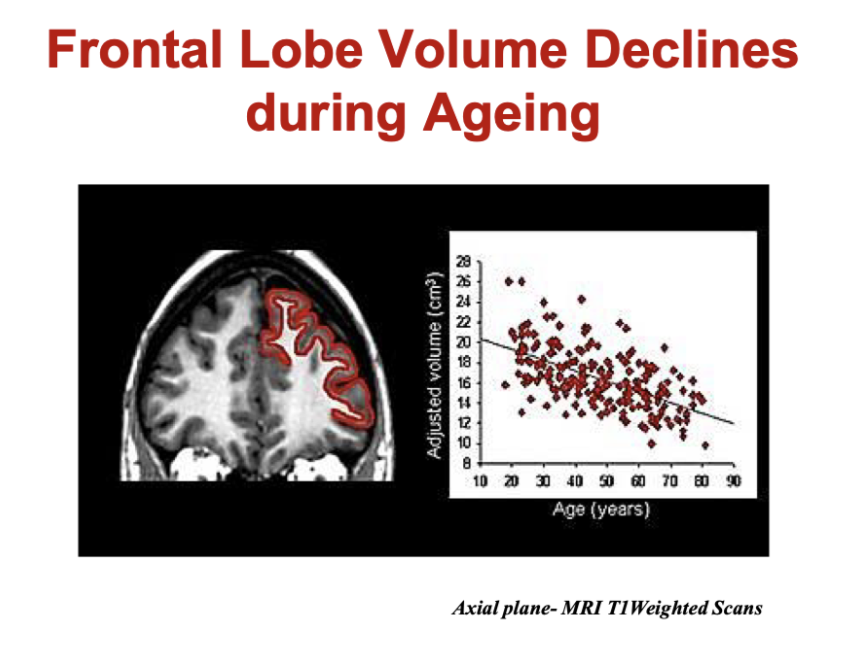
<p>frontal lobe volume declines during ageing </p>
14
New cards

what happens to brain structure in dementia

  • atrophy is greater in alzheimer’s diseases (AD) than in dementia with lewy bodies (DLB)

  • atrophy of medial temporal lobes structures most likely in AD
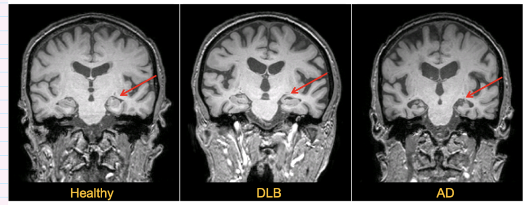
<ul><li><p>atrophy is greater in alzheimer’s diseases (AD) than in dementia with lewy bodies (DLB)</p></li><li><p>atrophy of medial temporal lobes structures most likely in AD</p></li></ul><p></p>
15
New cards

Which brain regions show early volume loss in Alzheimer’s disease?

  • Early atrophy occurs in:

    • Hippocampus

    • Entorhinal cortex

  • Compared to healthy ageing:

    • AD shows greater and earlier volume loss in these regions

  • Key idea:

    • These areas are critical for memory → explains early memory symptoms in AD
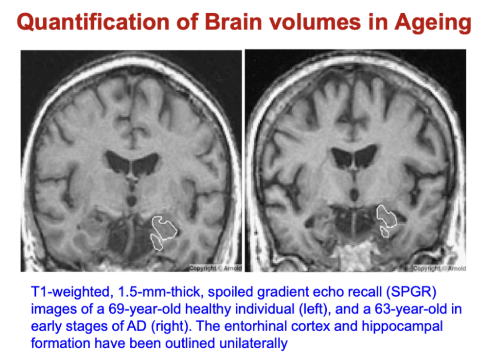
<ul><li><p>Early atrophy occurs in:</p><ul><li><p><strong>Hippocampus</strong></p></li><li><p><strong>Entorhinal cortex</strong></p></li></ul></li><li><p>Compared to healthy ageing:</p><ul><li><p>AD shows <strong>greater and earlier volume loss</strong> in these regions</p></li></ul></li><li><p>Key idea:</p><ul><li><p>These areas are critical for <strong>memory → explains early memory symptoms in AD</strong></p></li></ul></li></ul><p></p>
16
New cards

How does hippocampal atrophy differ between normal ageing and Alzheimer’s disease?

  • Normal ageing:

    • ~1.6% volume loss per year

  • Alzheimer’s disease (AD):

    • ~4.0% volume loss per year

    • 2× faster atrophy than controls

  • Key idea:

    • Ageing → gradual hippocampal loss

    • AD → accelerated medial temporal lobe atrophy
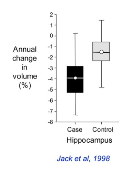
<ul><li><p><strong>Normal ageing:</strong></p><ul><li><p>~<strong>1.6% volume loss per year</strong></p></li></ul></li><li><p><strong>Alzheimer’s disease (AD):</strong></p><ul><li><p>~<strong>4.0% volume loss per year</strong></p></li><li><p>≈ <strong>2× faster atrophy</strong> than controls</p></li></ul></li><li><p>Key idea:</p><ul><li><p>Ageing → gradual hippocampal loss</p></li><li><p>AD → <strong>accelerated medial temporal lobe atrophy</strong></p></li></ul></li></ul><p></p>
17
New cards

what are the patterns of hippocampal neuronal loss in ageing and AD

  • most distinctive AD related neuron loss was seen in the CA1 region (hippocampus subfield)- in normal ageing pr MCI almost no neuron loss

  • - neurodegenerative processes associated with AD qualitatively different- AD is not accelerated by ageing but a distinct pathological process
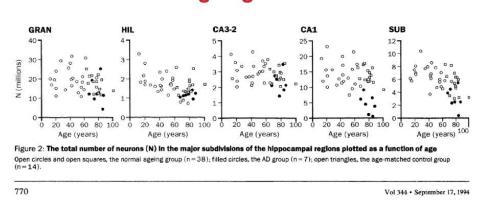
<ul><li><p>most distinctive AD related neuron loss was seen in the CA1 region (hippocampus subfield)- in normal ageing pr MCI almost no neuron loss</p></li><li><p>- neurodegenerative processes associated with AD qualitatively different- AD is not accelerated by ageing but a distinct pathological process</p></li></ul><p></p>
18
New cards

how does the appearance of hippocampal neurones change during ageing

With ageing:

  • Reduced dendritic length and branching

  • Simpler neuronal structure (less connectivity)
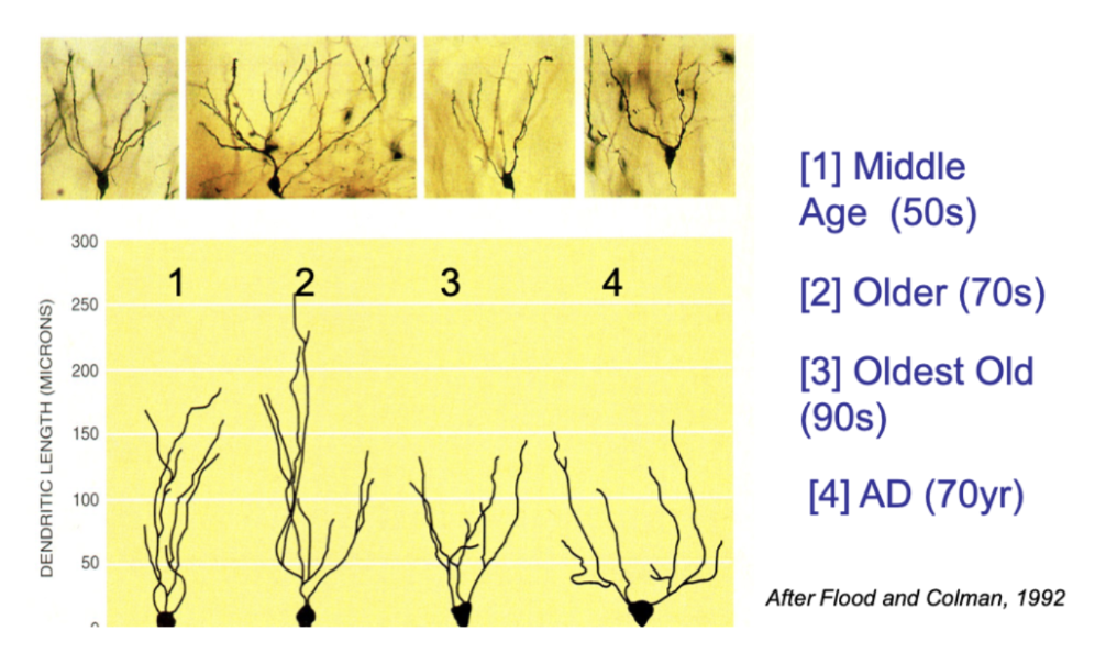
<p>With ageing:</p><ul><li><p><strong>Reduced dendritic length and branching</strong></p></li><li><p>Simpler neuronal structure (less connectivity)</p></li></ul><p></p>
19
New cards

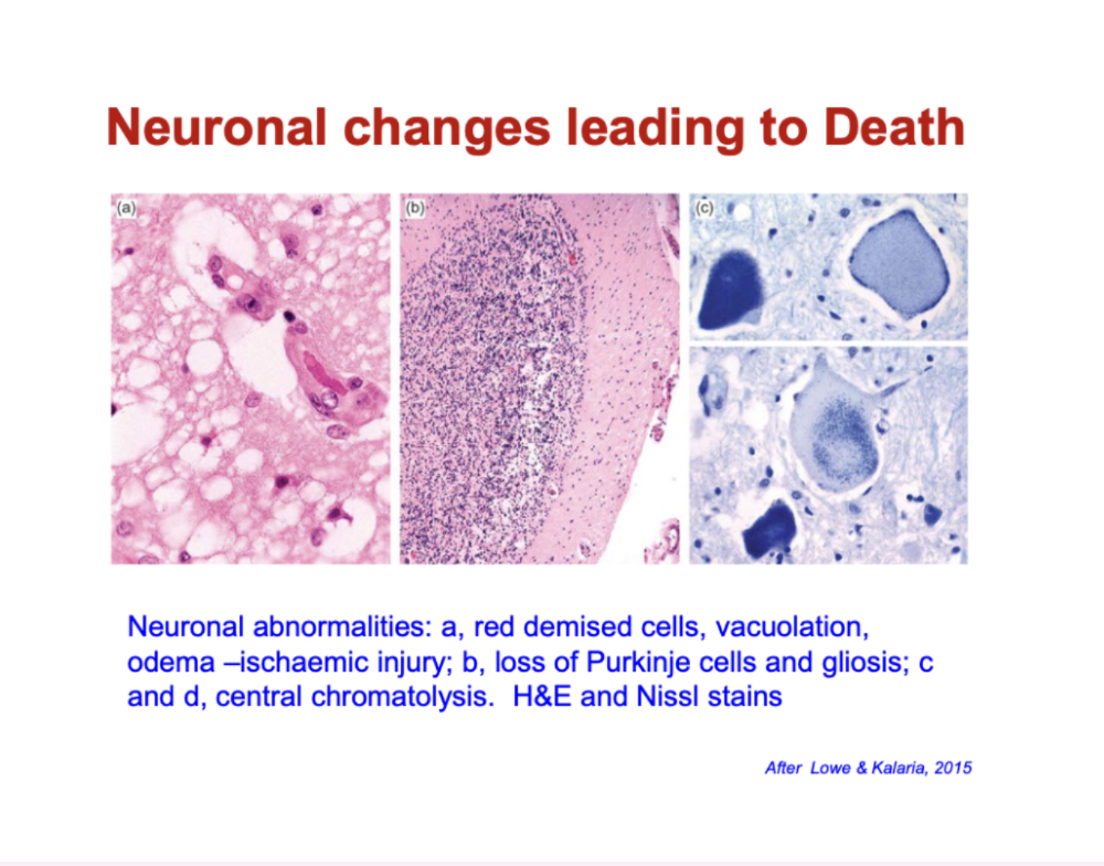
What histological changes indicate neuronal injury and death?

  • Early injury (e.g. ischaemia):

    • “Red neurons” (shrunken, intensely stained)

    • Vacuolation & oedema (cell swelling/damage)

  • Progression:

    • Loss of neurons (e.g. Purkinje cells)

    • Gliosis (increase in supporting glial cells after injury)

  • Cellular breakdown:

    • Chromatolysis (loss/disruption of Nissl substance → impaired protein synthesis)
<ul><li><p><strong>Early injury (e.g. ischaemia):</strong></p><ul><li><p><strong>“Red neurons”</strong> (shrunken, intensely stained)</p></li><li><p><strong>Vacuolation &amp; oedema</strong> (cell swelling/damage)</p></li></ul></li><li><p><strong>Progression:</strong></p><ul><li><p><strong>Loss of neurons</strong> (e.g. Purkinje cells)</p></li><li><p><strong>Gliosis</strong> (increase in supporting glial cells after injury)</p></li></ul></li><li><p><strong>Cellular breakdown:</strong></p><ul><li><p><strong>Chromatolysis</strong> (loss/disruption of Nissl substance → impaired protein synthesis)</p></li></ul></li></ul><p></p>
20
New cards

what are the changes in mechanisms of neurodegeneration during ageing

↑ Oxidative stress mechanisms

↑ Neuroinflammatory responses

↑ Abnormal Protein-Protein Interactions

↓The Ubiquitin-Proteasome system

↑ The autophagy-lysosome system (autophagy)

↑ Apoptosis and delayed cell death

21
New cards

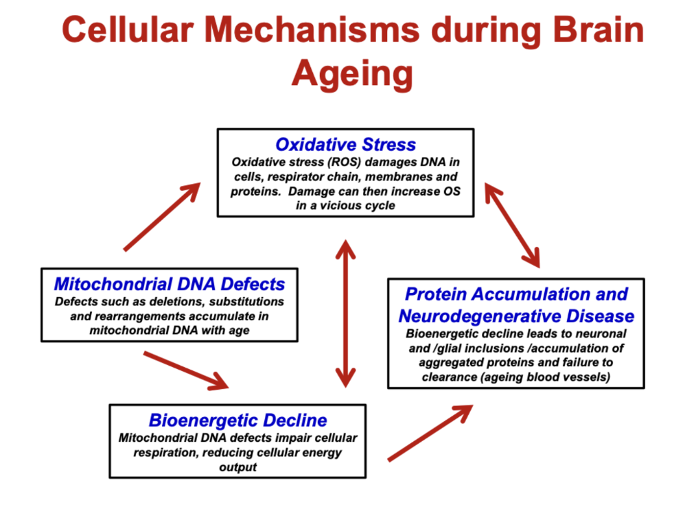
What are the key cellular mechanisms driving brain ageing?

  • Oxidative stress (ROS):

    • Damages DNA, proteins, membranes

    • Creates a vicious cycle of further damage

  • Mitochondrial DNA defects:

    • Accumulate with age

    • Impair mitochondrial function

  • Bioenergetic decline:

    • ↓ cellular respiration → ↓ energy (ATP)

  • Protein accumulation:

    • Misfolded/aggregated proteins build up

    • Impaired clearance → neurodegeneration

  • Key idea:

    • These processes are interconnected and amplify each other → neuronal dysfunction and disease
<ul><li><p><strong>Oxidative stress (ROS):</strong></p><ul><li><p>Damages DNA, proteins, membranes</p></li><li><p>Creates a <strong>vicious cycle</strong> of further damage</p></li></ul></li><li><p><strong>Mitochondrial DNA defects:</strong></p><ul><li><p>Accumulate with age</p></li><li><p>Impair mitochondrial function</p></li></ul></li><li><p><strong>Bioenergetic decline:</strong></p><ul><li><p>↓ cellular respiration → ↓ energy (ATP)</p></li></ul></li><li><p><strong>Protein accumulation:</strong></p><ul><li><p>Misfolded/aggregated proteins build up</p></li><li><p>Impaired clearance → <strong>neurodegeneration</strong></p></li></ul></li><li><p><strong>Key idea:</strong></p><ul><li><p>These processes are <strong>interconnected</strong> and amplify each other → neuronal dysfunction and disease</p></li></ul></li></ul><p></p>
22
New cards

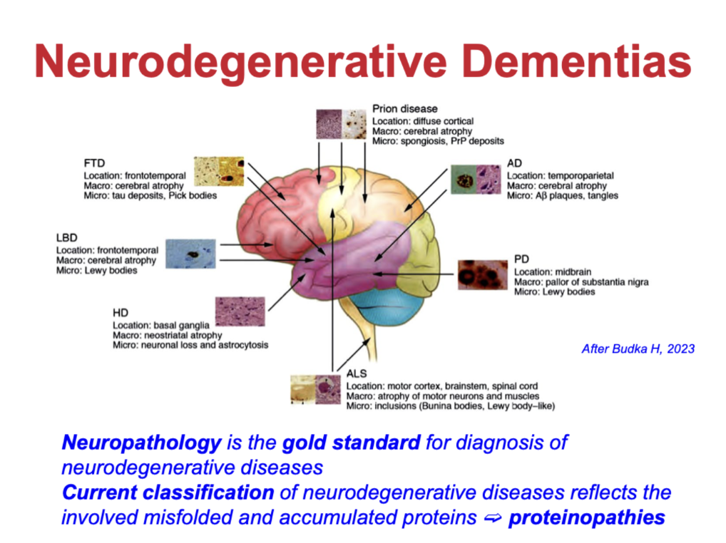
How are neurodegenerative dementias classified?

  • Classified by misfolded/accumulated proteins → “proteinopathies”

  • Examples:

    • Alzheimer’s disease (AD): Aβ plaques, tau tangles

    • Parkinson’s disease (PD) / Lewy body dementia: α-synuclein (Lewy bodies)

    • Frontotemporal dementia (FTD): tau or TDP-43

    • Prion diseases: misfolded prion protein

    • Huntington’s disease (HD): mutant huntingtin

    • ALS: protein inclusions (e.g. TDP-43)

  • Neuropathology = gold standard for diagnosis

    • Disease type depends on which protein accumulates and where in the brain
<ul><li><p>Classified by <strong>misfolded/accumulated proteins → “proteinopathies”</strong></p></li><li><p>Examples:</p><ul><li><p><strong>Alzheimer’s disease (AD):</strong> Aβ plaques, tau tangles</p></li><li><p><strong>Parkinson’s disease (PD) / Lewy body dementia:</strong> α-synuclein (Lewy bodies)</p></li><li><p><strong>Frontotemporal dementia (FTD):</strong> tau or TDP-43</p></li><li><p><strong>Prion diseases:</strong> misfolded prion protein</p></li><li><p><strong>Huntington’s disease (HD):</strong> mutant huntingtin</p></li><li><p><strong>ALS:</strong> protein inclusions (e.g. TDP-43)</p></li></ul></li><li><p><strong>Neuropathology = gold standard</strong> for diagnosis</p><ul><li><p>Disease type depends on <strong>which protein accumulates and where in the brain</strong></p></li></ul></li></ul><p></p>
23
New cards

how do we define dementia

impairment in any of

  • memory

  • language

  • visual processing and orientation

  • mood, personality and social skills

  • frontal executive function, including planning and problem solving

causes inability to function independently

24
New cards

how does normal cognitive state change over time
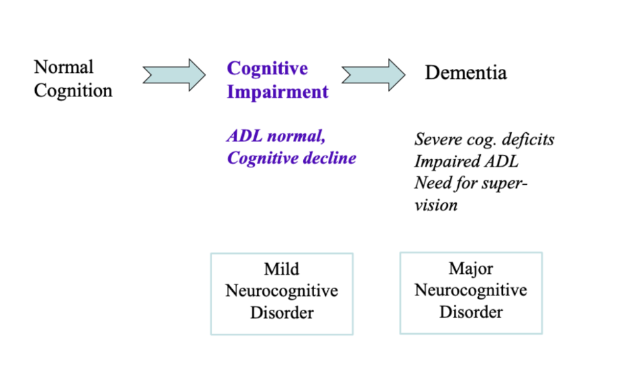
knowt flashcard image
25
New cards

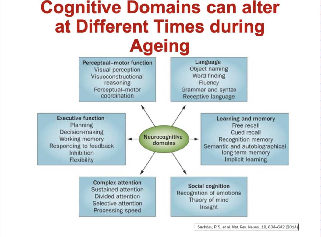
Which cognitive domains are affected during ageing?

  • Executive function: planning, decision-making, working memory, flexibility

  • Memory & learning: recall (free/cued), recognition, long-term memory

  • Language: word finding, fluency, grammar, comprehension

  • Attention: sustained, divided, selective attention, processing speed

  • Perceptual-motor: visual perception, coordination, visuospatial skills

  • Social cognition: emotion recognition, theory of mind, insight

    • Different domains decline at different rates and times during ageing
<ul><li><p><strong>Executive function:</strong> planning, decision-making, working memory, flexibility</p></li><li><p><strong>Memory &amp; learning:</strong> recall (free/cued), recognition, long-term memory</p></li><li><p><strong>Language:</strong> word finding, fluency, grammar, comprehension</p></li><li><p><strong>Attention:</strong> sustained, divided, selective attention, processing speed</p></li><li><p><strong>Perceptual-motor:</strong> visual perception, coordination, visuospatial skills</p></li><li><p><strong>Social cognition:</strong> emotion recognition, theory of mind, insight</p><ul><li><p>Different domains decline at <strong>different rates and times</strong> during ageing</p></li></ul></li></ul><p></p>
26
New cards

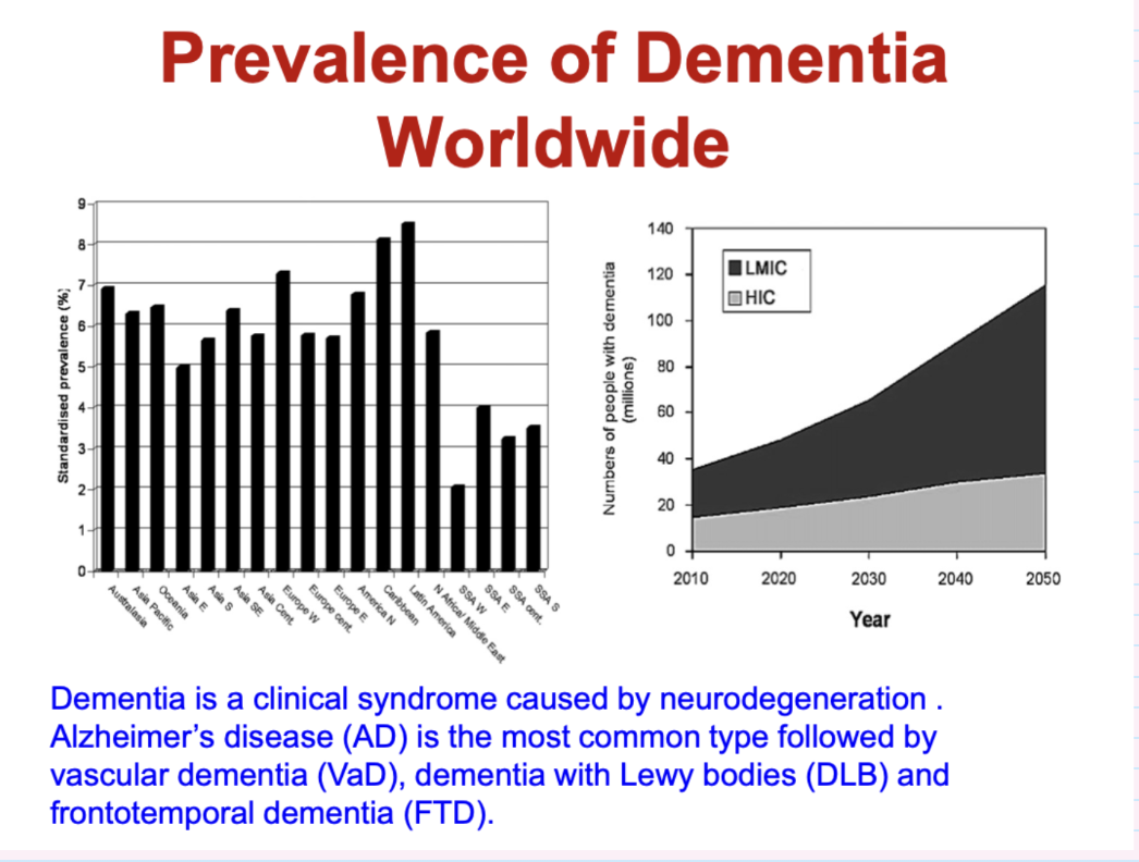
describe the prevelence if dementia worldwide

Dementia is a clinical syndrome caused by neurodegeneration . Alzheimer’s disease (AD) is the most common type followed by vascular dementia (VaD), dementia with Lewy bodies (DLB) and frontotemporal dementia (FTD).
<p>Dementia is a clinical syndrome caused by neurodegeneration . Alzheimer’s disease (AD) is the most common type followed by vascular dementia (VaD), dementia with Lewy bodies (DLB) and frontotemporal dementia (FTD).</p><p></p>
27
New cards

what are the most common causes of degenerative dementias

  • Alzheimer’s disease: ~55–70% (most common)

  • Vascular dementia: ~15–25%

  • Other dementias (10–30%):

    • Lewy body dementia

    • Parkinson’s disease dementia

    • Frontotemporal dementia

28
New cards

whatt are the 4 types of dementia

  • alzheimer’s (50-75%)

  • vascular (20-30%)

  • lewy body (10-25%)

  • frontotemporal (10-15%)

29
New cards

give 6 common ageing related brain disorders and dementias

  • Alzheimer’s Disease

  • Parkinson’s Disease

  • Dementia with Lewy Bodies

  • Frontotemporal Dementia

  • Prion Diseases

  • Vascular Dementia

30
New cards

what is alzheimer’s disease

a progressive degenerative brain disorder and the most common cause of dementia

31
New cards

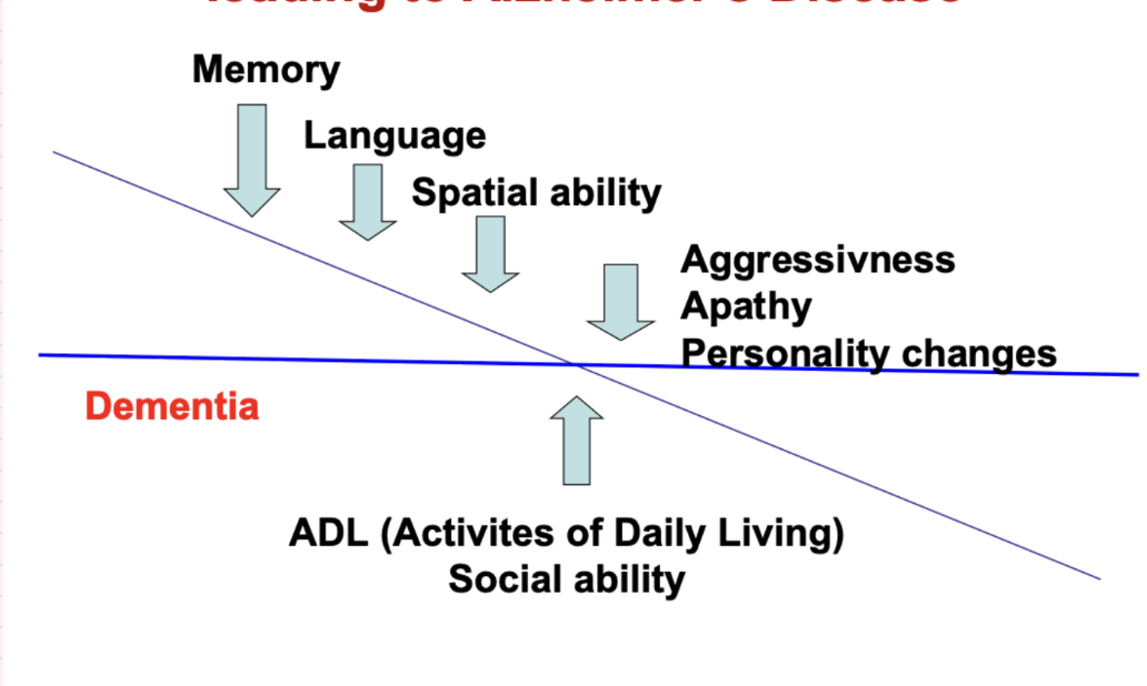
how do cognitive ageing related thresholds lead to alzheimer’s disease

once decline affects ADL (activities of daily living) and social ability- considered dementia
<p>once decline affects ADL (activities of daily living) and social ability- considered dementia </p>
32
New cards

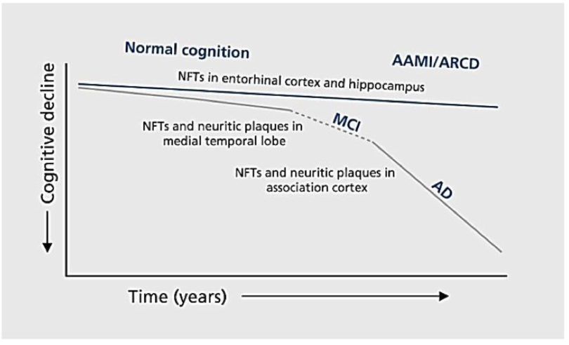
how does cognition and brain pathology cange during ageing

progressive accumulation of brain pathology increases damage and decreases cognitive functions
<p>progressive accumulation of brain pathology increases damage and decreases cognitive functions </p>
33
New cards

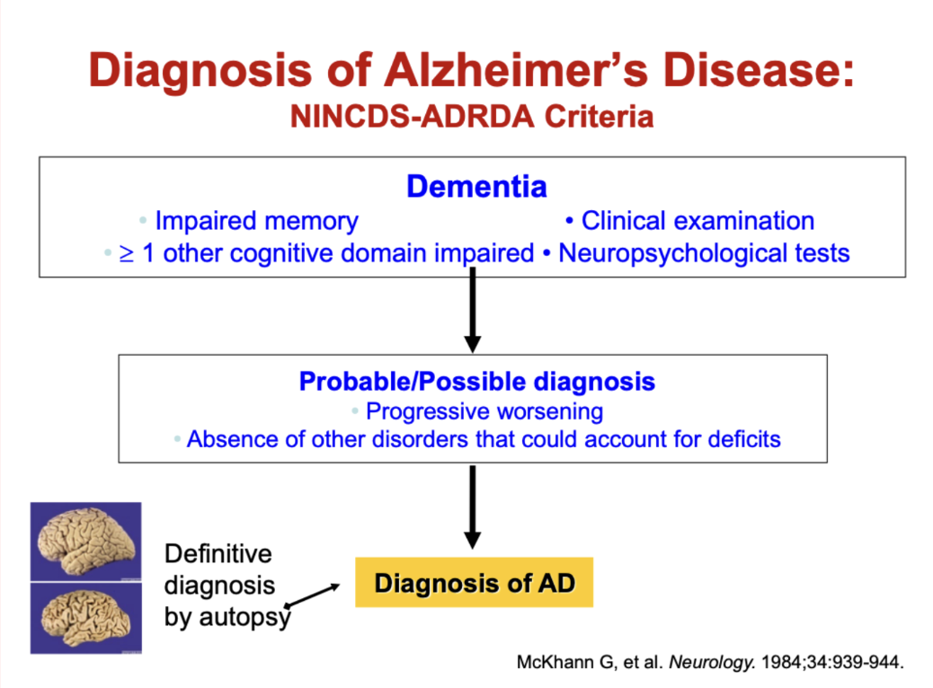
How is Alzheimer’s disease diagnosed (NINCDS-ADRDA criteria)?

  • Dementia required:

    • Impaired memory

    • ≥1 other cognitive domain impaired

    • Assessed by clinical exam + neuropsychological testing

  • Probable/Possible AD:

    • Progressive worsening over time

    • No other disorder explaining symptoms

  • Definitive diagnosis:

    • Confirmed by autopsy

<ul><li><p><strong>Dementia required:</strong></p><ul><li><p>Impaired memory</p></li><li><p>≥1 other cognitive domain impaired</p></li><li><p>Assessed by clinical exam + neuropsychological testing</p></li></ul></li><li><p><strong>Probable/Possible AD:</strong></p><ul><li><p>Progressive worsening over time</p></li><li><p>No other disorder explaining symptoms</p></li></ul></li><li><p><strong>Definitive diagnosis:</strong></p><ul><li><p>Confirmed by <strong>autopsy</strong></p></li></ul></li></ul><p></p>
34
New cards

what are the key pathological hallmarks od AD

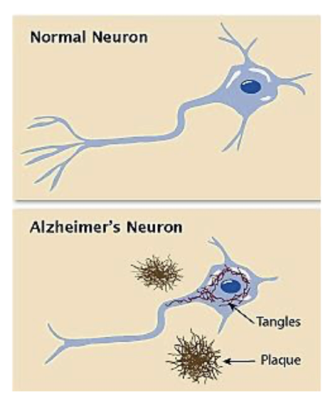
accumulation of brain pathology with age

  • amyloid or neuritic plaques (NP)

  • neurofibrillary tangles (NFT)

<p>accumulation of brain pathology with age </p><ul><li><p>amyloid or neuritic plaques (NP)</p></li><li><p>neurofibrillary tangles (NFT)</p></li></ul><p></p>
35
New cards

what are he pathological features of AD

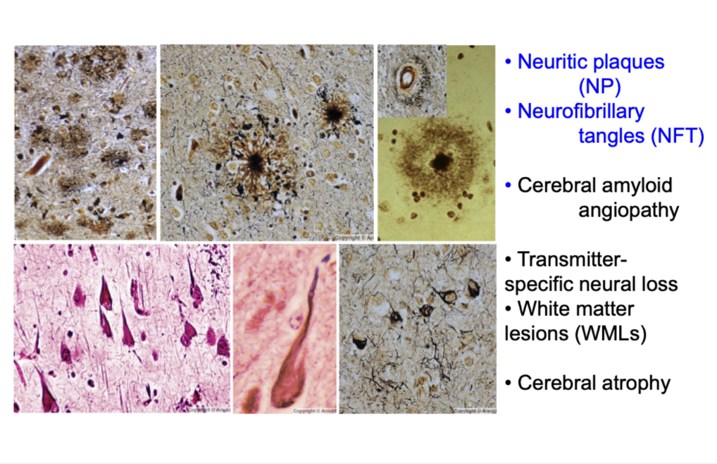
  • neuritic plaques (NP)

  • neurofibrillary tangles (NFT)

  • cerebral amyloid angiopathy

  • transmitter-specific neural liss

  • white matter lesions (WML)

  • cerebral atrophy

<ul><li><p>neuritic plaques (NP)</p></li><li><p>neurofibrillary tangles (NFT)</p></li><li><p>cerebral amyloid angiopathy</p></li><li><p>transmitter-specific neural liss</p></li><li><p>white matter lesions (WML)</p></li><li><p>cerebral atrophy</p></li></ul><p></p>
36
New cards

describe the change in brain amyloid plaques with ageing
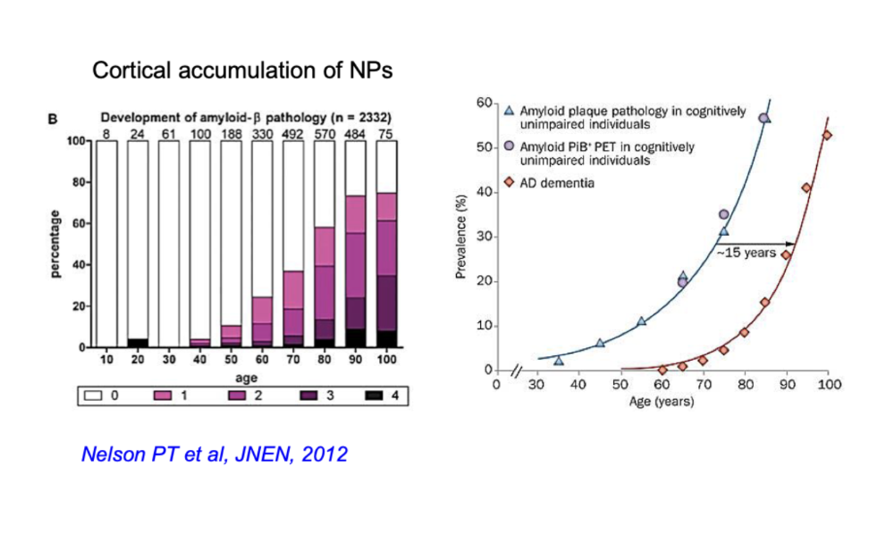
knowt flashcard image
37
New cards

what are amyloid plaques

Aβ protein deposits

  • Aβ in neocortex and hippocampal formation- EM shows fibrillae amyloid
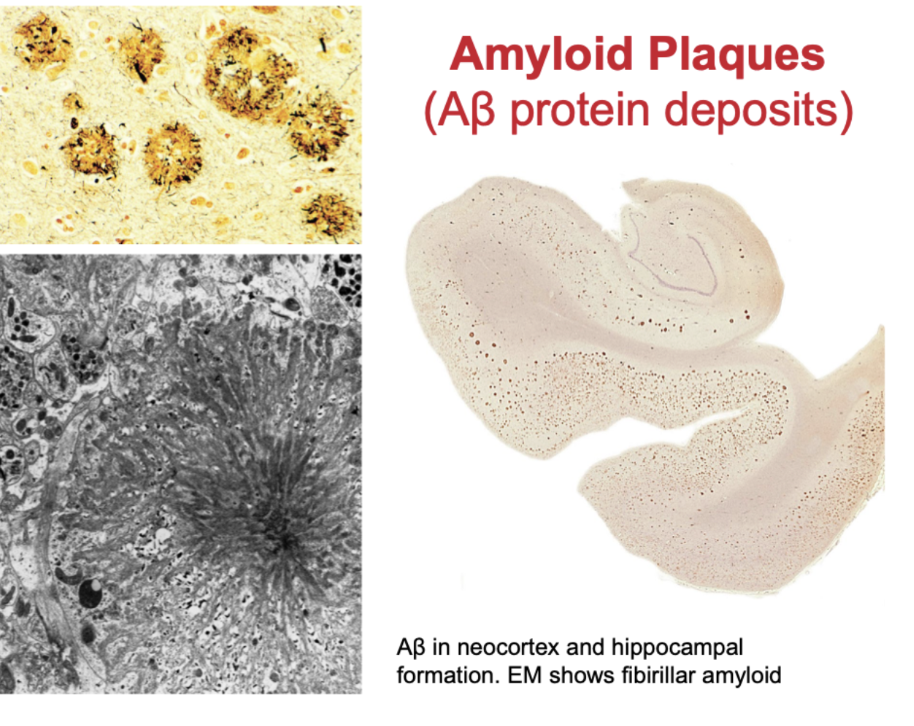
<p>Aβ protein deposits </p><ul><li><p>Aβ in neocortex and hippocampal formation- EM shows fibrillae amyloid</p></li></ul><p></p>
38
New cards

What initiates the amyloid β cascade in Alzheimer’s disease?

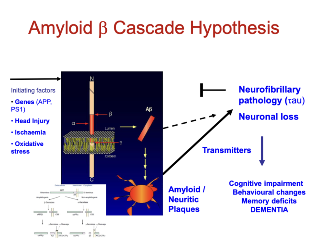
  • Triggers:

    • Genetic mutations (APP, PS1)

    • Head injury

    • Ischaemia

    • Oxidative stress

  • Key process:

    • Abnormal APP processing → amyloid-β (Aβ) production

    • Aβ aggregates → amyloid (neuritic) plaques
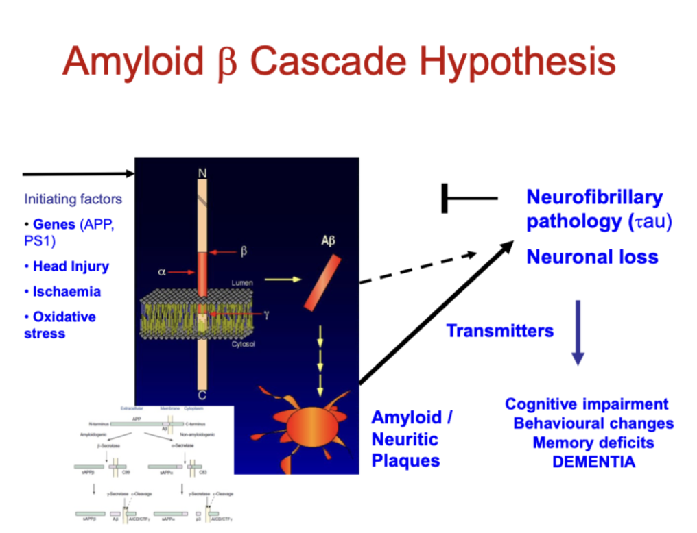
<ul><li><p><strong>Triggers:</strong></p><ul><li><p>Genetic mutations (APP, PS1)</p></li><li><p>Head injury</p></li><li><p>Ischaemia</p></li><li><p>Oxidative stress</p></li></ul></li><li><p><strong>Key process:</strong></p><ul><li><p>Abnormal APP processing → <strong>amyloid-β (Aβ) production</strong></p></li><li><p>Aβ aggregates → <strong>amyloid (neuritic) plaques</strong></p></li></ul></li></ul><p></p>
39
New cards

What are the downstream effects of the amyloid β cascade?

  • Pathological changes:

    • Tau pathology (neurofibrillary tangles)

    • Neuronal loss

    • Neurotransmitter deficits

  • Clinical outcome:

    • Cognitive impairment

    • Memory loss

    • Behavioural changes → Dementia

<ul><li><p><strong>Pathological changes:</strong></p><ul><li><p>Tau pathology (neurofibrillary tangles)</p></li><li><p>Neuronal loss</p></li><li><p>Neurotransmitter deficits</p></li></ul></li><li><p><strong>Clinical outcome:</strong></p><ul><li><p>Cognitive impairment</p></li><li><p>Memory loss</p></li><li><p>Behavioural changes → <strong>Dementia</strong></p></li></ul></li></ul><p></p>
40
New cards

What does the updated Amyloid β cascade hypothesis explain?

  • Aβ accumulation (genetic risk) initiates disease

  • Microglia become activated → inflammation + loss of normal function

  • Neuronal damage develops around plaques

  • Tau pathology spreads through the brain (prion-like)

  • Leads to progressive neuronal and synaptic loss

  • Key idea:
    Alzheimer’s is a multistep process involving Aβ, inflammation, and tau → ultimately causing dementia
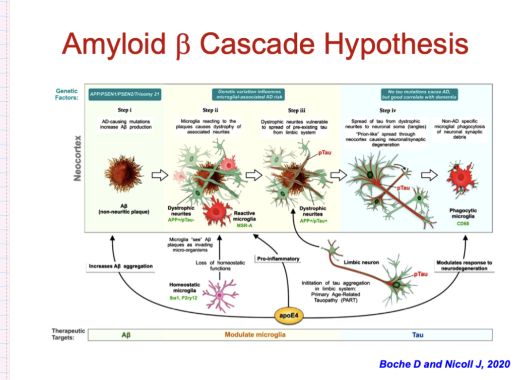
<ul><li><p><strong>Aβ accumulation (genetic risk)</strong> initiates disease</p></li><li><p><strong>Microglia become activated</strong> → inflammation + loss of normal function</p></li><li><p><strong>Neuronal damage develops around plaques</strong></p></li><li><p><strong>Tau pathology spreads through the brain</strong> (prion-like)</p></li><li><p>Leads to <strong>progressive neuronal and synaptic loss</strong></p></li><li><p><strong>Key idea:</strong><br>Alzheimer’s is a <strong>multistep process</strong> involving Aβ, inflammation, and tau → ultimately causing dementia</p></li></ul><p></p>
41
New cards

what are neurofibrillary tanges

Hyperphosphorylated tau protein

  • NFT in neocortex and hippocampal formation.

  • EM shows neurofibrillary twisted filaments (tangles)
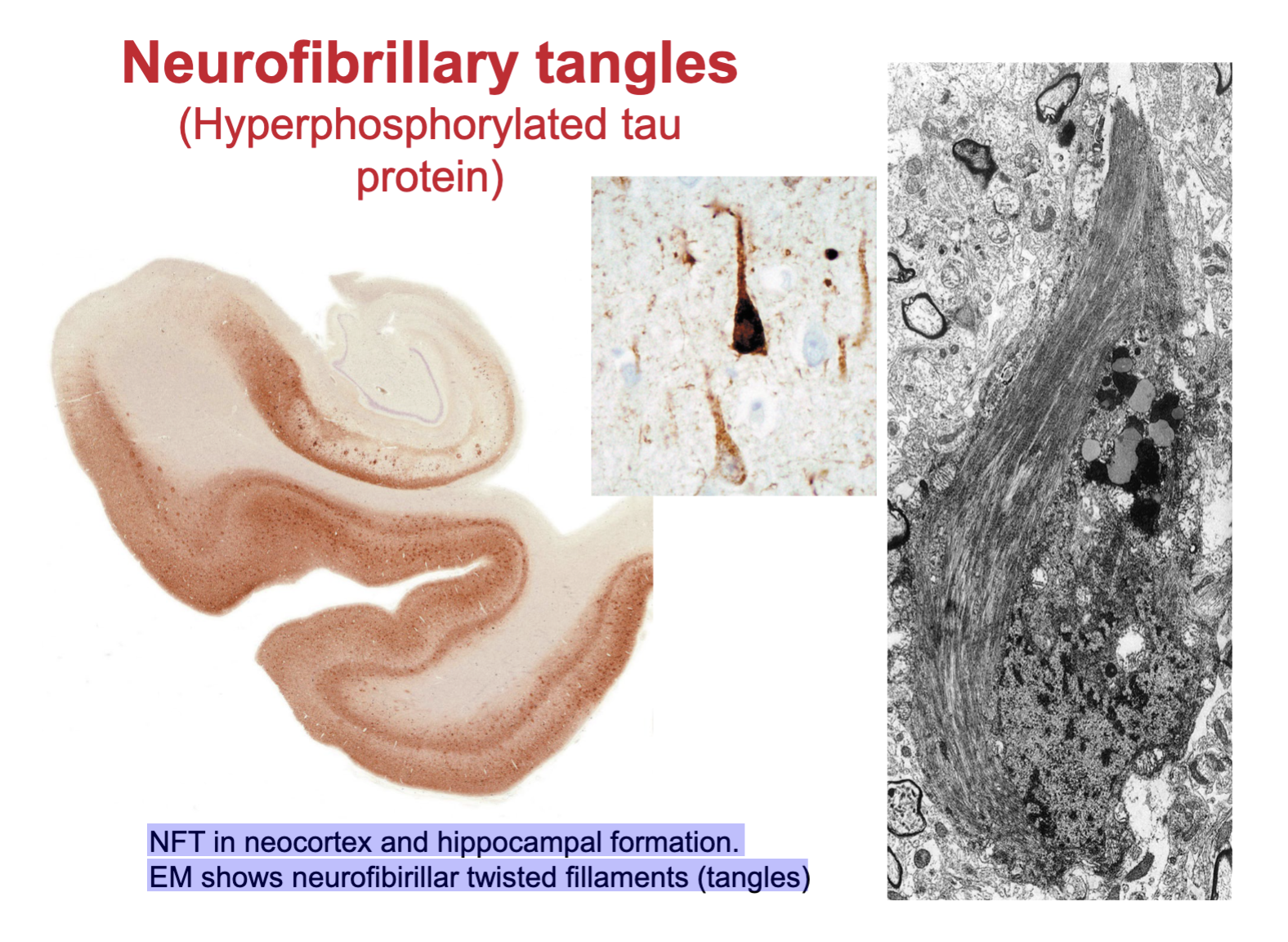
<p>Hyperphosphorylated tau protein</p><ul><li><p>NFT in neocortex and hippocampal formation. </p></li></ul><ul><li><p>EM shows neurofibrillary twisted filaments (tangles)</p></li></ul><p></p>
42
New cards

describe the progression of tau positive neurofbrillary pathology

  • Tau changes from normal → oligomers → filaments → neurofibrillary tangles (NFTs)

  • NFTs accumulate inside neurons → disrupt function

  • Neurons degenerate → release tau → extracellular “ghost” tangles

  • Associated with neuritic plaques and neuropil threads

  • Key idea:
    Tau pathology progressively damages neurons and spreads, driving neurodegeneration
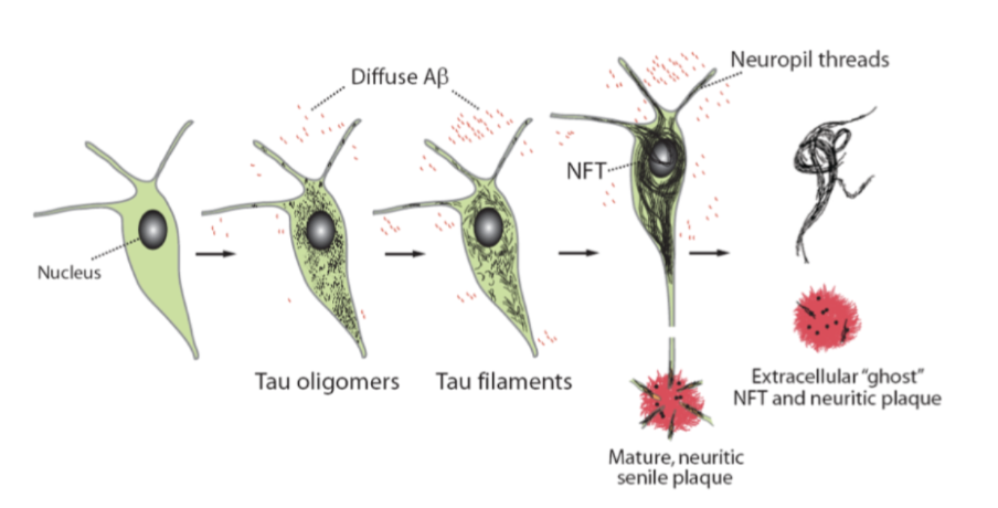
<ul><li><p>Tau changes from <strong>normal → oligomers → filaments → neurofibrillary tangles (NFTs)</strong></p></li><li><p>NFTs accumulate inside neurons → disrupt function</p></li><li><p>Neurons degenerate → release tau → <strong>extracellular “ghost” tangles</strong></p></li><li><p>Associated with <strong>neuritic plaques and neuropil threads</strong></p></li><li><p><strong>Key idea:</strong><br>Tau pathology <strong>progressively damages neurons and spreads</strong>, driving neurodegeneration</p></li></ul><p></p>
43
New cards

What are the main molecular classes of neurodegenerative dementias?

  • Alzheimer’s disease (Aβ + tau)

  • Synucleinopathies:

    • Dementia with Lewy bodies (DLB)

    • Parkinson’s disease dementia (PDD)

  • Tauopathies:

    • Frontotemporal dementia (FTD), PSP, CBD, Pick’s disease

44
New cards

What other disorders can cause neurodegenerative dementia?

  • Frontotemporal dementias (non-tau):

    • TDP-43, ubiquitin, progranulin

  • Prion diseases:

    • Creutzfeldt-Jakob disease, etc.

  • Trinucleotide repeat disorders:

    • Huntington’s disease, spinocerebellar ataxias

  • Motor neuron diseases:

    • ALS, PLS, SMA (with dementia)

  • Key idea:
    Dementias are classified by underlying protein pathology

45
New cards

What protein accumulations are associated with major neurodegenerative diseases?

  • Alzheimer’s disease: Aβ plaques, tau

  • Parkinson’s / DLB: α-synuclein (Lewy bodies)

  • Tauopathies (FTD, PSP, CBD, Pick’s): tau (3R/4R)

  • FTD (non-tau): TDP-43, ubiquitin, progranulin

  • Prion diseases: PrP plaques

46
New cards

What is the key pathological mechanism in neurodegenerative proteinopathies?

  • Diseases involve misfolded proteins that:

    • Form intracellular inclusions or extracellular deposits

    • Are insoluble and aggregate

  • Spread via “seeding” mechanism (prion-like propagation)

  • Examples:

    • Synuclein (Parkinson’s, MSA)

    • Tau (FTD, AD)

    • Polyglutamine (Huntington’s)

  • Key idea:
    Neurodegeneration is driven by toxic protein accumulation and spread

47
New cards

How do Aβ, tau, and α-synuclein spread in dementias?

  • Spread progressively in stages across the brain

  • Aβ: cortex early

  • Tau: limbic → cortex

  • α-synuclein: brainstem → cortex

  • Higher stage = more severe disease / diagnostic pattern
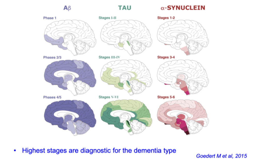
<ul><li><p>Spread <strong>progressively in stages across the brain</strong></p></li><li><p><strong>Aβ:</strong> cortex early</p></li><li><p><strong>Tau:</strong> limbic → cortex</p></li><li><p><strong>α-synuclein:</strong> brainstem → cortex</p></li><li><p><strong>Higher stage = more severe disease / diagnostic pattern</strong></p></li></ul><p></p>
48
New cards

what is programmed cell death (PCD) and apoptosis- mechanisms of degeneration

  • PCD, a process where cell plays an active role in its own demise

  • Critical role in the development of NS and in its response to insult

  • Both anti-PCD and pro-PCD modulators play prominent roles, e.g. ischaemic (stroke) injury

  • Non-apoptotic forms of PCD e.g. autophagy

  • PCDs that do not fit the criteria for either apoptosis or autophagy – suggest other pathways?

49
New cards

what is necrosis vs apoptosis- mechanisms of neurodegeneration

Apoptosis is distinguished from necrosis; mechanism that allows cells to self- destruct when stimulated by the appropriate trigger

  • Initiated if cell is no longer needed or becomes a threat to health

  • Aberrant inhibition or initiation of apoptosis contributes to many disease processes

  • Process of programmed cell death (PCD) has had broader recognition since 1972
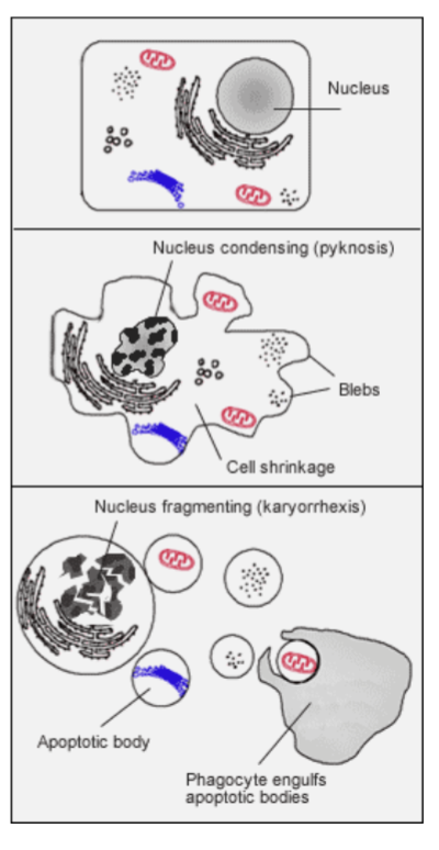
<p>Apoptosis is distinguished from necrosis; mechanism that allows cells to self- destruct when stimulated by the appropriate trigger</p><ul><li><p>Initiated if cell is no longer needed or becomes a threat to health</p></li><li><p>Aberrant inhibition or initiation of apoptosis contributes to many disease processes</p></li><li><p>Process of programmed cell death (PCD) has had broader recognition since 1972</p></li></ul><p></p>
50
New cards

what is autophagy in neurodegeneration

  • Autophagy clears misfolded/aggregate-prone proteins

  • Process: phagophore → autophagosome → lysosome → degradation

  • Protective role:

    • Reduces toxic protein aggregates

    • Limits neuronal damage

  • Key idea:
    Impaired autophagy → protein accumulation → neurodegeneration
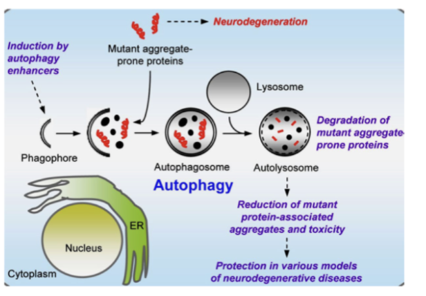
<ul><li><p>Autophagy <strong>clears misfolded/aggregate-prone proteins</strong></p></li><li><p>Process: phagophore → autophagosome → lysosome → degradation</p></li><li><p><strong>Protective role:</strong></p><ul><li><p>Reduces toxic protein aggregates</p></li><li><p>Limits neuronal damage</p></li></ul></li><li><p><strong>Key idea:</strong><br>Impaired autophagy → <strong>protein accumulation → neurodegeneration</strong></p></li></ul><p></p>
51
New cards

what are the processes influencing neuron health

  • genetically determined disease process

  • ageing related decline

  • environmental risk factors/comorbidity

  • neuron stress and repair mechanisms

additional opportunities for interventions