Journey of the gametes
 Call Kai
 Learn
Learn Practice Test
 Spaced Repetition
 Match
Match Flashcards
 Knowt Play
1/10

Earn XP
Description and Tags
1. Identify the head (caput), body (corpus) and tail (cauda) segments of epididymis according to their cellular structural variations. 2. Relate the histology of the accessory glands and spermatic cord to their functions. 3. Identify the histological features of the uterine tube, uterus and cervix and describe how they change during the oestrous cycle.
Name | Mastery | Learn | Test | Matching | Spaced | Call with Kai |
|---|
No analytics yet
Send a link to your students to track their progress
11 Terms
epididymis head structure and how it relates to function
lumen
epithelium type
head end: tubules have relatively small lumen and thick wall. have visible stereocilia (seen as pink strands in histology) and has pesudostratified epithelium with very crowded nuclei in its tall cells
so head resorbs fluid bathing the sperm
fluid originate from sertoli cells and moved sperm by bulk flow
but sperm has to be concentrated by the removal of liquid
stereocilia increase surface area for resorption
tail area of the epididymis
lumen
epithelium
function
muscle and why
larger lumen
thinner epithelium
sperm are being stored here so there is less need for resorption
so less stereocilia
smooth muscle around the epithelium is thicker than in the head reflecting the need to actively move the sperm to the ductus deferens
what are the structures around the ductus deferens
large number of blood vessels
pampiniform plexus
testicular blood supply and a heat exchanger
skeletal muscle
external cremaster muscle
raise or lower the testes bringing them closer or further from the body and providing further temperature regulation
what are the 2 accessory glands and what do they do
what type of glands are they
what type of epithelium do they have
seminal and prostate
produce the majority of seminal fluid which carry, norish and supprt the sperm during and after ejaculatio
both are compound alveolar tubular glands
both have pseudostratified columnar or cuboidal epithelia
in seminal vesicles the epithelium is classed as pseudostratified as a result of very rare basal cells which lie out of line with all the other epithelial nuclei
fornix
arch shaped structures resulting from part of the cervx extending into the vagina
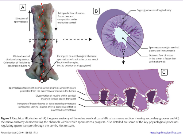
why is artificial insemination in a ewe difficult
highy folded surface of the cervical lumen
create branch points where the central canal turns and a canula can be misdirected into a large fold
mucus in anoestrus vs oestrus
in anoestrus the mucus is produced in low volumes but is thick restricting movement of sperm or pathogens
in oestrs it is thinner but produced in larger volumes
how are pathogens removed whilst protecting sperm
fluid flows faster in the main lumen and slower in grooves
how does the ampulla and isthmus structure relate to function
ampulla has larger lumen and less muscle
isthmus can restrict dimensions to prevent egg from entering
sperm can pass through to the egg at the ampullary isthmic junction for fertilisation but may also be protected by constrained environment of the isthmus is they arrive before the egg
under hormonal control the muscle of the isthmus relaxes when the uters is ready to recieve the fertilised egg
what is the function of the endometrial glands in anoestrus, oestrus and pregnancy and strutural changes
contribution to cervical mucus before pregnancy and will support embryo afterwards
in anoestrus they are relatively sparse becoming more prominent as straight glands in oestrus
driven by the increase in oestrogen levels
proliferative phase
within pregnancy they expand much further becoming coiled to accomodate the amount of secretory epithelium required
driven by progesterone
secretory phase
dr