H3 Toth- Cytoskeleton II - actin network

0.0(0)
Studied by 0 people
call kaiCall Kai
learnLearn
examPractice Test
spaced repetitionSpaced Repetition
heart puzzleMatch
flashcardsFlashcards
GameKnowt Play
Card Sorting

1/8

encourage image

There's no tags or description

Looks like no tags are added yet.

Last updated 4:31 PM on 2/12/26
Name
Mastery
Learn
Test
Matching
Spaced
Call with Kai

No analytics yet

Send a link to your students to track their progress

9 Terms

1
New cards

describe the structure and assembly of actin filaments

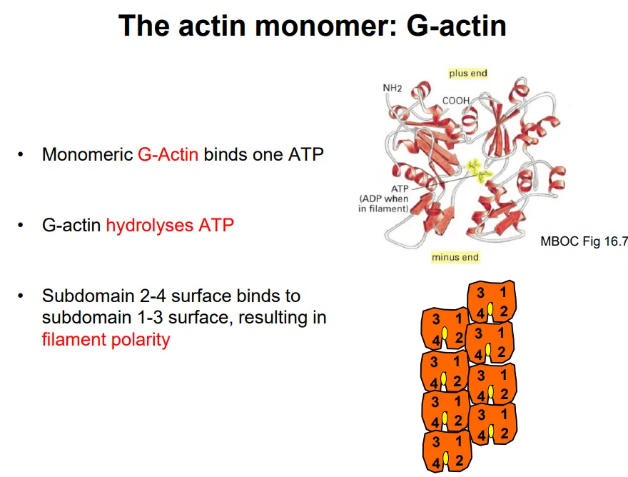
  • F-actin (filamentous actin) is a linear polymer of a single globular monomer, G-actin

  • it is a left-handed helix with 13 monomers per turn (37nm)

  • each G-actin binds and hydrolyses one ATP to spontaneously self-assemble (once three monomers have bound together)

  • one surface (2-4 subdomain) binds preferentially to the opposite surface (1-3 subdomain), so the filament is polar

  • polymerisation happens at the plus end, which has a greater binding affinity to ATP-bound G-actin

  • after addition, the G-actin monomers hydrolyse the ATP slowly, so that the minus end is made up of ADP-bound G-actin, which can dissociate (this is called treadmilling)

however the rate of filament assembly and disassembly is normally controlled by actin-binding proteins, by:

  • nucleation- holding the three monomers together to allow for self-assembly

  • capping- binding to the plus end to prvent further polymerisation

  • severing- cutting the filament to induce depolymerisation

  • sequestering- binding to monomers to prevent incorporation

  • bundling- bind to actin filaments to connect them

<ul><li><p><strong>F-actin</strong> (filamentous actin) is a <strong>linear </strong>polymer of a <strong>single </strong>globular monomer, <strong>G-actin</strong></p></li><li><p>it is a <strong>left-handed helix </strong>with 13 monomers per turn (37nm)</p></li><li><p>each G-actin binds and hydrolyses <strong>one ATP</strong> to <strong>spontaneously self-assemble </strong>(once three monomers have bound together)</p></li><li><p>one surface (2-4 subdomain) binds <strong>preferentially </strong>to the <strong>opposite </strong>surface (1-3 subdomain), so the filament is <strong>polar</strong></p></li><li><p><strong>polymerisation </strong>happens at the <strong>plus </strong>end, which has a<strong> greater binding affinity to ATP-bound G-actin</strong></p></li><li><p>after addition, the G-actin monomers <strong>hydrolyse </strong>the ATP <strong>slowly</strong>, so that the <strong>minus </strong>end is made up of <strong>ADP-bound </strong>G-actin, which can <strong>dissociate </strong>(this is called <strong>treadmilling</strong>) </p></li></ul><p>however the rate of filament assembly and disassembly is normally controlled by <strong>actin-binding proteins</strong>, by:</p><ul><li><p><strong>nucleation</strong>- holding the three monomers together to allow for self-assembly</p></li><li><p><strong>capping</strong>- binding to the plus end to prvent further polymerisation</p></li><li><p><strong>severing</strong>- cutting the filament to induce depolymerisation</p></li><li><p><strong>sequestering</strong>- binding to monomers to prevent incorporation </p></li><li><p><strong>bundling</strong>- bind to actin filaments to connect them</p></li></ul><p></p>
2
New cards

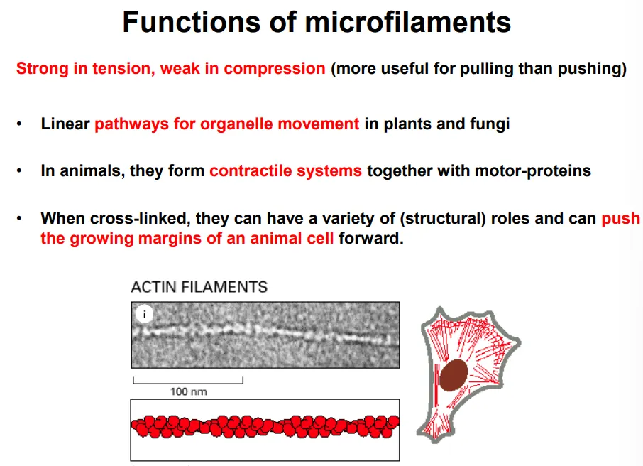
what are the functions of actin filaments?

  • stronger in tension than compression (used for pulling)

in conjunction with myosin motor proteins, they are used for:

  • organelle movement in plants and fungi

  • changing the cell shape in contractile systems in animals, eg:

    • sarcomeres in muscle cells

    • contractile rings in cytokinesis

    • stress fibres anchored to focal adhesions

  • cell movement

<ul><li><p>stronger in tension than compression (used for pulling)</p></li></ul><p>in conjunction with myosin motor proteins, they are used for:</p><ul><li><p>organelle movement in plants and fungi</p></li><li><p>changing the cell shape in contractile systems in animals, eg:</p><ul><li><p>sarcomeres in muscle cells</p></li><li><p>contractile rings in cytokinesis</p></li><li><p>stress fibres anchored to focal adhesions</p></li></ul></li><li><p>cell movement </p></li></ul><p></p>
3
New cards

how are organelles moved around the cell by actin filaments?

  • in plants and fungi, the large vacuole means organelles and vesicles must be moved around by myosin motor proteins, causing the cytoplasm to also move by cytoplasmic streaming (overcomes diffusion barrier)

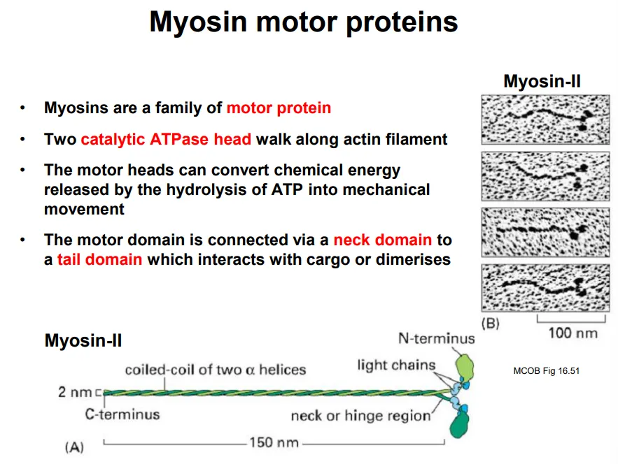
  • two catalytic heads walk along the actin filament in steps by conformational changes from hydrolysing ATP

  • this is connected to a neck domain, with a tail domain on the end to bind to cargo

<ul><li><p>in plants and fungi, the large vacuole means organelles and vesicles must be moved around by <strong>myosin motor proteins</strong>, causing the cytoplasm to also move by <strong>cytoplasmic streaming </strong>(overcomes diffusion barrier)</p></li></ul><ul><li><p><strong>two catalytic heads</strong> walk along the actin filament in steps by conformational changes from <strong>hydrolysing ATP</strong></p></li><li><p>this is connected to a <strong>neck </strong>domain, with a <strong>tail </strong>domain on the end to bind to <strong>cargo</strong></p></li></ul><p></p>
4
New cards

how are actin filaments organised in skeletal muscle cells?

  • in animal skeletal muscle tissue, actin and myosin filaments (myofibrils) form contractile arrays in multinucleated syncitial cells (myoblasts fused together)

  • muscle myosin proteins assemble into bipolar myosin filaments (thick), where the heads project out along the length in specific radial positions (43 nm repeat)

each repeating unit is a sarcomere, which is striated:

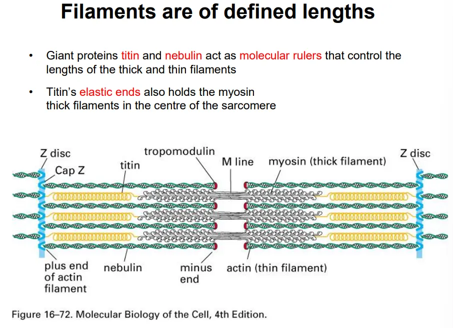
  • the plus ends of the actin filaments (thin) are anchored at the z disc, and the minus end is capped by tropomodulin (can’t polymerise/depolymerise)- these are all the same length due to attachment to a long nebulin protein

  • elastic titin proteins attach to the z disc and connect to myosin thick filaments to keep them centred along the m line

this shortens according to the sliding filament model

  • the thick filaments are hexagonally packed very regularly

<ul><li><p>in animal skeletal muscle tissue, <strong>actin and myosin filaments </strong>(myofibrils) form contractile arrays in multinucleated <strong>syncitial </strong>cells (myoblasts fused together)</p></li><li><p>muscle myosin proteins <strong>assemble </strong>into bipolar myosin filaments (<strong>thick</strong>), where the <strong>heads project </strong>out along the length in specific radial positions (43 nm repeat)</p></li></ul><p>each repeating unit is a <strong>sarcomere</strong>, which is <strong>striated</strong>:</p><ul><li><p>the <strong>plus </strong>ends of the <strong>actin </strong>filaments (thin) are <strong>anchored </strong>at the <strong>z disc</strong>, and the <strong>minus </strong>end is <strong>capped </strong>by <strong>tropomodulin </strong>(can’t polymerise/depolymerise)- these are all the <strong>same length </strong>due to attachment to a long <strong>nebulin </strong>protein</p></li><li><p><strong>elastic titin </strong>proteins attach to the<strong> z disc </strong>and connect to <strong>myosin </strong>thick filaments to keep them centred along the <strong>m line</strong></p></li></ul><p></p><p>this shortens according to the <strong>sliding filament model</strong></p><ul><li><p>the thick filaments are hexagonally packed very regularly</p></li></ul><p></p>
5
New cards

describe how sarcomeres shorten

each sarcomere in skeletal muscle cells contracts according to the sliding filament model:

  • when relaxed, the titin proteins (connected to the z disc and myosin filaments) are stretched, so the actin and myosin only just overlap

  • when the muscle contracts, the myosin filaments walk along the actin filaments, pulling the z discs on either side towards the m line in the centre

this occurs by the myosin cross-bridge cycle, driven by ATP hydrolysis:

  • the two myosin heads of each protein act independently

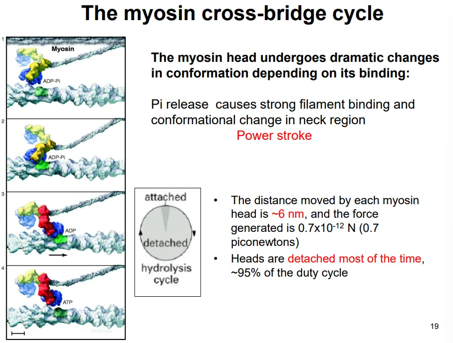
  • they bind to the actin and release a phosphate (from previous hydrolysis), causing the power stroke, which pulls the actin filament 6nm

  • the ADP dissociates, and the next ATP binds

  • this causes the head to detach from the actin (detached 95% of the time)

  • the hydrolysis of this ATP causes the head to move back to its original conformation

the spacing of the myosin heads doesn’t match up with that of the myosin binding sites of actin, and the nine radial positions of the myosin heads doesn’t match up with the hexagonal packing of actin around each thick filament

  • this means that not all the myosin heads can bind to actin at one time- this means there are always some heads attached to maintain tension

<p>each <strong>sarcomere </strong>in skeletal muscle cells contracts according to the <strong>sliding filament model:</strong></p><ul><li><p>when <strong>relaxed</strong>, the <strong>titin </strong>proteins (connected to the z disc and myosin filaments) are <strong>stretched</strong>, so the actin and myosin only just overlap</p></li><li><p>when the muscle <strong>contracts</strong>, the myosin filaments walk along the actin filaments, <strong>pulling the z discs</strong> on either side <strong>towards the m line</strong> in the centre</p></li></ul><p></p><p>this occurs by the <strong>myosin cross-bridge cycle</strong>, driven by ATP hydrolysis:</p><ul><li><p>the two myosin heads of each protein act independently</p></li><li><p>they <strong>bind </strong>to the actin and <strong>release </strong>a <strong>phosphate </strong>(from previous hydrolysis), causing the <strong>power stroke</strong>, which pulls the actin filament 6nm</p></li><li><p>the ADP <strong>dissociates</strong>, and the next <strong>ATP binds</strong></p></li><li><p>this causes the head to <strong>detach </strong>from the actin (detached 95% of the time)</p></li><li><p>the <strong>hydrolysis </strong>of this ATP causes the head to move back to its <strong>original </strong>conformation</p></li></ul><p></p><p>the spacing of the myosin heads doesn’t match up with that of the myosin binding sites of actin, and the nine radial positions of the myosin heads doesn’t match up with the hexagonal packing of actin around each thick filament</p><ul><li><p>this means that not all the myosin heads can bind to actin at one time- this means there are always some heads attached to maintain tension</p></li></ul><p></p>
6
New cards

how is muscle contraction controlled?

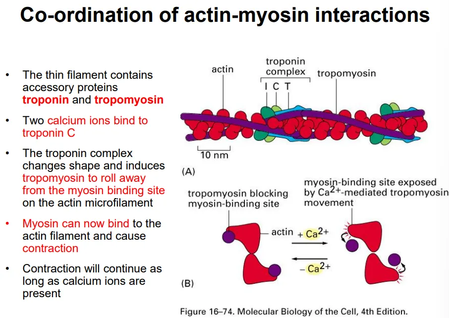
  • tropomyosin proteins are wrapped around the thin actin filament and cover the myosin binding sites

  • when an action potential arrives at the cell, it is transmitted through invaginations of the plasma membrane

  • this causes the voltage-gated release of calcium ions from the sarcoplasmic reticulum

  • two calcium ions bind to a troponin complex, which changes shape and causes the tropomyosin to detach

  • this allows myosin to bind to the actin filament, causing contraction by ATP hydrolysis as long as the calcium ions are present

<ul><li><p><strong>tropomyosin </strong>proteins are wrapped around the thin actin filament and <strong>cover </strong>the <strong>myosin binding sites</strong></p></li><li><p>when an <strong>action potential </strong>arrives at the cell, it is transmitted through <strong>invaginations </strong>of the plasma membrane</p></li><li><p>this causes the <strong>voltage-gated</strong> release of <strong>calcium ions</strong> from the <strong>sarcoplasmic reticulum</strong> </p></li><li><p><strong>two</strong> calcium ions bind to a <strong>troponin </strong>complex, which changes shape and causes the tropomyosin to <strong>detach</strong></p></li><li><p>this allows <strong>myosin </strong>to <strong>bind </strong>to the actin filament, causing contraction by <strong>ATP hydrolysis</strong> as long as the calcium ions are present</p></li></ul><p></p>
7
New cards

what are the three actin filament organisations in crawling cells?

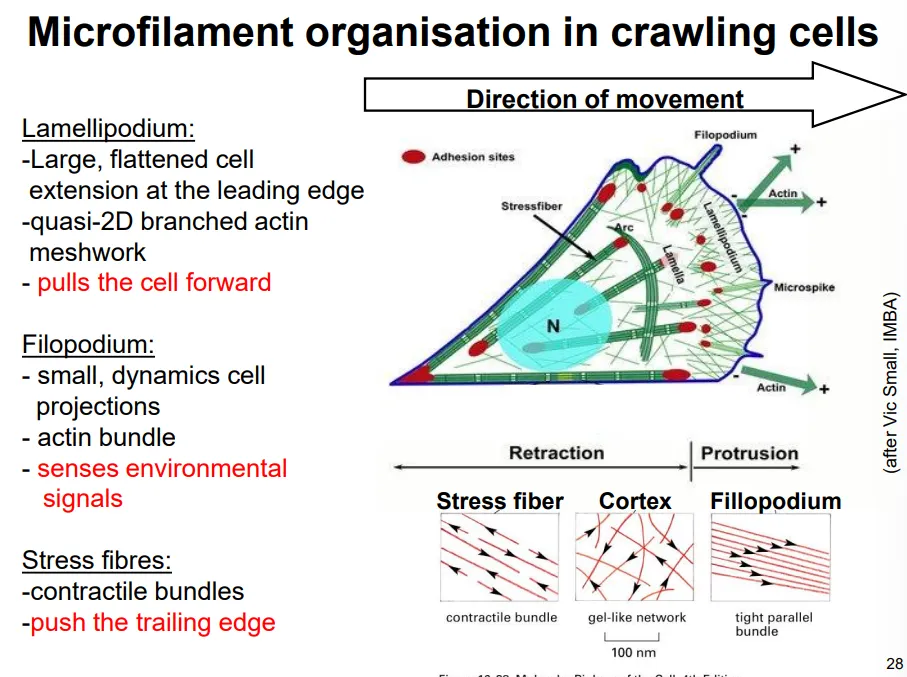
  • in the lamellipodia, the actin is branched at 70 degree angles due to the binding of actin-related proteins (ARPs) that facilitate new filaments forming, by holding together a trimer

  • this extension occurs via a treadmilling model, where the actin filaments grow a short distance before being capped, at which point they act as a site for new filaments to be assembled, while the older end of the network is disassembled by severing proteins

  • new focal adhesion points will be made and stress fibres attached, which will contract when this becomes the trailing end

  • this protrusion force is provided purely by actin polymerisation, not by myosin II

<ul><li><p>in the <strong>lamellipodia</strong>, the actin is <strong>branched </strong>at <strong>70 degree </strong>angles due to the binding of <strong>actin-related proteins</strong> (ARPs) that facilitate new filaments forming, by holding together a <strong>trimer</strong></p></li><li><p>this extension occurs via a <strong>treadmilling mode</strong>l, where the actin filaments grow a short distance before being capped, at which point they act as a site for new filaments to be assembled, while the older end of the network is disassembled by severing proteins</p></li><li><p>new<strong> focal adhesion</strong> points will be made and <strong>stress fibres </strong>attached, which will <strong>contract </strong>when this becomes the <strong>trailing </strong>end</p></li><li><p>this protrusion force is provided purely by actin polymerisation, not by myosin II</p></li></ul><p></p>
8
New cards

how are the different kinds of actin structures initiated?

GTP-binding proteins (G proteins) spatially organise actin polymerisation:

  • Rho stimulates stress fibre production

  • Rac stimulates lamellipodia extension

  • Cdc-42 stimulates filopodia formation

  • these are spatially separated around the cell in the relevant areas, to promote each actin structure

  • these act as molecular switches, active when bound to GTP, which they hydrolyse to become inactive

9
New cards

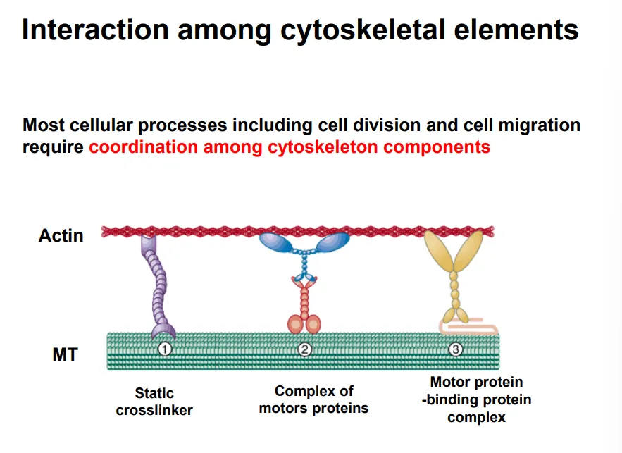
how are cytoskeletal elements interconnected?

  • many processes require the coordination of multiple cytoskeletal components

they can be connected by different crosslinkers to coordinate them:

  • static crosslinkers- neither can move

  • motor protein complexes- both proteins can move along their elements

  • motor protein-binding protein complexes- one is fixed, one can slide

<ul><li><p>many processes require the coordination of multiple cytoskeletal components</p></li></ul><p>they can be connected by different crosslinkers to coordinate them:</p><ul><li><p><strong>static crosslinkers</strong>- neither can move</p></li><li><p><strong>motor protein complexes</strong>- both proteins can move along their elements</p></li><li><p><strong>motor protein-binding protein complexes</strong>- one is fixed, one can slide</p></li></ul><p></p>