PODI3113 EXAM
 Call Kai
 Learn
Learn Practice Test
 Spaced Repetition
 Match
Match Flashcards
 Knowt Play
1/289

There's no tags or description
Looks like no tags are added yet.
Name | Mastery | Learn | Test | Matching | Spaced | Call with Kai |
|---|
No analytics yet
Send a link to your students to track their progress
290 Terms
Hindfoot
Includes Talus (connects to the leg) and Calcaneus (heel bone).
Midfoot bones
Includes Navicular, Cuboid, and three Cuneiforms (medial, intermediate, lateral).
Forefoot
Composed of 5 Metatarsals and 14 Phalanges (toes).
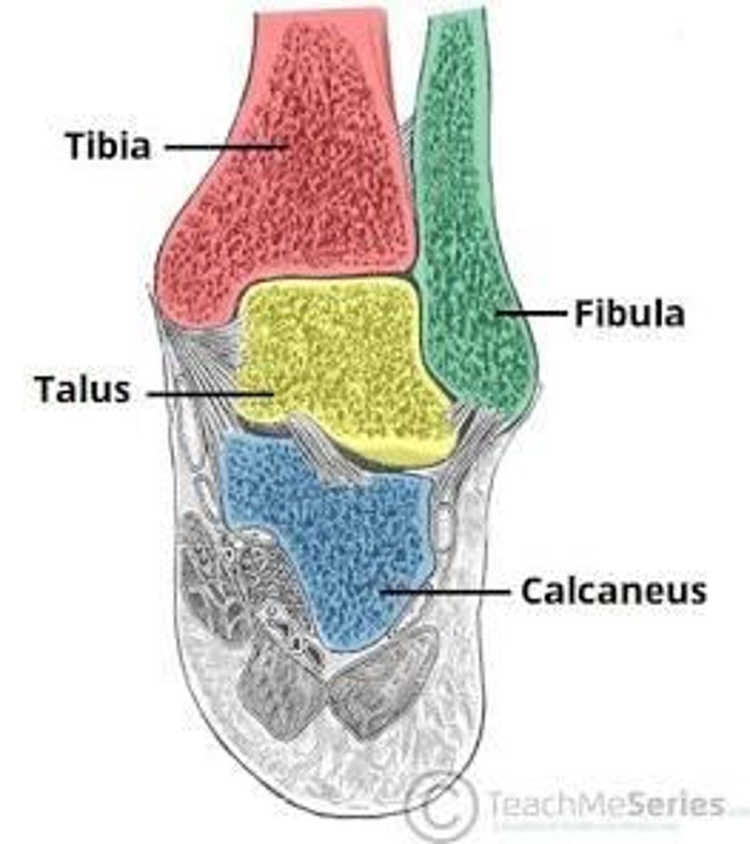
Talocrural (Ankle) Joint
Formed by the tibia, fibula, and talus; responsible for up (dorsiflexion) and down (plantarflexion) movement.
Subtalar Joint
Between the talus and calcaneus; allows for side-to-side tilting (inversion and eversion).
Metatarsophalangeal (MTP) Joints
Connect the metatarsals to the toes.
Lateral Complex
Includes the ATFL, CFL, and PTFL; often strained in ankle sprains.
Deltoid Ligament
A strong, fan-shaped ligament on the medial (inner) side.
Plantar Fascia
The longest ligament in the foot, supporting the arch.
Achilles Tendon
The body's strongest tendon; connects calf muscles to the heel.
Tibialis Posterior Tendon
Crucial for supporting the foot's arch.
Peroneal Tendons
Run behind the lateral ankle to help turn the foot outward.
Sciatic Nerve
The largest nerve in the body; bifurcates above the knee into the Tibial and Common Fibular nerves.
Tibial Nerve
Supplies the posterior compartment (calf muscles) and travels behind the medial malleolus into the sole of the foot.
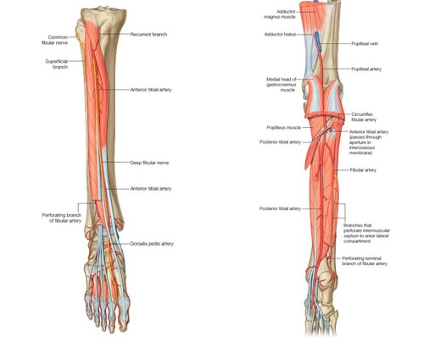
Common Fibular (Peroneal) Nerve
Divided into Deep Fibular Nerve and Superficial Fibular Nerve.
Deep Fibular Nerve
Supplies innervation to the anterior compartment (dorsiflexors) and the skin between the first and second toes.
Superficial Fibular Nerve
Supplies innervation the lateral compartment (evertors) and most of the skin on the top (dorsum) of the foot.
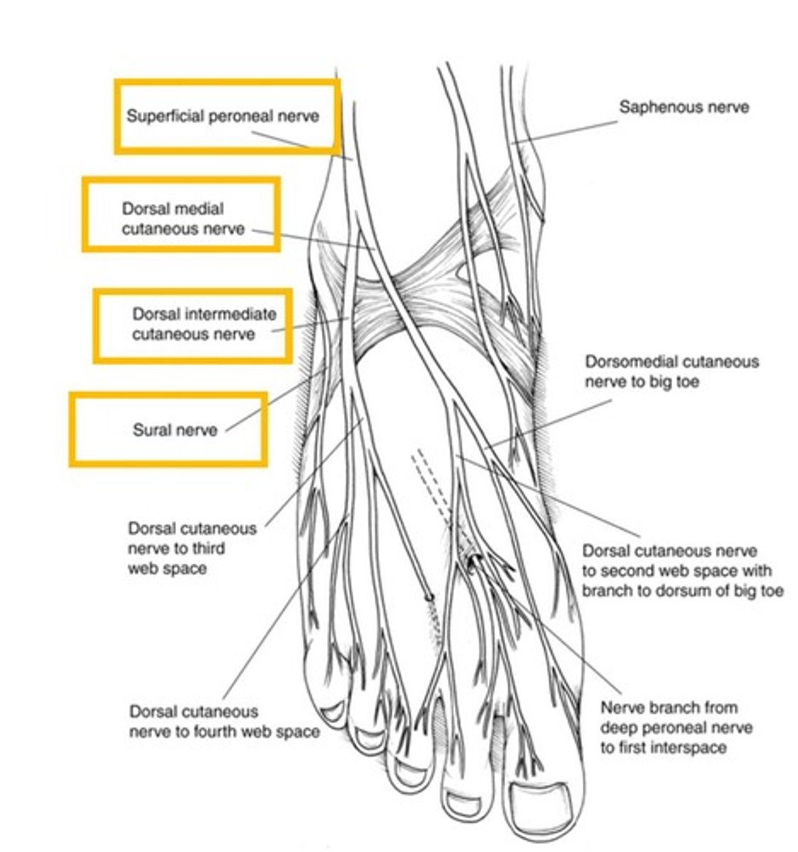
Saphenous Nerve
A purely sensory branch of the femoral nerve that supplies the medial aspect of the leg and ankle.
Popliteal Artery
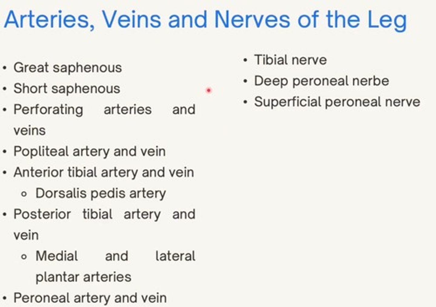
The continuation of the femoral artery behind the knee; divides into the anterior and posterior tibial arteries.
Anterior Tibial Artery
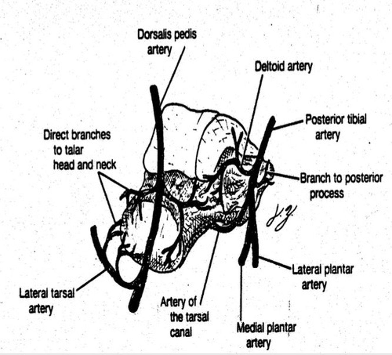
Supplies the anterior muscles and continues onto the top of the foot as the Dorsalis Pedis artery.
Posterior Tibial Artery
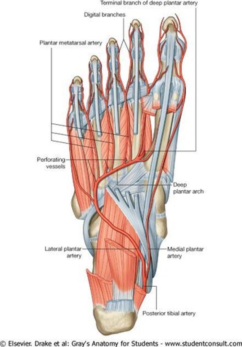
Supplies the posterior leg and enters the sole of the foot to form the medial and lateral plantar arteries.
Fibular (Peroneal) Artery
A branch of the posterior tibial artery that supplies the lateral compartment of the leg.
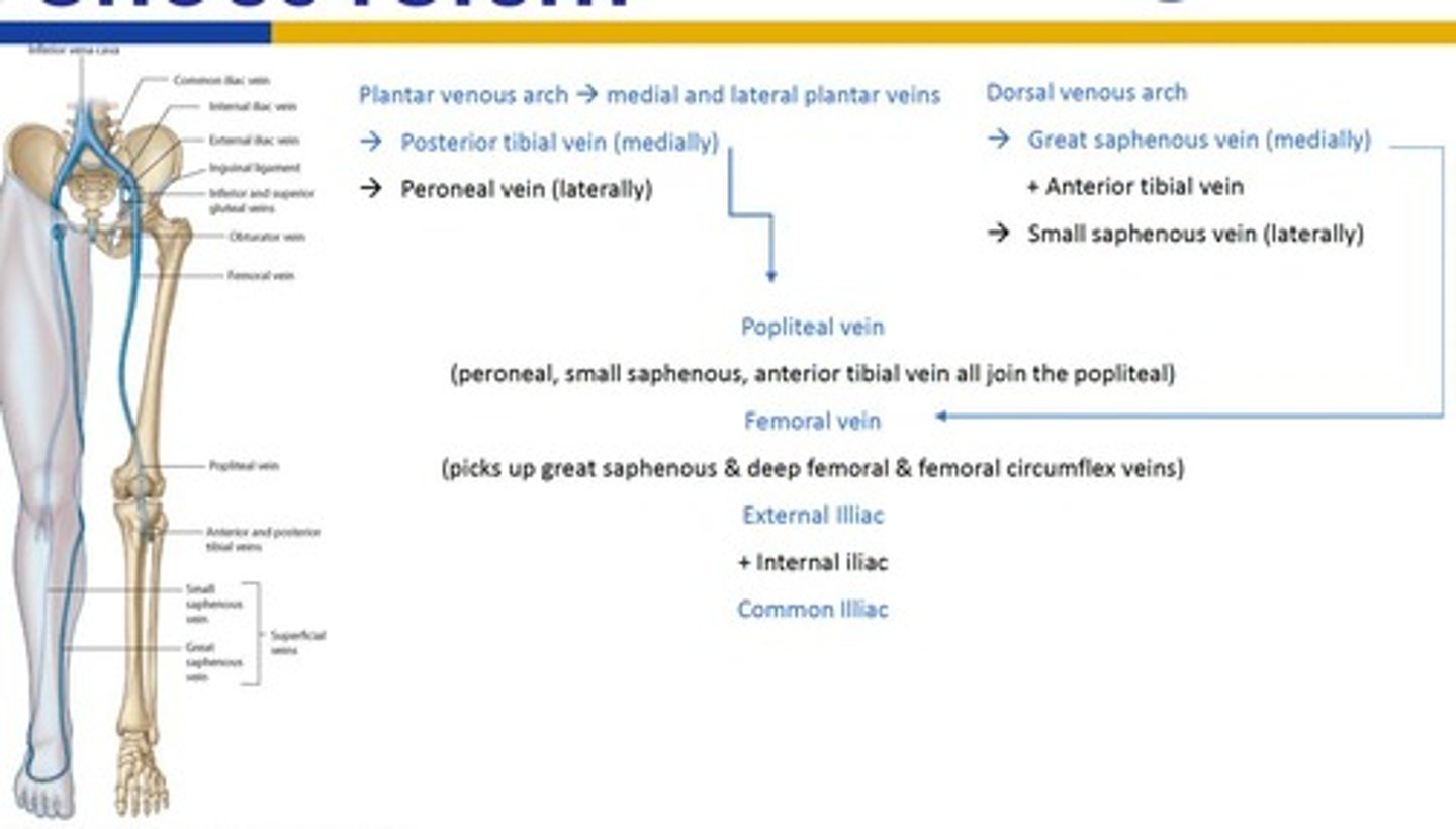
Deep Veins
Generally follow the path of the arteries and drain into the Popliteal Vein.
Great Saphenous Vein
Drains blood from the dorsal venous arch of the foot toward the thigh.
Sesamoid
A bony structure that is round with bone cortex & cancellous bone embedded in a tendon.
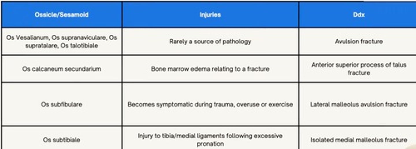
Ossicle
An anatomical variant that has no known function and is free-floating.
Primary Ossification Centre
Where the bone first forms, visible at birth.
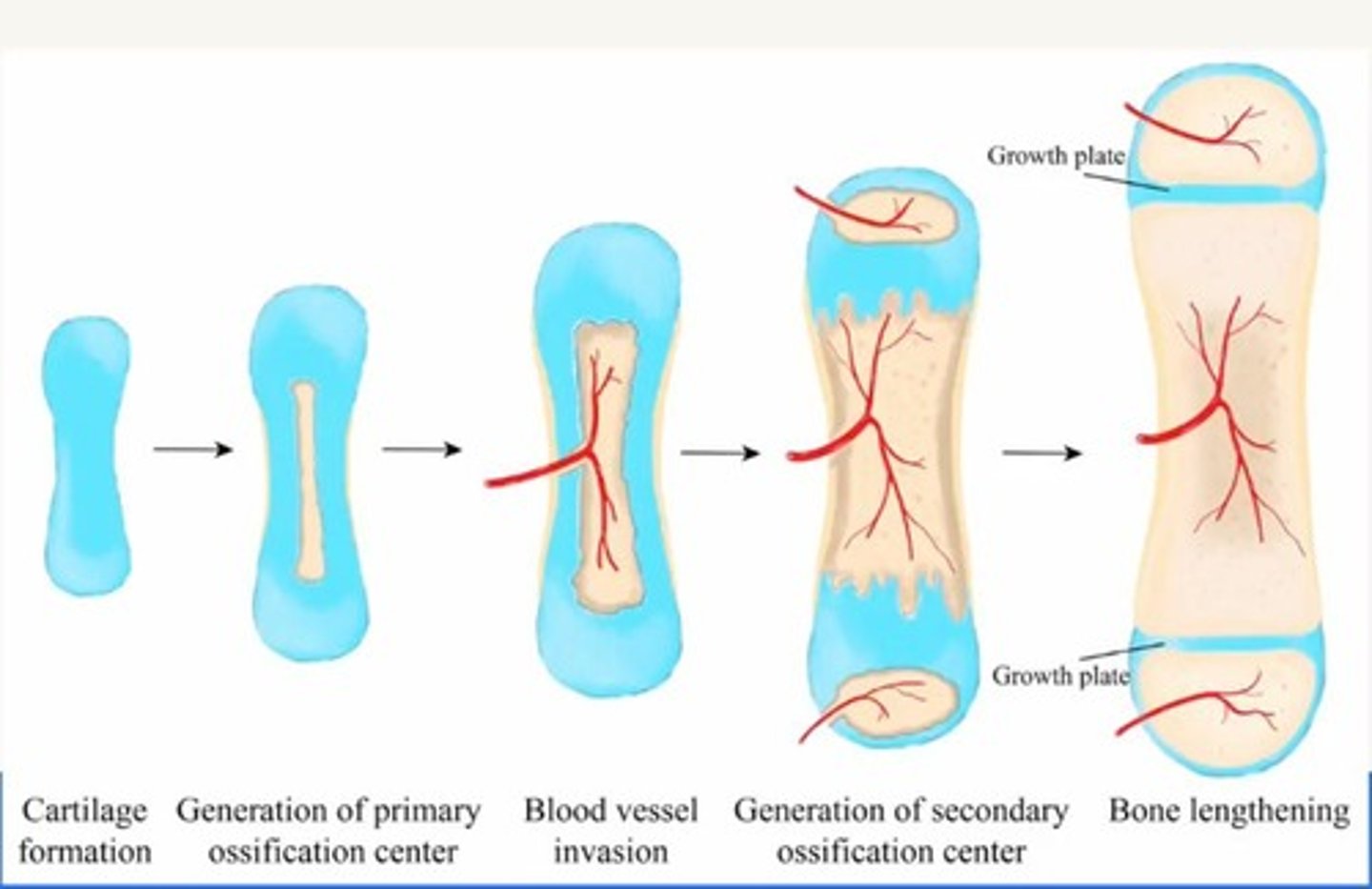
Secondary Ossification Centre
Additional sites of bone formation in a single bone with its own blood supply.
Epiphyseal plate
Contains hyaline cartilage
Tarsal bones ossification centres
Have 1 ossification centre (main body), except for the calcaneus (it has 2; main body and posterior process)
Sever's disease
Inflammation of the posterior epiphysial region
Metatarsals ossification centres
Have 2 centres: 1 for the shaft and 1 at the base of the 1st and heads of 2-5
Phalanges ossification centres
Have 2: 1 at the shaft and 1 at the base
Sever's & Osgood Schlatters
Both are inflammation
Osgood-Schlatter's disease
Occurs in the anterior part of the tibia (tibial tuberosity) where the quadriceps tendon attaches to
High risk activities for Sever's and Osgood-Schlatter's
Children who play soccer, football, and volleyball
MSK pathology
Linked with Neurodevelopmental pathology -> ADHD 70% increase in getting the disease
Imaging Characteristics of Sever's and Osgood-Schlatter's
Small, smooth and round
Imaging Characteristics of ossicles and sesamoids
Has an apparent cortical bone region
Hallucal Sesamoids
Location: plantar aspect of the first metatarsal head and the lateral slips of the flexor hallucis longus tendon 100% prevalence (6.65% bipartite and 0.16% multipartite)
Hallucal Interphalangeal sesamoids
Location: on the plantar aspect of the interphalangeal joint of the first digit of the foot (Hallux) Prevalence 2-13%
Lesser Metatarsal Sesamoids
Location: Plantar aspect of the metatarsal heads (2-5). Prevalence: Second: 0.4-0.315%, Third: 0.2-0.4%, Fourth: 0.1-0.72%, Fifth: 4.3-9.06%
Os Peroneneum
Location: at the cuboid tunnel near the calcaneocuboid joint, embedded in the peroneus longus tendon Prevalence 26%-28%
Os Trigonum
Location: posterior to the talus. Prevalence 7-25%
Os Intermetatarseum
Location: between the first and second metatarsal (lisfranc joint) Prevalence 1-13%
Os Naviculare
Prevalence: 2-14%. Types: type 1: os tibiale, free floating(20%); Type 2: os tibiale externum, linked by a cartilaginous link to the body of the navicular (50%); type 3: naviculare secundarium, the ossicle fused with bone to the navicular body (30%)
Os Vesalianum
Location: proximal to the base of the 5th metatarsal, within the tendon of the peroneus brevis. Prevalence 0.1-1%. can be a sesamoid or ossiclele
Os Supranaviculare
Other names: os talonaviculares dorsalis, talonavicular ossicle, Pirries bone, dorsal to the talonavicular joint. Prevalence: 1-3.5%
Os Supratalare
Location: over the ridge of the head/neck of the talus, may be fused with the talus or free-floating. Prevalence: 0.2-2.4%
Os Talotibiale
Location: anterior to the tibiotalar joint. Can cause ankle impingement syndrome and its prevalence is 0.5%
Os Calcaneus Secundarium
Location: adjacent to the anterior calcaneal process. Prevalence 0.6-7%
Os Subfibulare
Location: inferior to the lateral malleolus or fibula. Prevalence 1%
Os subtibiale
Location: inferior to the medial malleolus or tibia. Prevalence of 1.2%
Pathological conditions
May result in neighbouring bones fracturing or ossicle fracture only
Hallucal sesamoids fracture
Can be fractured from large force trauma (falling from great heights)
Os trigonum fracture
Can be caused by forced plantar flexion
Os peroneum fracture
Can be caused by inversion injury and its proximal migration -> peroneus longus tendon tear
Os subfibulare fracture
Can lead to anterior talofibular ligament (ATFL) from trauma
Sesamoiditis
Chronic pain (specifically in hallucal sesamoids) caused by stress/fracture/OA/osteonecrosis/tendonitis/capsular inflammation
Infection
Osteomyelitis, the sesamoids and ossicles are infected -> secondary infection
Dislocations
Hyperextension of 1st MTPJ -> dislocation of proximal phalanx -> hallucal sesamoids dislocated laterally.
Degenerative Diseases
Due to OA (hallucal sesamoids), symptoms: loss of joint space, osteophyte formation, sclerosis
Ankle impingement
Posterior ankle pain associated with os trigonum degenerative changes, anterior ankle impingement may be associated with degenerative changes to os talotibiale
Clinical pearls
Often recognised as secondary findings, ossicle/sesamoid injury can cause injuries to other parts of the foot
Ossicles and sesamoids
Often asymptomatic but are sometimes associated with local pain or can be mistaken for pathological conditions (fractures)
Ossicle/sesamoid diagnosis
Gold standard: 2 views on radiographic imaging
Incidence of accessory ossicles in the foot
18-36% of the general population
Lymph capillaries
Absent in: bone marrow, CNS, tissues that lack blood flow (e.g. epidermis, cornea)
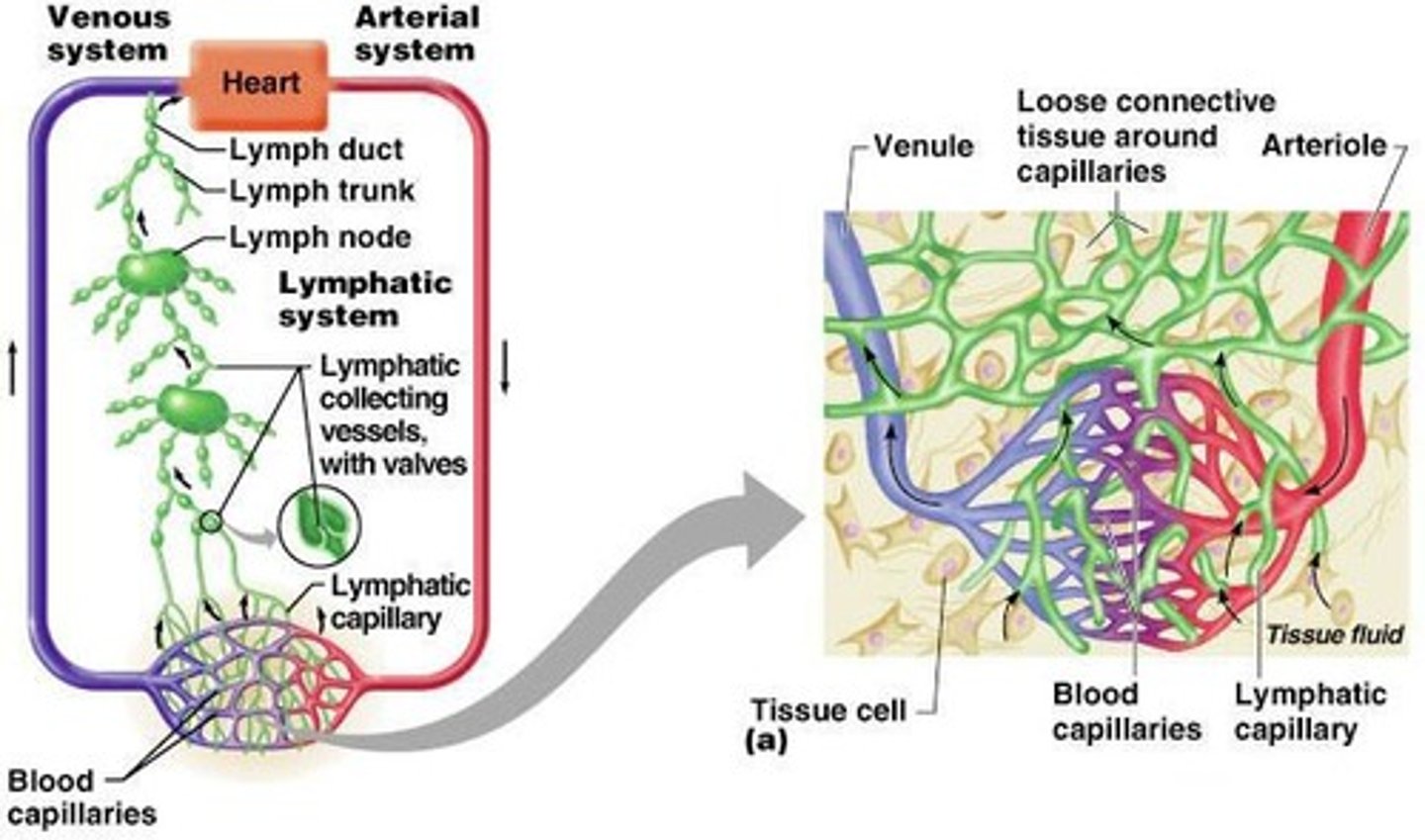
Right Lymphatic Duct
Drains the upper arm and right side of the head and thorax
Thoracic duct
Arises from the cisterna chyli and drains the rest of the body
Lower Limb Lymph Drainage
Starts from the femoral triangle; Major groups: superficial vessels and nodes + deep vessels and nodes
Superficial lymphatic vessel (Medial)
Follows the great saphenous vein and is larger
Superficial lymphatic vessel (Lateral)
Follows the small saphenous vein and is smaller
Deep Lymphatic vessels
Accompany the deep veins; Sets: anterior tibial lymph vessel, posterior tibial lymph vessel, peroneal lymph vessel
Popliteal Lymph Nodes
Embedded in the fat of the popliteal fossa; Small and few
Inguinal Lymph Nodes
Superficial inguinal glands: below the inguinal ligament and forms a chain
Obturator nerve
One of the nerves supplying the lower limb
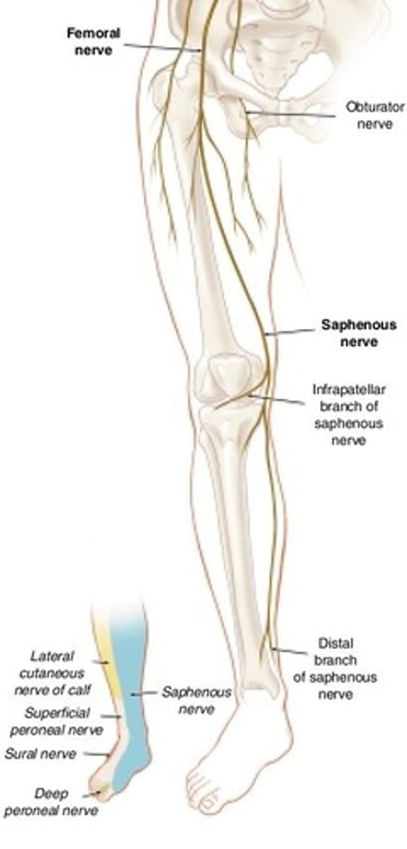
Femoral nerve
One of the nerves supplying the lower limb
Common fibular/peroneal nerve
Originates from the sciatic nerve
Sural nerve
From the tibial nerve and common peroneal nerve; Travels with small saphenous vein
Medial plantar nerve
Largest and most anterior terminating branch of tibial nerve
Lateral plantar nerve
Lies between flexor digitorum brevis and quadratus plantae
Medial calcaneal nerve
Innervates medial and posterior parts of the heel
Lateral calcaneal nerve
Innervates the lateral and posterior parts of the heel
Intermetatarsal Neuroma
Condition where the nerves are enlarged due to friction or sustained compression of a nerve for a long time
Common iliac artery
Branches into internal iliac and external iliac arteries
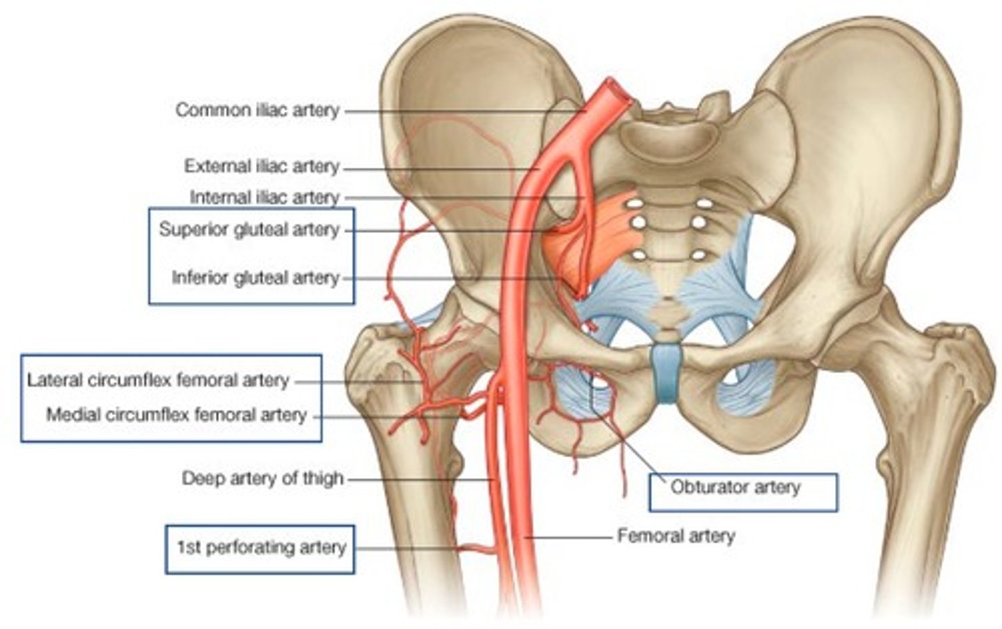
Femoral artery
Branches include medial circumflex, lateral circumflex, profunda, and 4 perforating branches
Peroneal artery
Branches from the posterior tibial artery
Cruciate Anastomoses
Arterial network located around the proximal femur or hip joint.
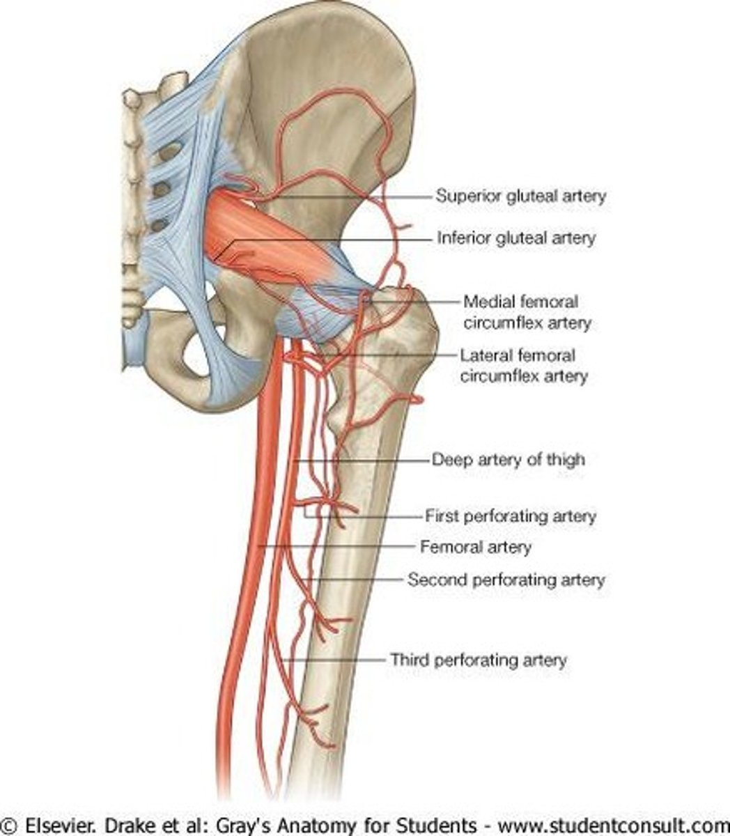
Inferior gluteal artery
One of the four main arteries in the cruciate anastomoses.
Lateral and medial circumflex femoral arteries
Two of the four main arteries in the cruciate anastomoses.
First perforating artery of the profunda femoris artery
One of the four main arteries in the cruciate anastomoses.
Anastomotic branch of the posterior branch of the obturator artery
One of the four main arteries in the cruciate anastomoses.
Obturator artery branch
Supplies the artery of the head of the femur.
Nutrient artery of the femoral shaft
Only reaches the head in adults after epiphysis fusion.
Retinacular arteries of the femoral neck
Arteries that arise from the cruciate anastomoses.
Deep Penetrating Arteries
Starts from the femoral artery and continues to the perforating artery.
Periarticular Genicular Anastomosis
Arteries that surround the knee and provide collateral circulation during full knee flexion.
Superior lateral genicular artery
One of the branches of the popliteal artery in the knee.
Superior medial genicular artery
One of the branches of the popliteal artery in the knee.