Other Vascular Disorders, Including Carotid Occlusive Disease, Anemias, & Leukemias - Posterior Segment & Ocular Disease Spring 2026
1/120
There's no tags or description
Looks like no tags are added yet.
Name | Mastery | Learn | Test | Matching | Spaced | Call with Kai |
|---|
No analytics yet
Send a link to your students to track their progress
121 Terms
blockage of carotid or ophthalmic artery
What is the cause of hypoperfusion syndrome?
dull ache in or around the eye (ocular pain), TIA symptoms, light induced amaurosis fugax, carotid bruit, decreased carotid pulse
What are the systemic signs of hypoperfusion syndrome?
ophthalmoscopy shows unilateral venous dilation, narrowed arteries, venous beading, non-tortuous vessels, mid-peripheral hemes, retinal emboli
What are the ocular signs of hypoperfusion syndrome?
carotid and heart
Most common origin of retinal emboli?
ocular ischemic syndrome (OIS)
Hypoperfusion syndrome is a precursor to what?
-hypoperfusion
-signs of hypoxia-induced inflammation
**chronic condition
What is ocular ischemic syndrome (OIS) a combo of?
-uveitis
-slugglish pupils
-conj congesiton
-corneal edema
-NVD/NVE/NVI
Hypoxia-induced inflammation can include what conditions?
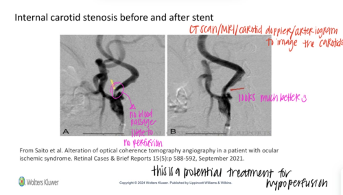
Internal Carotid Stenosis Before and After a Stent (Pic)
Internal Carotid Stenosis Before and After a Stent (Pic)

carotid stenosis
Ocular Ischemic Syndrome (OIS) is most significant in patients with what?
10; 70
Ocular Ischemic Syndrome (OIS) only occurs in ____% of eyes with ____% or more blocked carotids
-Decrease VA (inflammation in cornea and AC)
-Ocular pain (d/t inflammation & blockage of TM increasing IOP)
-Unilateral red or injected eye
-Corneal Edema with potential folds in Descemets membrane
-Anterior chamber inflammation/uveitis
-Neovascularization
What are the ocular signs of Ocular Ischemic Syndrome (OIS)?
Mid Peripheral Retinal Hemorrhages (Pic)
Mid Peripheral Retinal Hemorrhages (Pic)
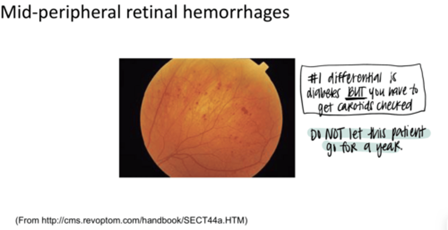
Diabetes
What is the #1 differential of this?

80
REVIEW: OIS is _____% unilateral
bilateral
REVIEW: Diabetic Retinopathy is more likely to be (unilateral/bilateral)
-non-tortuous vessels in OIS
-tortuous vessels with diabetic retinopathy
REVIEW: OIS demonstrates what signs in the blood vessels compared to diabetic retinopathy?
mid-peripheral
REVIEW: OIS often shows a preponderance of _______ hemes
anywhere
REVIEW:
Diabetic hemes appear ______
-Systemic vascular workup (FBS, BP, CBC, ESR or CRP, lipid profile)
-Carotid evaluation (Doppler or Duplex ultrasound and/or angiogram)
-Refer to ophthalmology
What is the management of OIS?
It would be difficult to control the development of neovascular glaucoma by ourselves
Why do you refer patients with OIS to ophthalmology?
-increase in size of the foveal avascular zone
-reduced vessel area density (VAD)
What are the signs that may be present on OCT-A with OIS?
inherited disorder with abnormal erythrocyte function -- RBCs are rigid and elongated
What are sickle hemoglobinopathies?
d/t substitution of single amino acids
Why is there abnormal hemoglobin structure with sickle hemoglobinopathies?
distorted Hb decreases RBC O2-transport capability
What is the consequence of abnormal hemoglobin structure with sickle hemoglobinopathies?
-hemolytic anemia
-RBCs are eliminated early because the body recognizes them as foreign
Sickle cell hemoglobinopathies are sometimes considered a ______ anemia. What does this mean?
malaria
Sickle cell hemoglobinopathies are common where _____ is endemic
protects against malarial infections
Why is sickle cell endemic in areas of malaria?
10
Sickle cell hemoglobinopathy is present in about ___% of African Americans
normal adult
Sickle Hemoglobinopathies
HbA
normal fetal
Sickle Hemoglobinopathies
HbF
sickled -- substitution of valine for glutamate
Sickle Hemoglobinopathies
HbS
sickled -- substitution of lysine for glutamate
Sickle Hemoglobinopathies
HbC
diminished production of one or two subunits of the hemoglobin molecule
What is thalassemia?
sickle cell anemia (SS)
Inheritance Patterns of Sickle Hemoglobinopathies
HbS + HbS
HbS + HbS
Inheritance Patterns of Sickle Hemoglobinopathies
What is the most severe form?
sickle cell trait (SA)
Inheritance Patterns of Sickle Hemoglobinopathies
HbS + HbA
Asymptomatic; no anemia
Inheritance Patterns of Sickle Hemoglobinopathies
Symptoms of HbS + HbA
sickle cell C disease
Inheritance Patterns of Sickle Hemoglobinopathies
HbS + HbC
moderate severity
Inheritance Patterns of Sickle Hemoglobinopathies
What is the severity of HbS + HbC
Sickle cell thalassemia (SThal)
Inheritance Patterns of Sickle Hemoglobinopathies
HbS + Thal
moderate severity
Inheritance Patterns of Sickle Hemoglobinopathies
What is the severity of HbS + Thal
Hemoglobin C trait
Inheritance Patterns of Sickle Hemoglobinopathies
HbA + HbC
rare systemic problems
Inheritance Patterns of Sickle Hemoglobinopathies
What is the consequence of HbA + HbC
SS (sickle cell anemia), SC (sickle cell disease), and possibly SThal (sickle cell thalassemia)
Sickle cell ocular changes are mainly talking about what forms of sickle cell hemoglobinopathies?
if they have other associated systemic conditions
A patient with SA (sickle cell trait), AC (hemoglobin C trait) could manifest ocular problems is what?
alteration of retinal circulation by sickled hemoglobin coupled with hypoxia particularly in the peripheral retina
Sickle Cell Ocular Changes result from what?
-conj sickling
-iris atrophy
-hyphema
What are the anterior segment changes from sickle cell anemia?
Sickle cell testing should be ordered
If a hyphema appears in a child of African Descent, what do you have to do?
rigid red erythrocytes cannot get through the TM
Any hyphema with sickle cell hemoglobinopathy carries increased risk for severe IOP increase. Why?
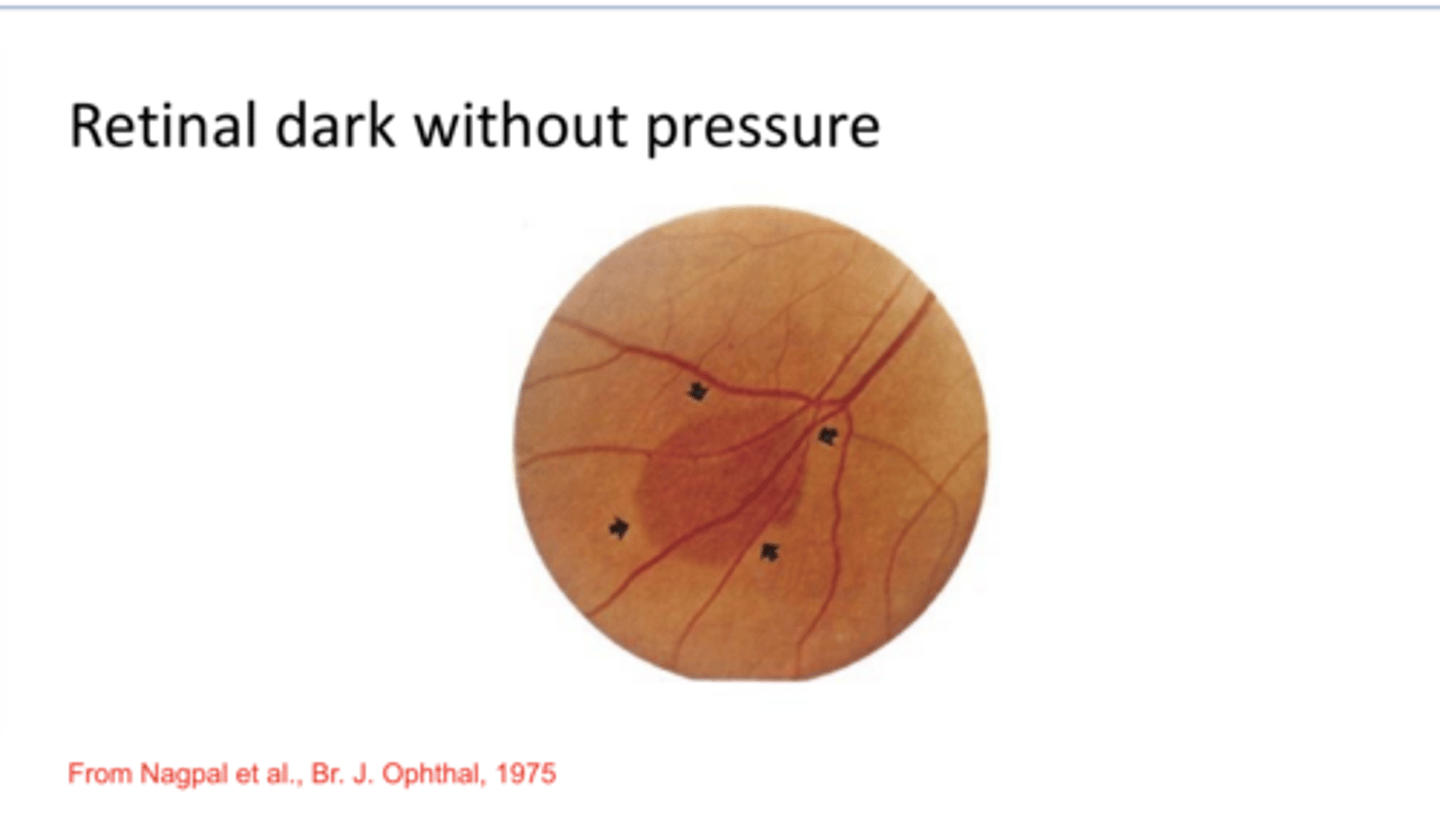
-retinal dark without pressure
-salmon patch intraretinal hemorrhages
-black sunburst lesion (RPE hyperplasia in the areas of old hemorrhages)
-Tortuous veins
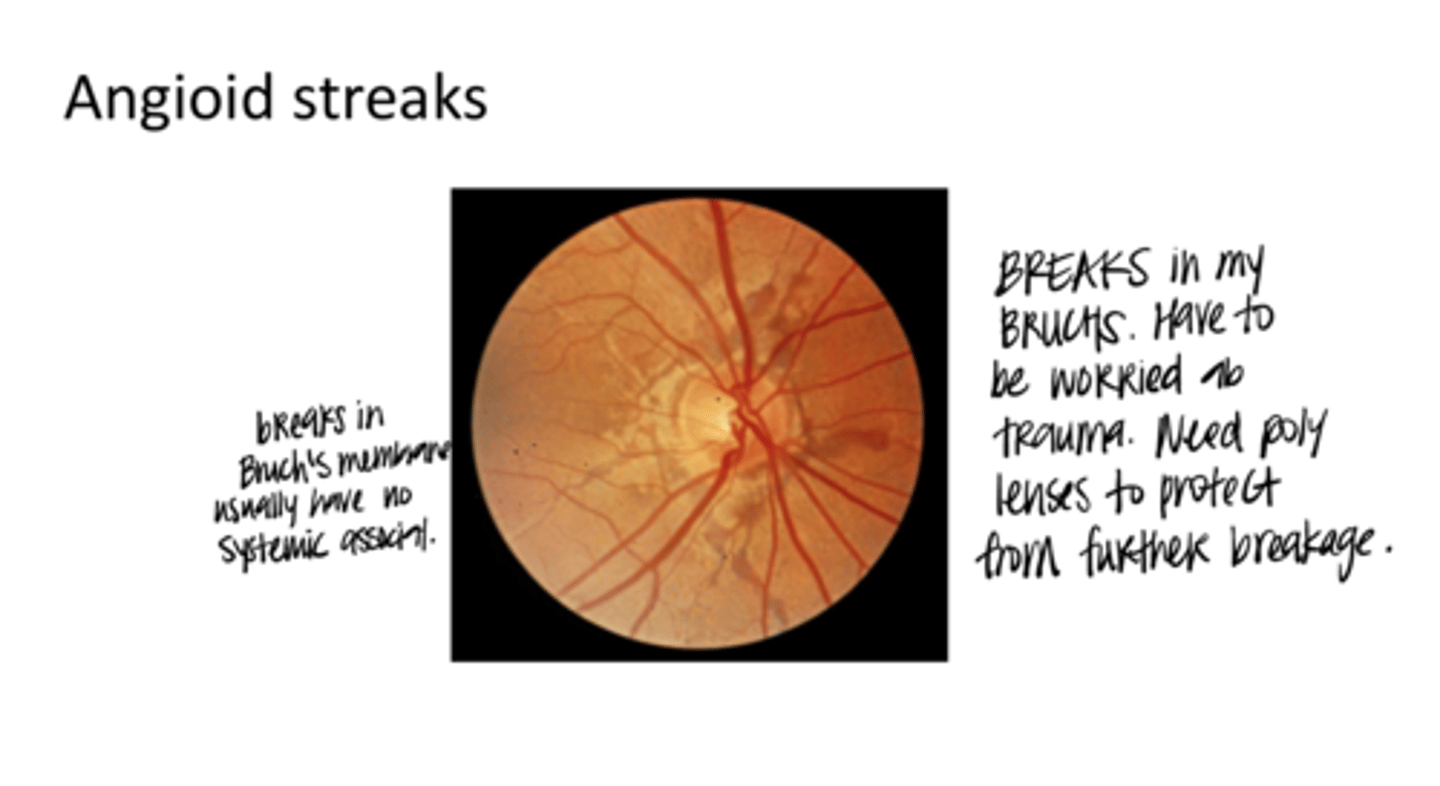
-Angioid streaks (cracks in Bruch's membrane)
What are the nonproliferative retinal signs of sickle cell anemia?
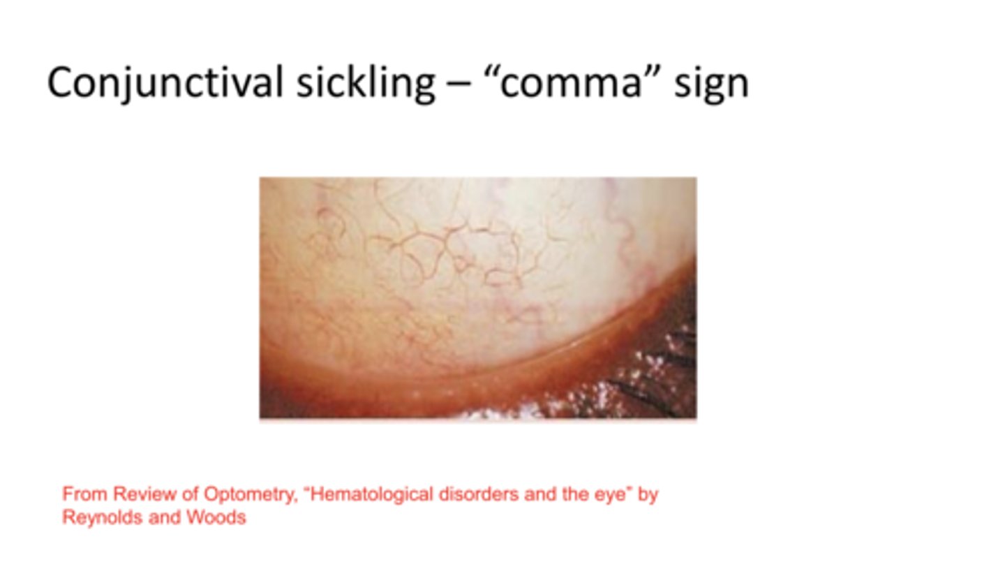
Conjunctival Sickling -- Comma Sign (Pic)
Conjunctival Sickling -- Comma Sign (Pic)
Retinal Dark w/o Pressure (Pic)
Retinal Dark w/o Pressure (Pic)

No -- most patients with this DO NOT have sickle cell
Is retinal dark w/o pressure an indication to order sickle cell testing?
Salmon Patch Hemorrhages and Black Sunburst (Pic)
Salmon Patch Hemorrhages and Black Sunburst (Pic)
**RBCs deoxygenated in salmon patch
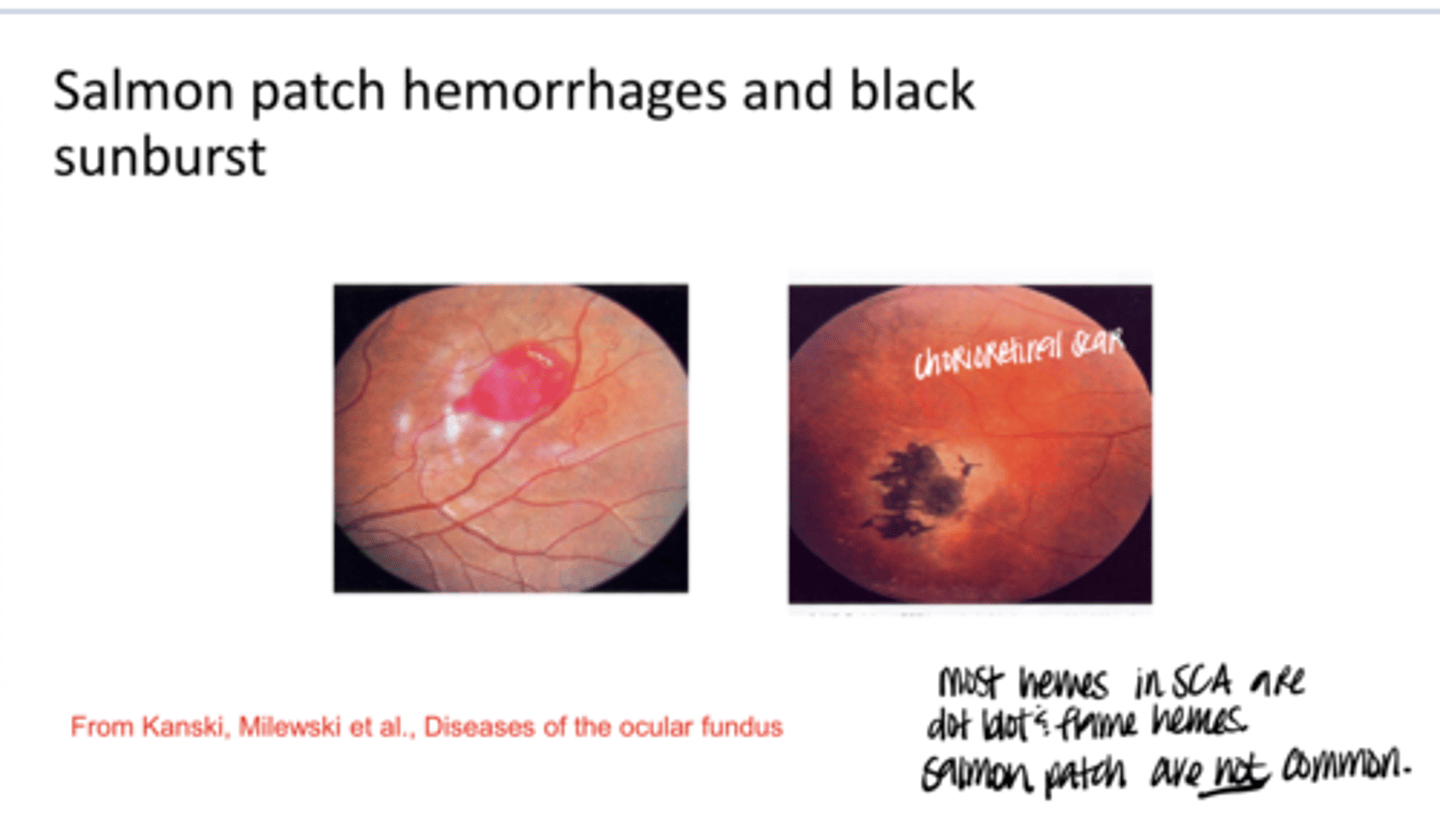
Salmon Patch Hemorrhages -- most of the times the hemes look like dot-blot or flame hemes
What is BY FAR the least common heme in sickle cell disease?

Angioid Streaks (Pic)
Angioid Streaks (Pic)

choroidal neovasc
Whenever you compromise Bruch's membrane, what are you at risk for 70% of the time?

protective lenses (polycarb)
What should you recommend for patients with Angioid streaks?

-PEPSI-HAM
-Pseudoxanthoma elasticum
-Ehlers-Danlos syndrome
-Paget's disease of bone
-Sickle cell disease and other hemoglobinopathies
-Idiopathic
-Homocystinuria
-Acromegaly
-Marfan syndrome
EXAM QUESTION: What are the systemic associations with Angioid streaks?
-sea fan neovascularization -- possibly in the periphery
-fibrotic scaffolding (white)
-vitreous hemorrhage
-traction retinal detachment
What are the proliferative signs of sickle cell retinopathy?
Sea Fan Neovascularization (Pic)
Sea Fan Neovascularization (Pic)
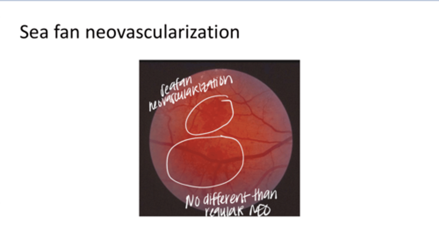
proliferative sickle-cell retinopathy (PSR)
What is the most common cause of vision loss d/t sickle cell retinopathy?
-S thal
-SC
_____ and _____ patients consistently show higher rates of visual impairment from proliferative chanes
true
True or False:
Sickle cell disease can be detected through newborn screening
-Hb studies (HbS)
-CBC
What sickle cell bloodwork needs to be ordered for patients with unaccounted for hemes?
No -- relatively unlikely
Is diagnosis of sickle cell disease common for optometrists?
-sickle cell trait (SA)
-hemoglobin C trait (AC)
In what cases may it be possible to diagnose sickle cell anemia from an eye exam?
1) Decrease in red blood cells (Erythrocytes) OR
2) Decrease in hemoglobin
What are the general definitions of anemia?
Yes
Is it possible to have a normal erythrocyte count and low hemoglobin (iron deficiency)?
hemolysis -- where the erythrocytes are removed from the blood leaving unbound hemoglobin
If the erythrocytes are low and hemoglobin is normal, what is one thing to consider?
-Under production of blood in bone marrow, kidney, diet, absorption, liver
-Hemolytic
-Bleeding
-Anemia of chronic disease (kidney disease, cancer, autoimmune disorders, chronic infections)
What are the 4 major mechanisms of anemias?
iron deficiency anemia
What is the most common anemia in the US?
bleeding
What is the most common etiology for iron deficiency anemia?
hemoglobin
____ decreases as iron deficient anemia progresses
-cells normochromic (normal hemoglobin content)
-Normocytic (normal mean cell volume)
Characteristics of early stage iron deficient anemia
Normochromic and microcytic
Characteristics of middle stage iron deficient anemia
hypochromic and microcytic
Characteristics of final stage iron deficient anemia
-probably lower
-smaller and fewer RBCs
-less blood volume filled by the RBCs
Hematocrit in Iron Deficient anemia
yes
Are there specific tests to run for iron deficiency?
B12 deficiency -- crucial for the synthesis of nucleic acids which are involved in forming blood precursors
What is the etiology of pernicious anemia?
1) Hereditary autoimmune disorder to gastric mucosa secondary to surgery, infection, drugs, and digestive disorders
2) Nutritional deficiency
What are the 2 ways in which you can develop pernicious anemia?
-macrocytic (mean cell volume increased)
-normochromic
Characteristics of pernicious anemia
-elderly of northern European ancestry
-African Americans
-Latin-Americans
Who does pernicious anemia typically present in?
-conj pallor
-anemic retinopathy
-optic nerve head pallor from lack of perfusion
-blue sclera
-subconj hemorrhage
-retinal arterial or venous occlusions
-anterior ischemic optic neuropathy
Ocular findings of pernicious anemia
Not really, in a lot of cases of pernicious anemia this does not present
Is conj pallor useful in dx of pernicious anemia?
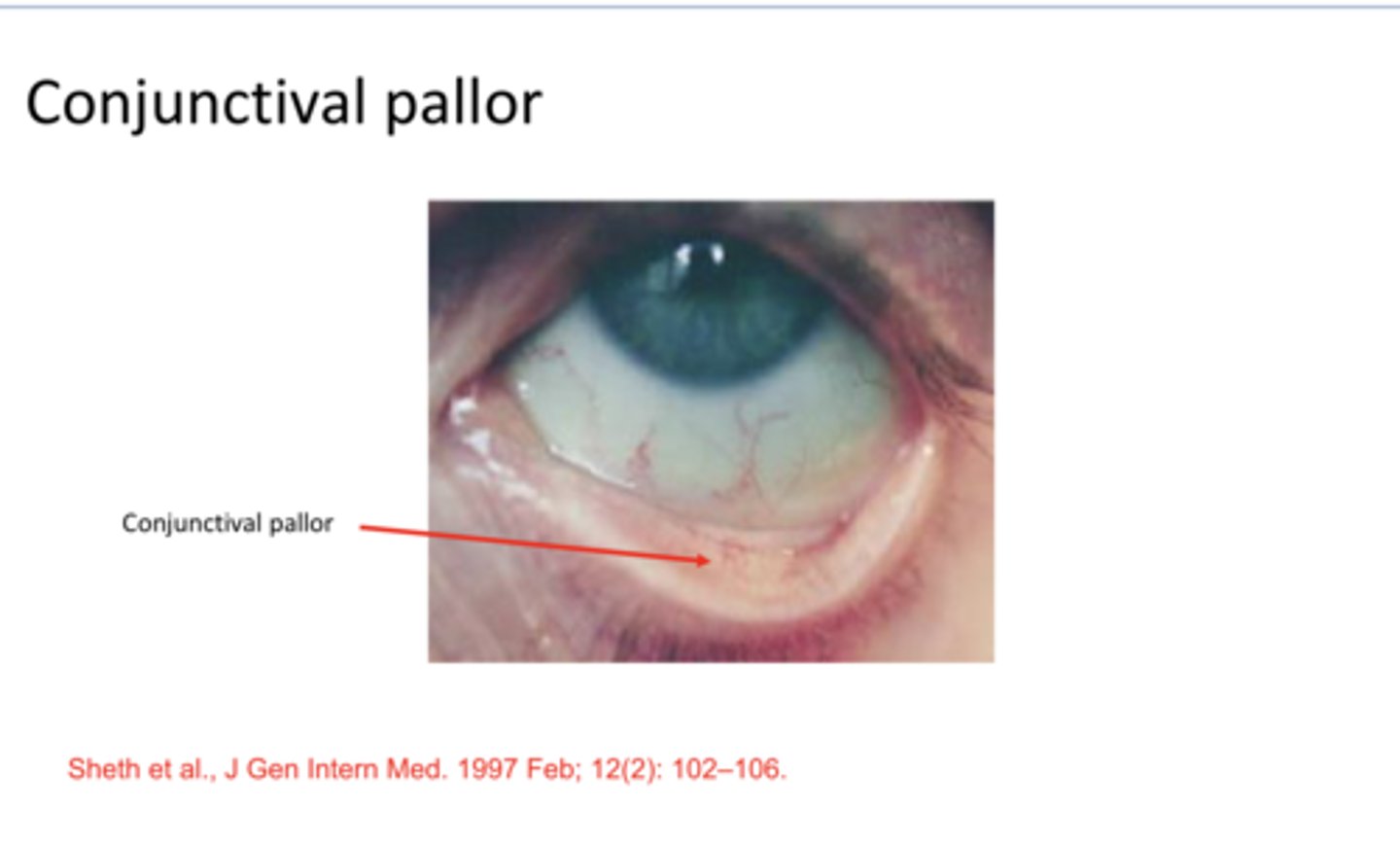
Conjunctival Pallor (Pic)
Conjunctival Pallor (Pic)

Iron supplements
What is the "cure" for iron deficient anemia?
yes
If a patient starts to take iron supplements for their iron deficient anemia, will the eye manifestations likely go away?
1) severe anemia
2) thrombocytopenia at the same time as the anemia
Under what 2 conditions would anemic retinopathy present?
Scleral thinning; can see the melanin in the choroid
What is a blue sclera d/t?
No -- there are usually no manifestations
Are retinal arterial or venous occlusions common with anemia?
Not high
EXAM QUESTION: What is the likelihood of ocular manifestations d/t anemia?
bone marrow disorder
What is aplastic anemia?
issues with red blood cells, white blood cells and platelets
What does aplastic anemia d/t?
more
EXAM QUESTION: Systemic and ocular manifestations (retina/optic nerve head involvement) are (more/less) likely with aplastic anemia than with iron deficiency/pernicious anemia
neoplatic disease of the bone marrow characterized by abnormal, uncontrolled proliferation of one or more hematopietic or lymphoid cells
EXAM QUESTION: What is leukemia?
13/100,000
What is the incidence of leukemia?
-Acute v chronic (time course)
-Myelogenous v Lymphocytic (cell type)
What is the classification of leukemia?
acute and rapidly fatal (immature cells are affected -- blasts)
Acute leukemia definition
More prolonged course (involve more differentiated -- mature cells)
Chronic leukemia definition