Lab 2 Exam Review
 Note
 Call Kai
 Learn
 Practice Test
 Spaced Repetition
 Match
 Flashcards
 Knowt Play
1/299

There's no tags or description
Looks like no tags are added yet.
300 Terms
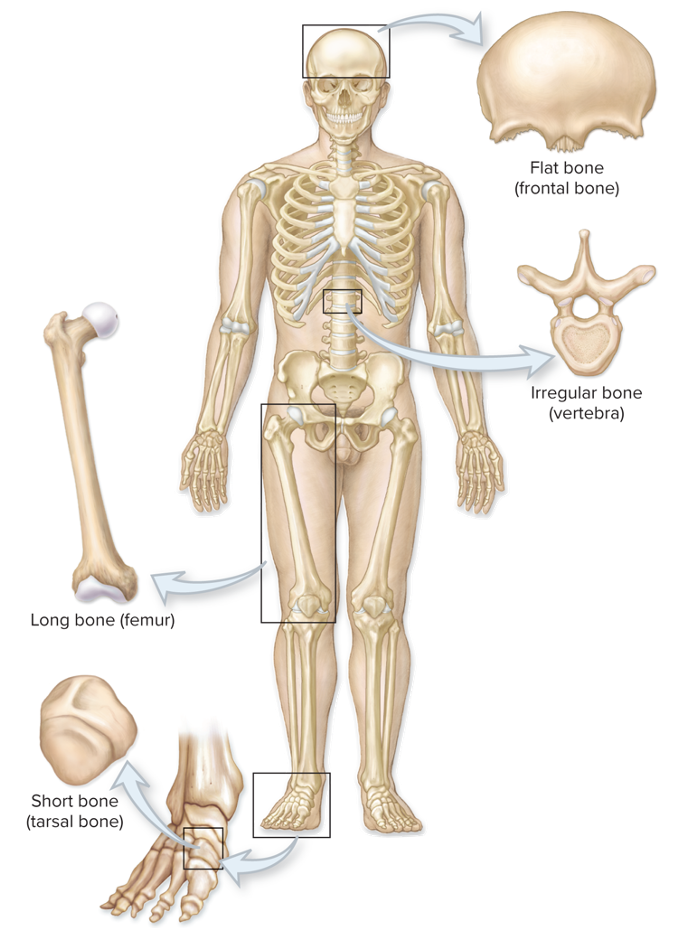
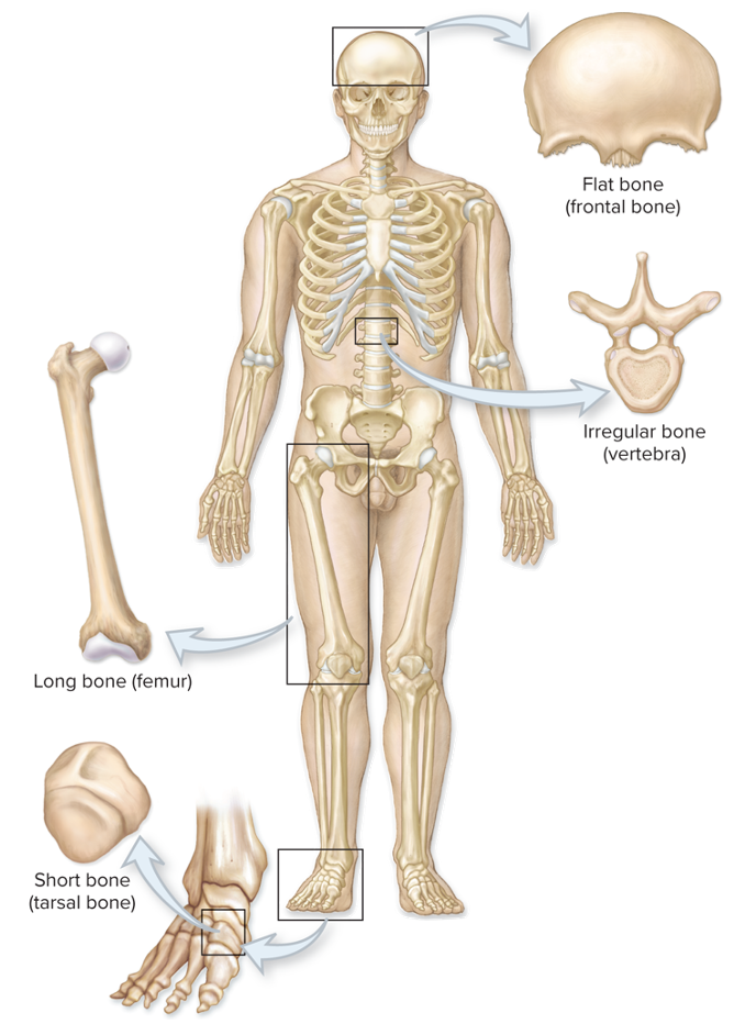
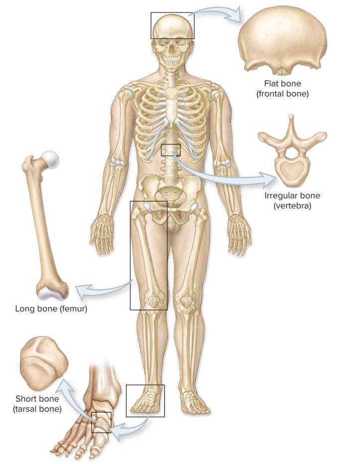
Long Bones
Greater in length than width (femur, humerus)
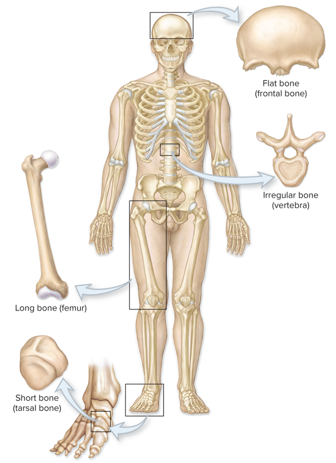
Short Bones
Length nearly equal to width (carpals + tarsals)
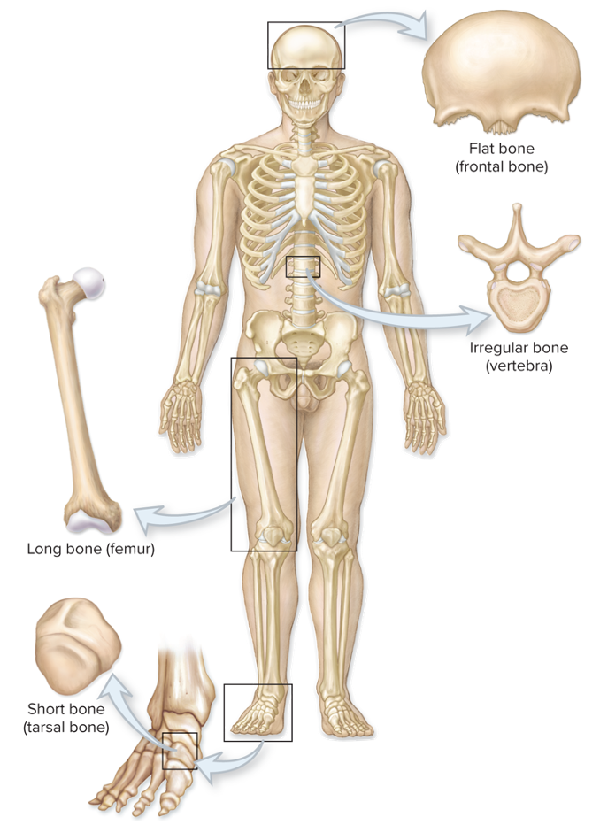
Flat Bones
Flat, thin surfaces, may be slightly curved (cranial bones)
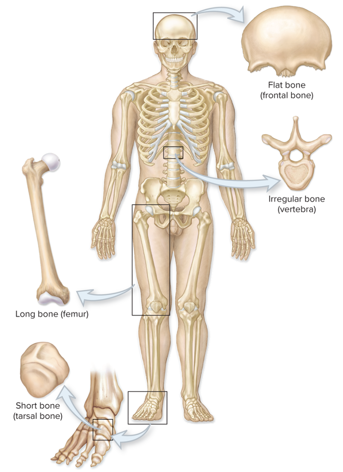
Irregular Bones
Elaborate, sometimes complex shapes (vertebrae)
Sesamoid Bones
Small, sesame seed-shaped bones along the tendons of some muscles, classified as short bones. (Patella is the largest sesamoid bone)
Sutural Bones
Small bones located within sutures of the skull, vary in number and shape among individuals
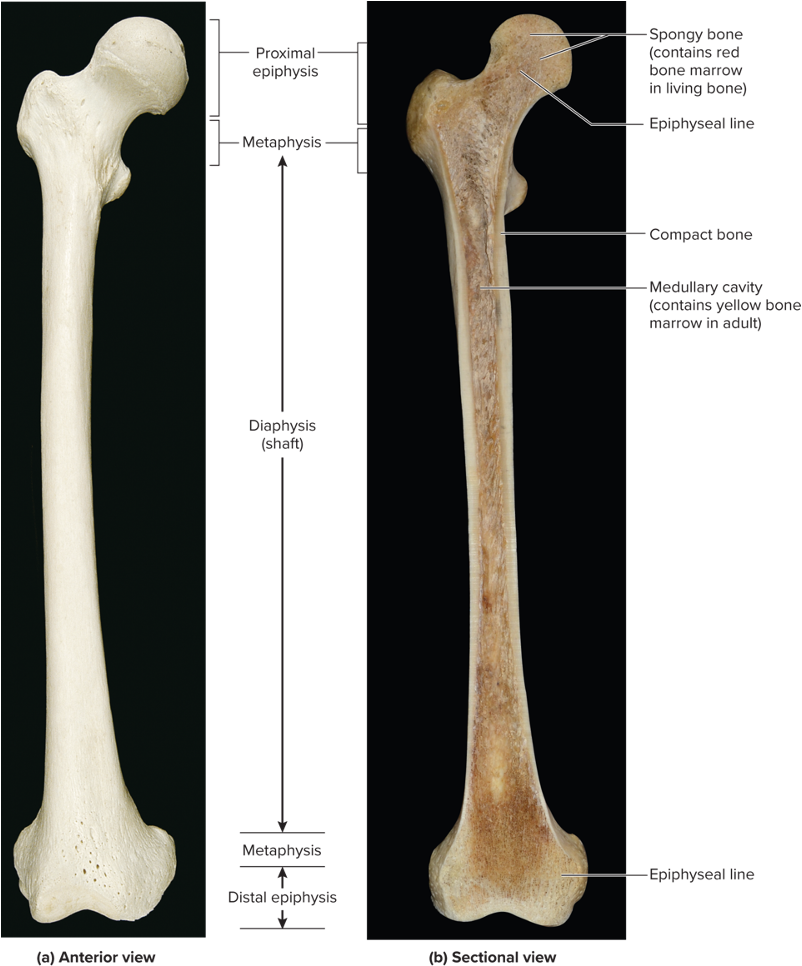
Long Bone Structures - Diaphysis/shaft
Elongated, usually cylindrical shaft
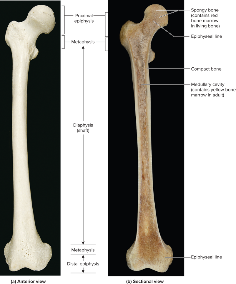
Long Bone Structures - Medullary Cavity
Hollow, cylindrical space within the diaphysis
Contains red bone marrow in children and yellow bone marrow in adults
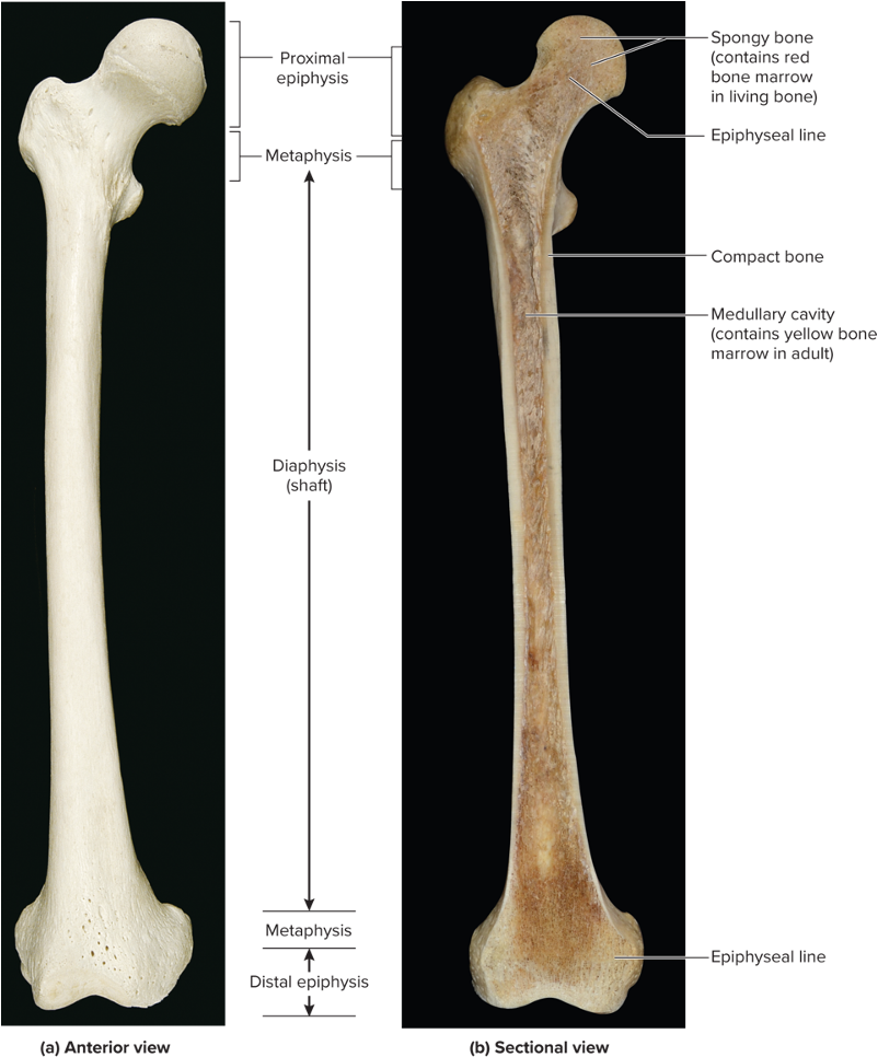
Long Bone Structures - Metaphysis
Region between the diaphysis and epiphysis
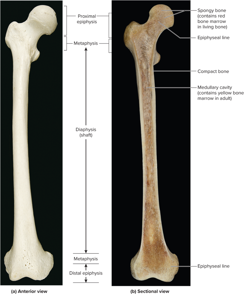
Long Bone Structures - Epiphysis
Knobby region at each end of the long bone
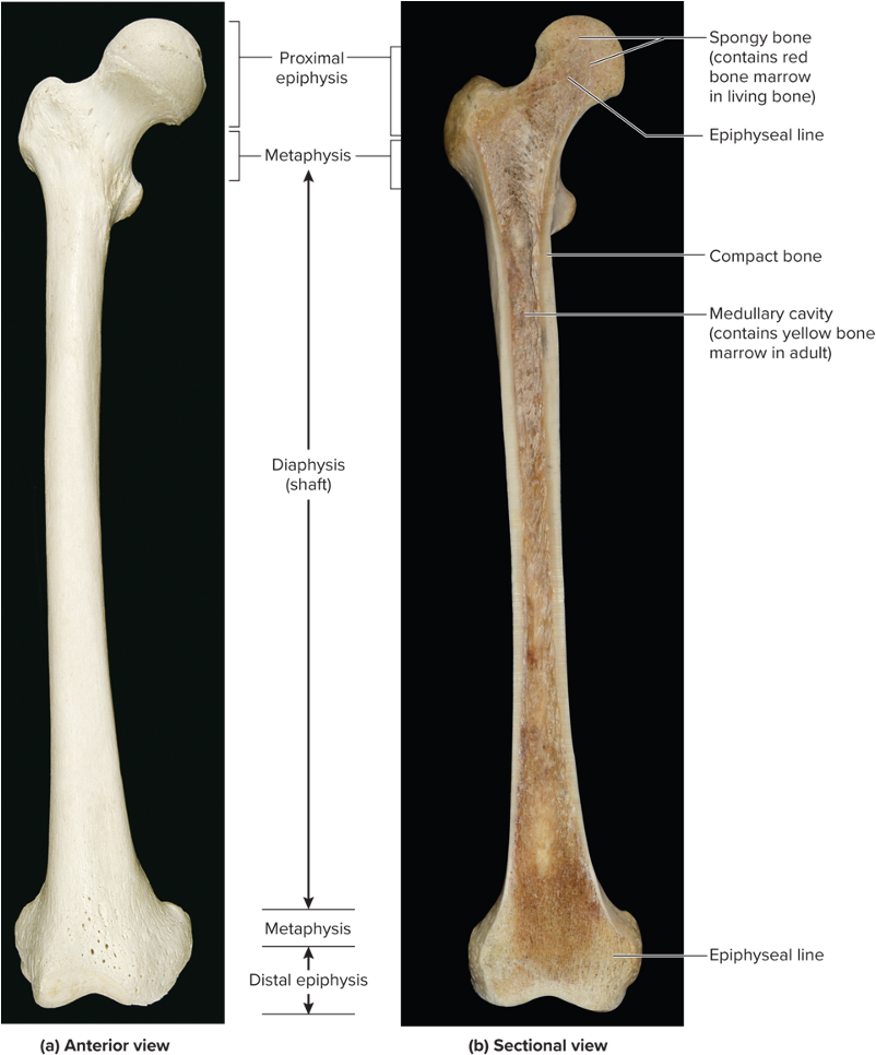
Long Bone Structures - Epiphyseal Plate/Line
Thin layer of hyaline cartilage located within metaphysis
In adults, the epiphyseal line, is the remnant of the epiphyseal plate
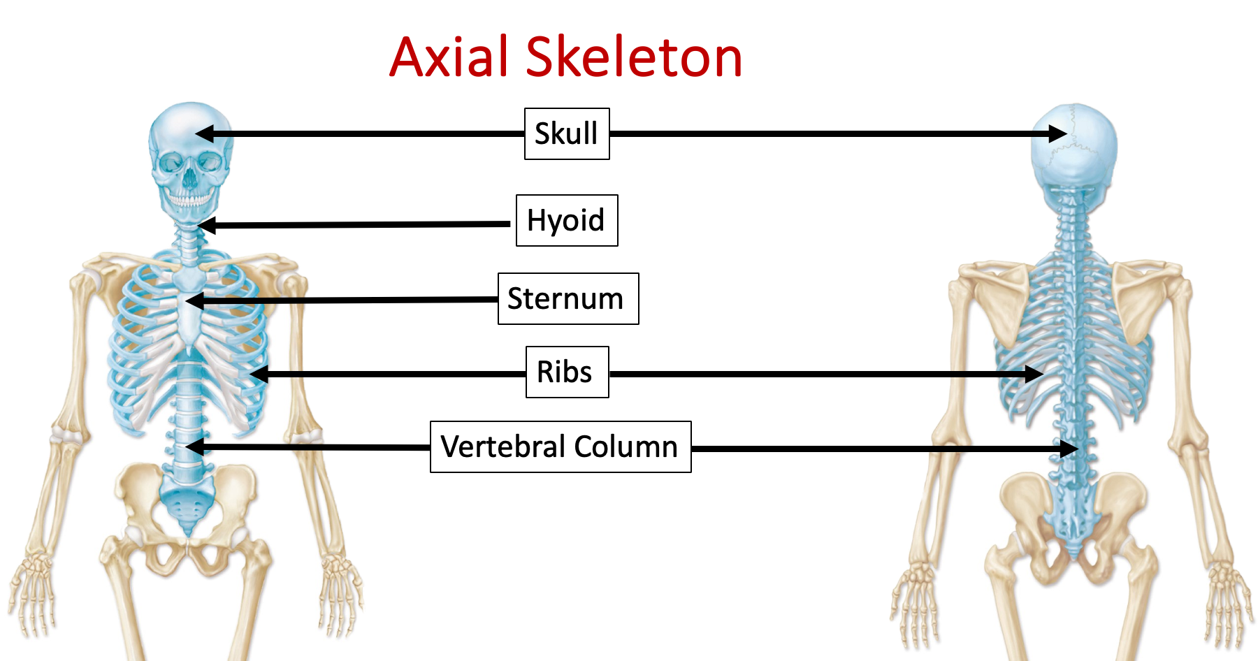
What structures are located in the axial skeleton?
The skull, hyoid, sternum, ribs, and vertebral column
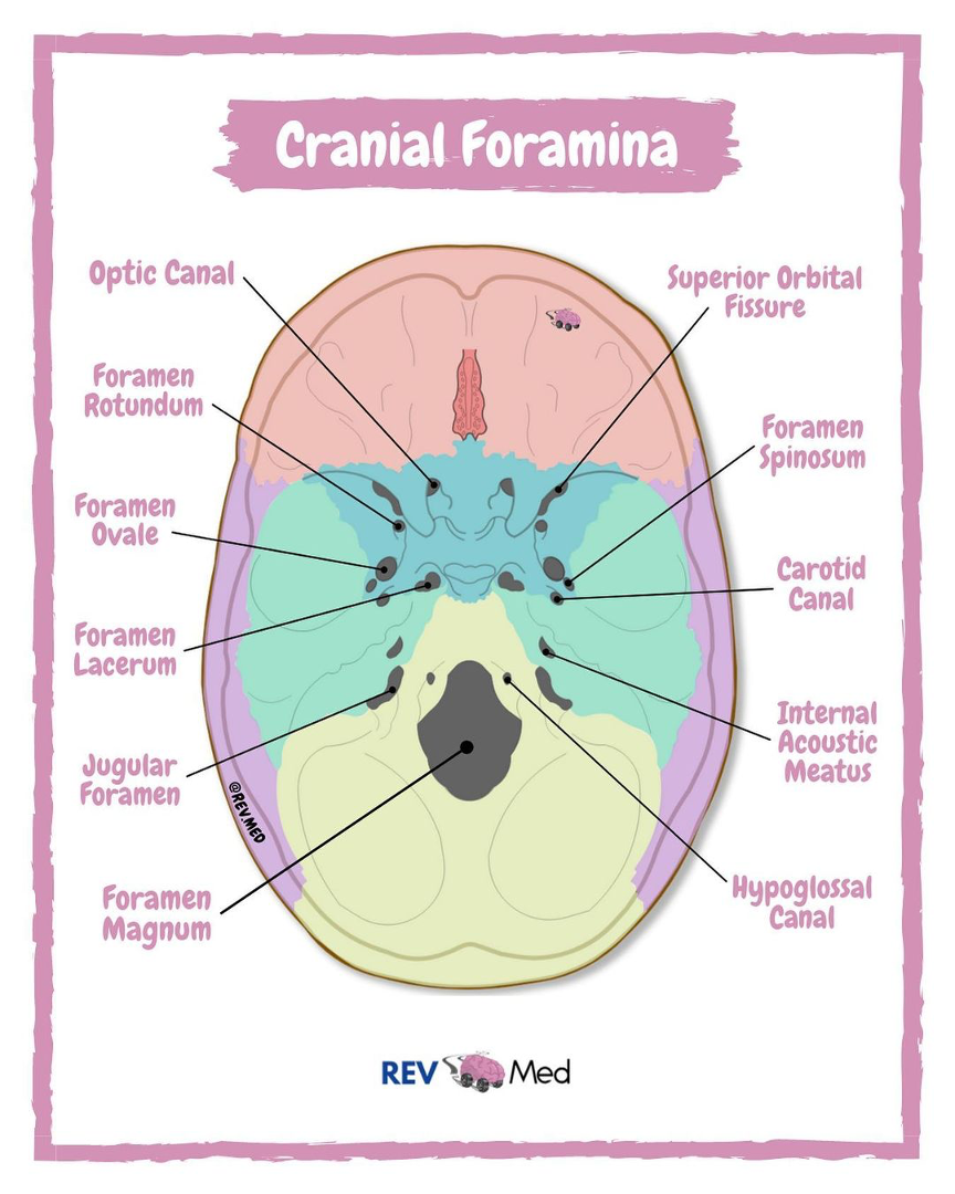
What is the mnemonic for the foramina of the skull?
“Old Rotund Owls Spin Lazily Across Jugs”
Optic
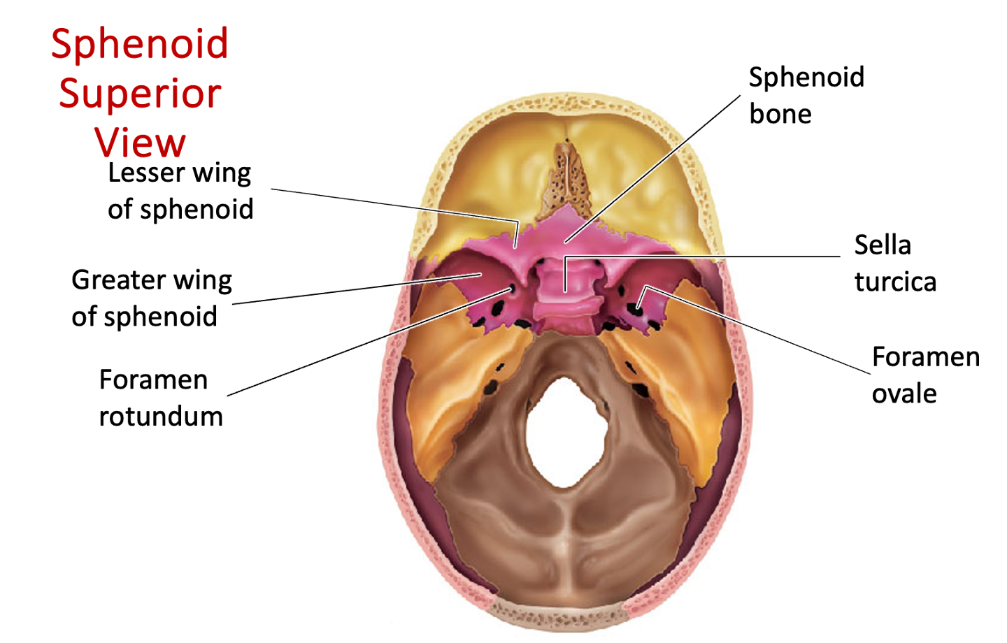
Rotundum
Ovale
Spinosum
Lacerum
Acoustic
Jugular
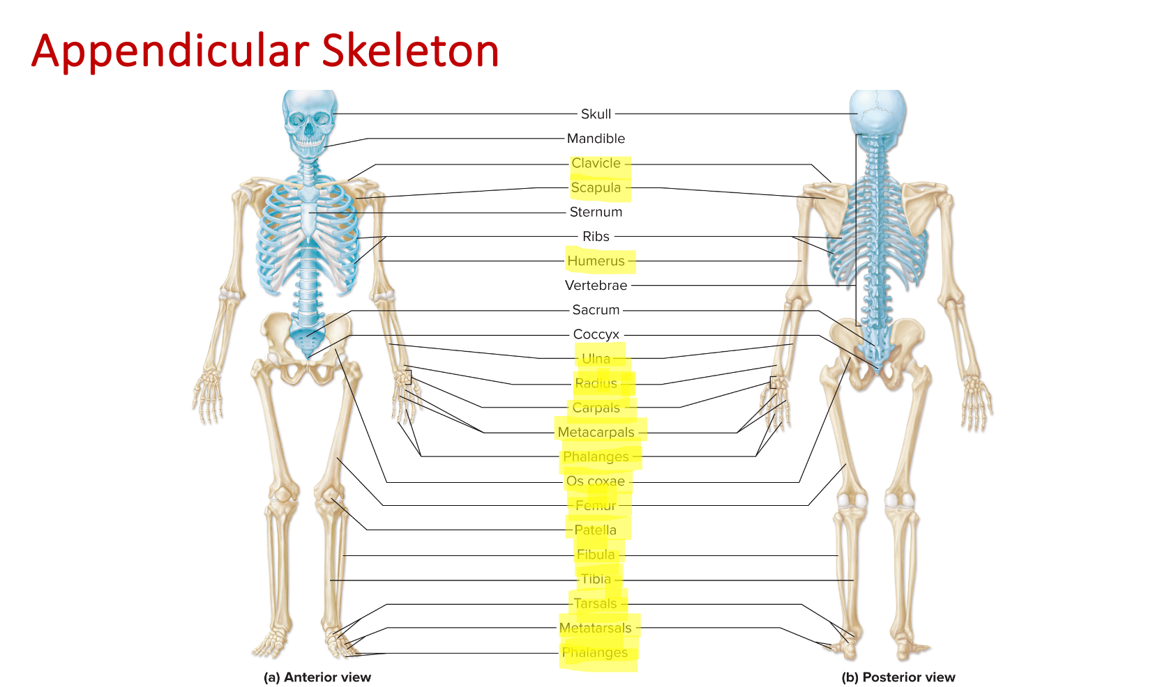
Appendicular Skeleton Bones
Clavicle, scapula, humerus, ulna, radius, carpals, metacarpals, phalanges, os coxae, femur, patella, fibula, tibia, tarsals, metatarsals, phalanges
Synarthroses
Immobile joints, can be fibrous or cartilaginous joints
Amphiarthrosis
Slightly mobile joints, can be fibrous or cartilaginous joints
Diarthroses
Freely mobile joints, all synovial joints
Fibrous Joints
Bones connected by dense regular CT
Have no joint cavity
Immobile or only slightly mobile
Three most common types:
Gomphoses
Sutures
Syndesmoses
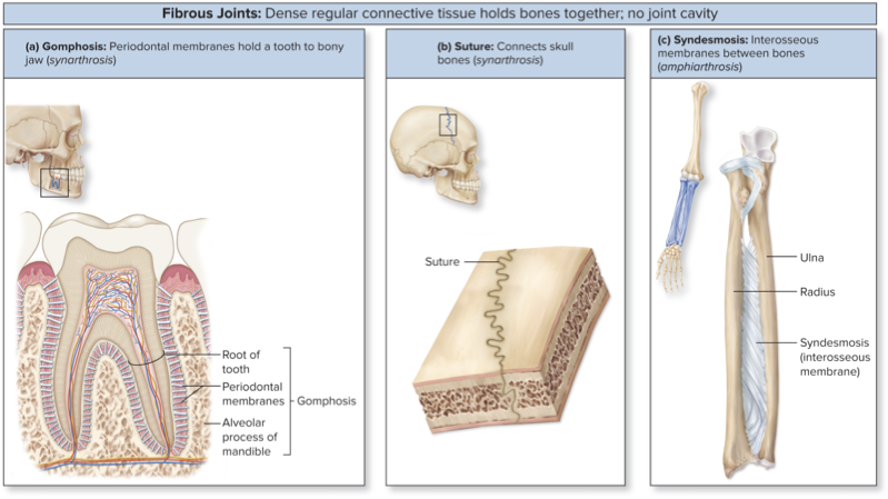
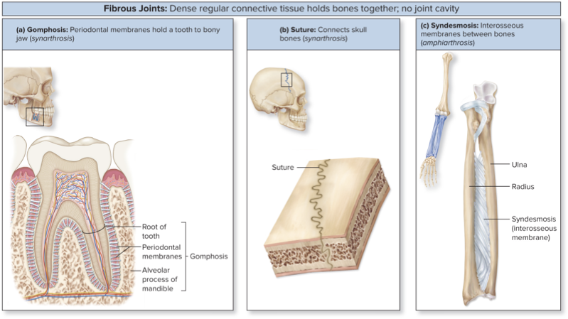
Gomphosis
“Peg in a socket”
Articulations of teeth with sockets of mandible + maxillae
Tooth held in place by fibrous periodontal membranes
Function as synarthroses (very stable)
Suture
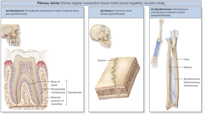
Found between some skull bones
Very short fibers
Interlocking, irregular edges
Increases stability + decreases risk of fracture
Function as synarthroses
Allow growth in childhood
Become ossified synostoses in older adults
Syndesmoses
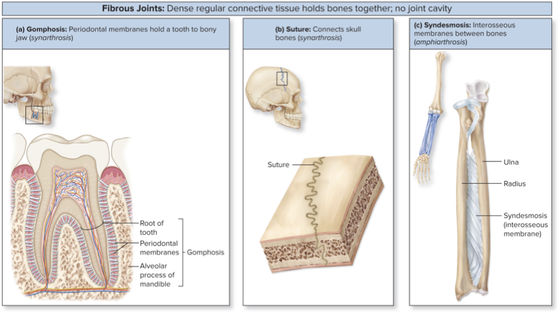
Bound by interosseous membrane, broad ligamentous sheet
Found between radius + ulna and between tibia + fibula
Function as amphiarthroses
Provides pivot for two long bones
Cartilaginous Joints
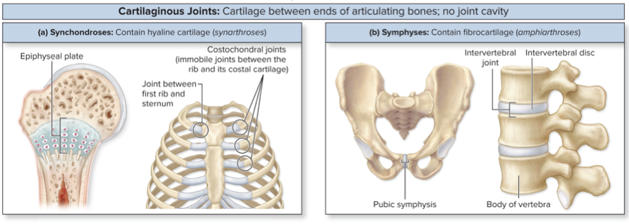
Either hyaline cartilage or fibrocartilage between bones
Lack a joint cavity
Immobile or slightly mobile
Synchondroses or symphyses
Synchondroses
Bones joined by hyaline cartilage
Immobile (synarthroses)
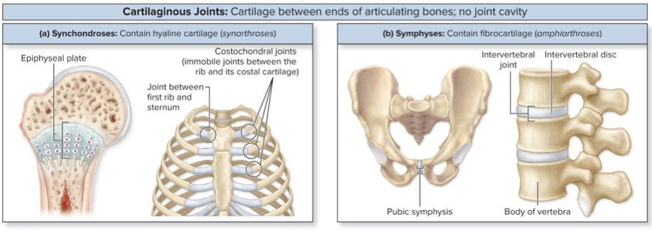
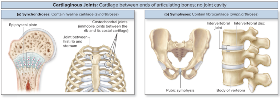
Symphyses
Pads of fibrocartilage between articulating bones
Resist compression and act as shock absorbers
Allow slight mobility (amphiarthroses)
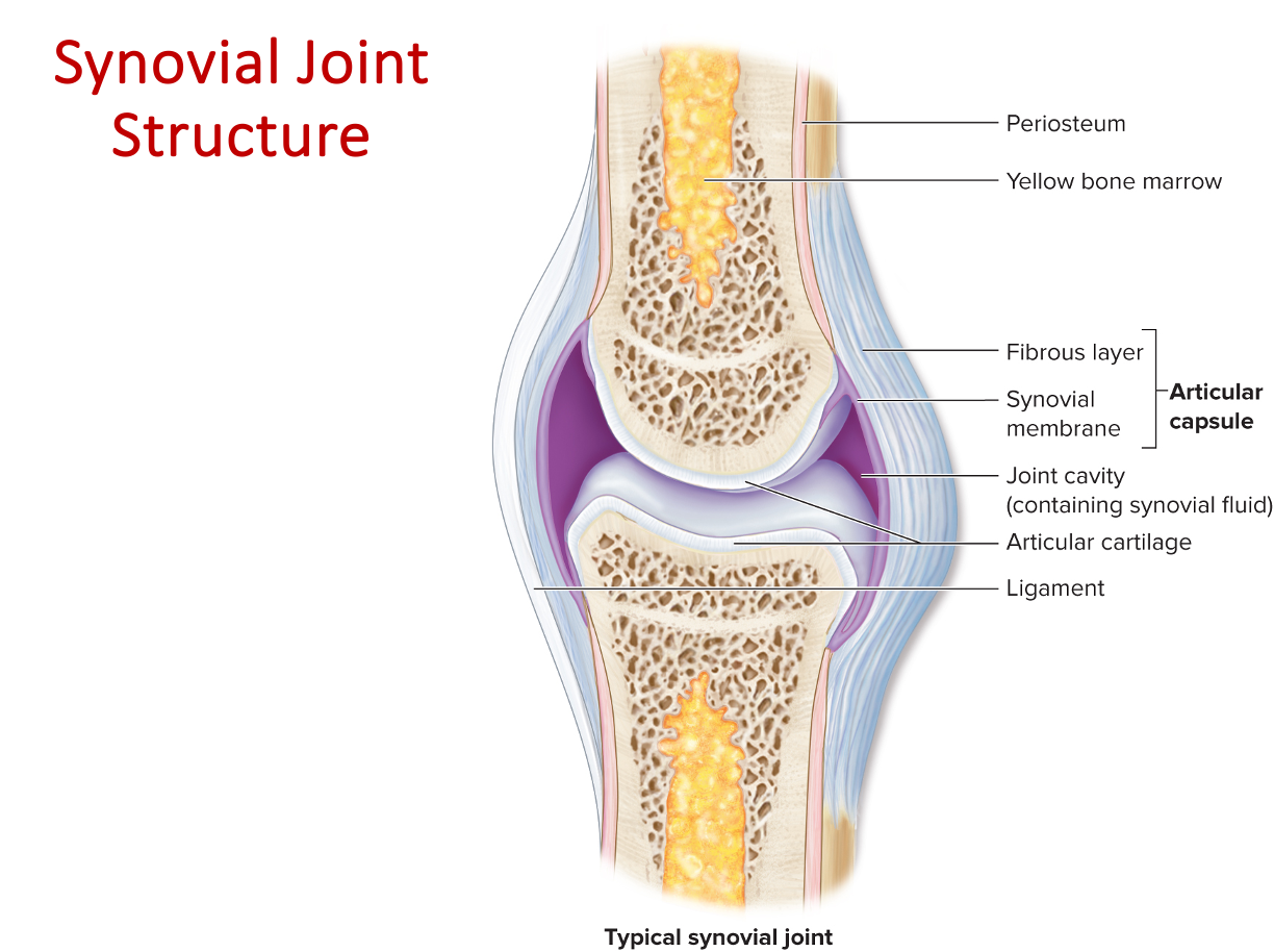
Synovial Joints
Bones separated by a joint cavity
Included most joints in the body
Diarthroses (freely mobile)
All with several basic features:
Articular capsule + joint cavity
Synovial fluid
Articular cartilage
Ligaments, nerves, + blood vessels
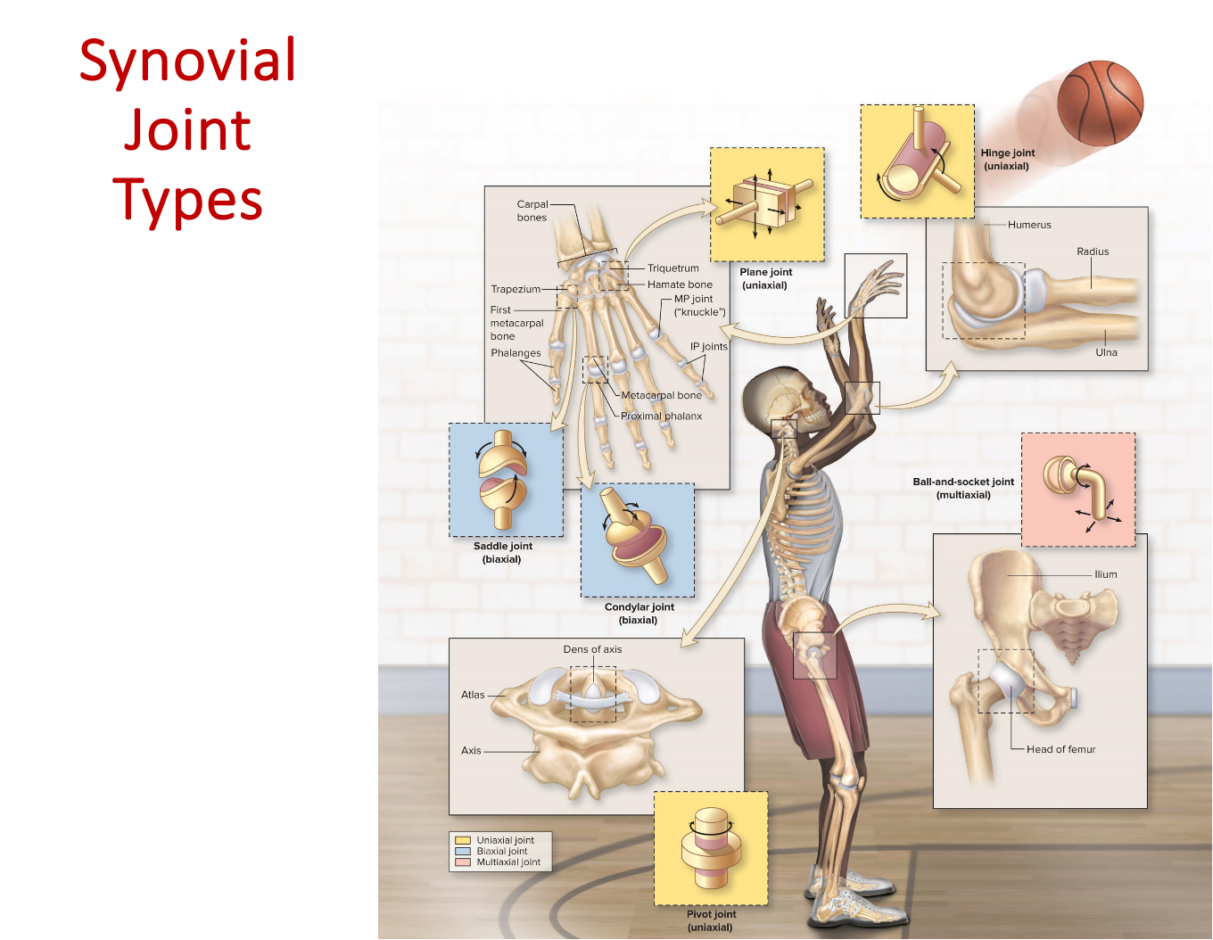
Planar Joint
Articular surfaces flat
Simplest, least mobile synovial articulation
Uniaxial: limited to side-to-side gliding movement in a single plane
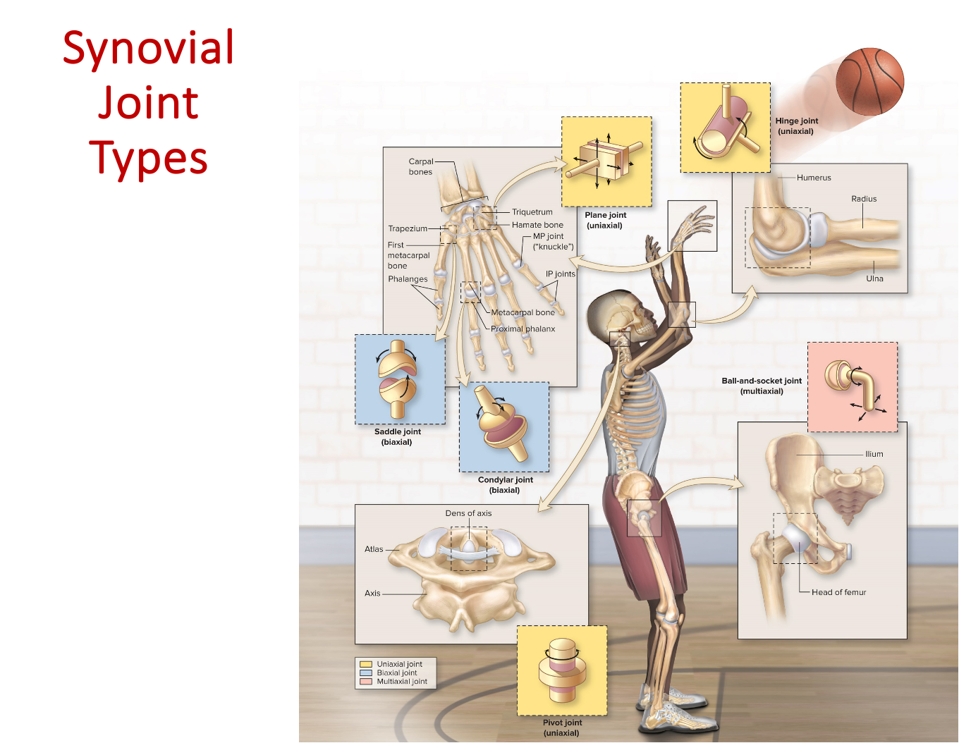
Hinge Joint
Convex surface within concave depression
Uniaxial: like the hinge of a door
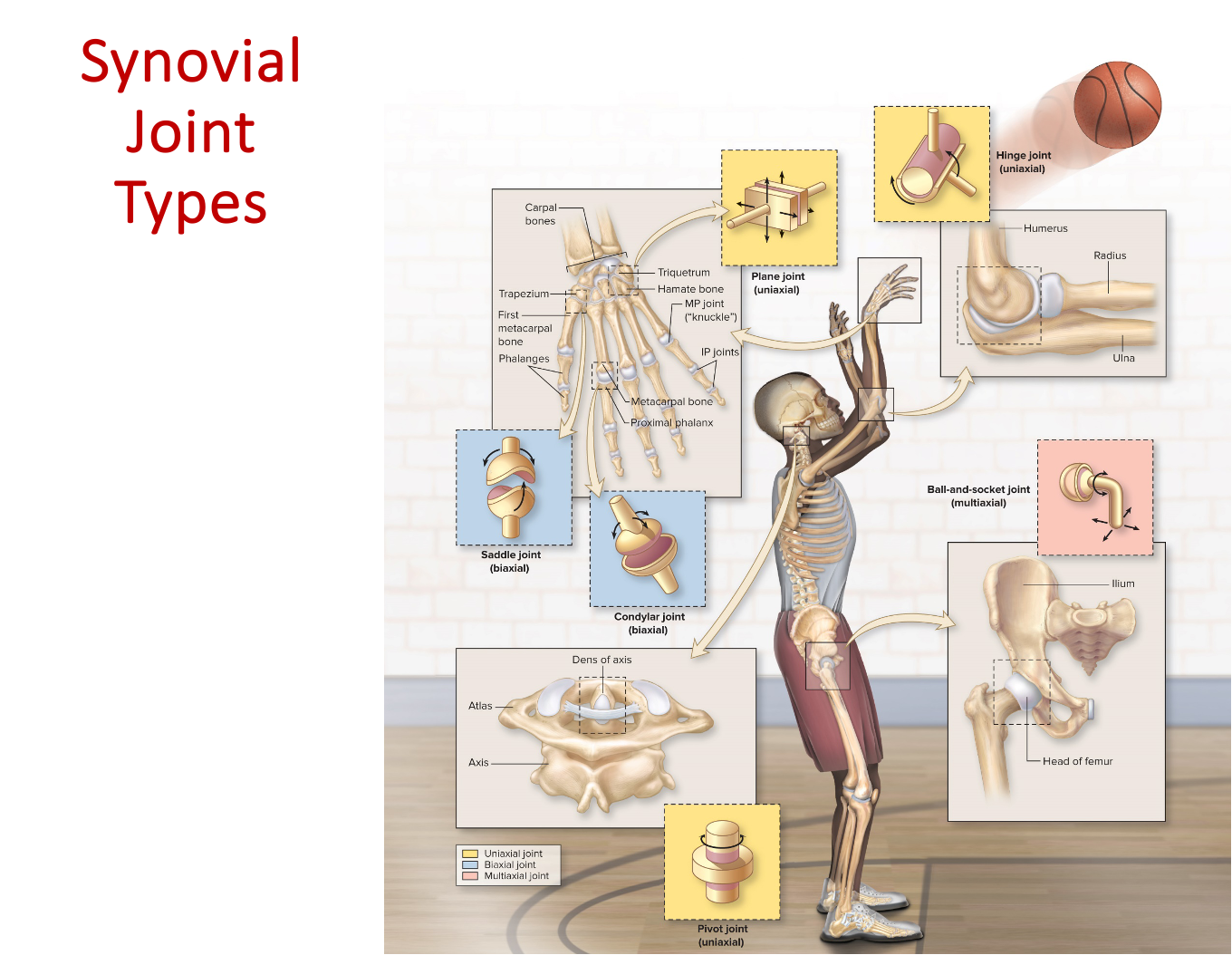
Pivot Joint
Bone with rounded surface fits into ligament ring
Uniaxial joint: rotation on longitudinal axis
EX: Rotation of head
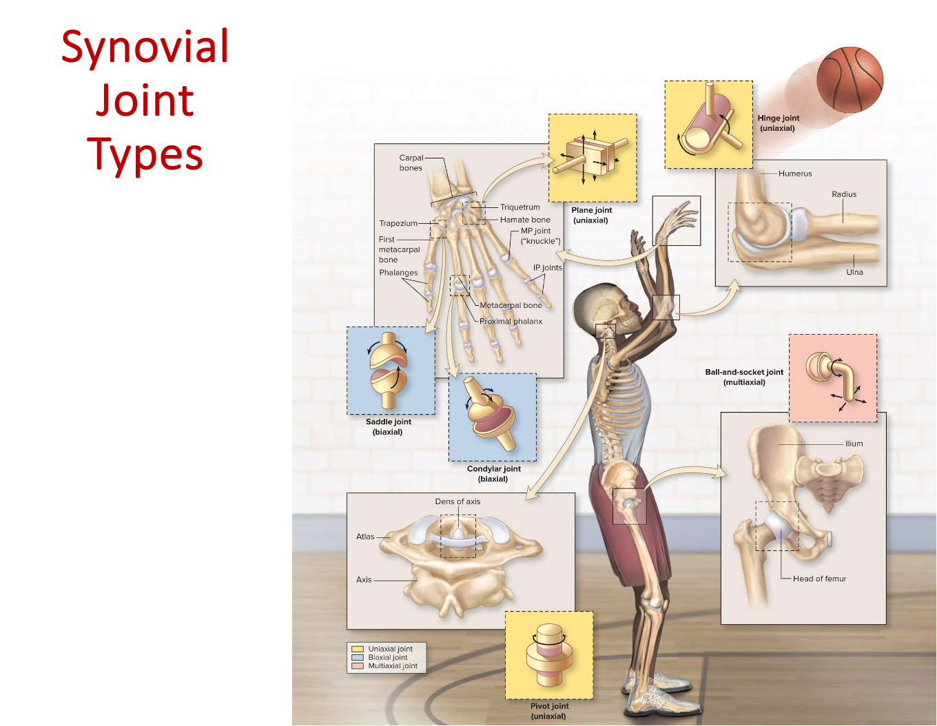
Condylar Joint
Oval, convex surface articulating with concave surface
Biaxial
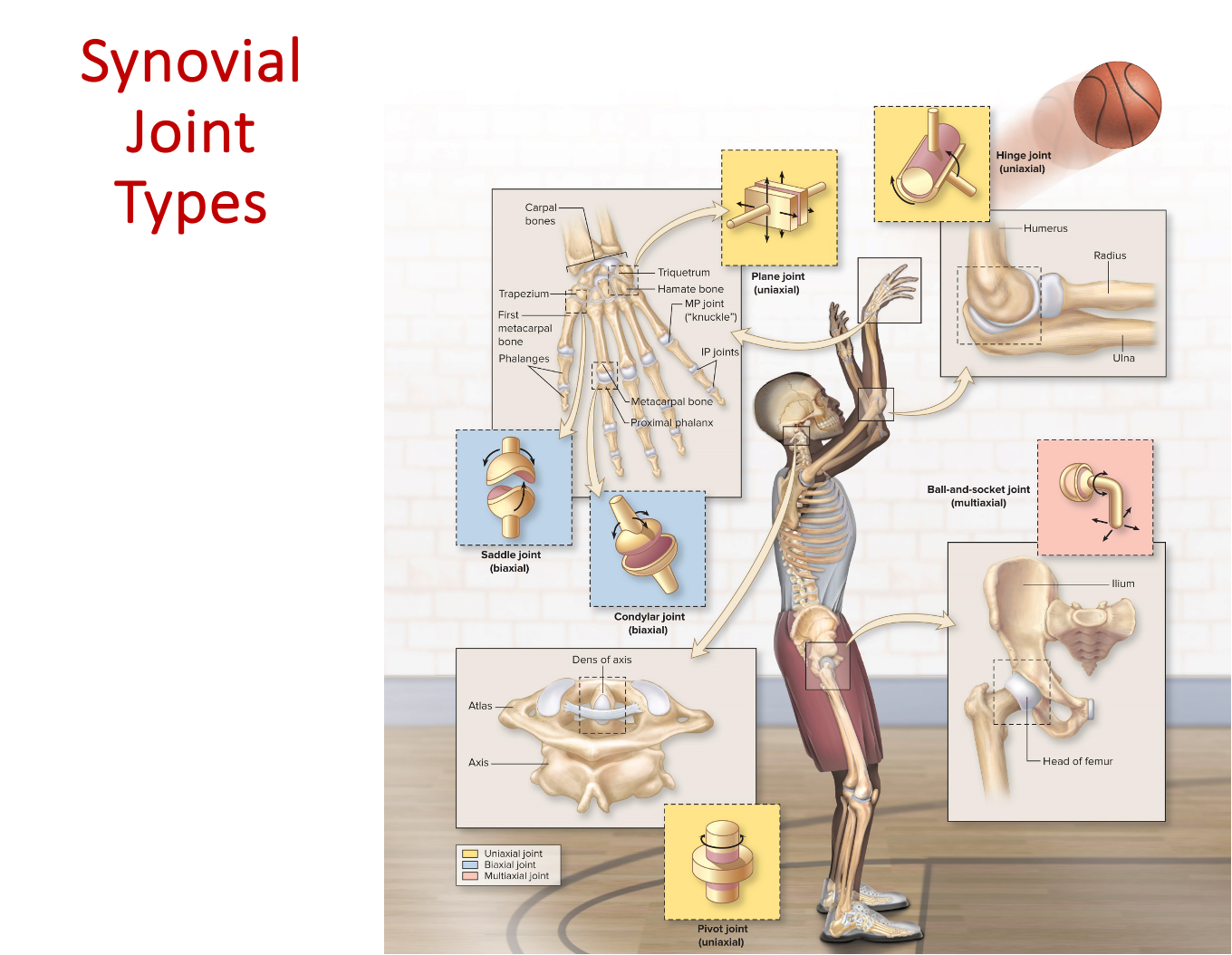
Saddle Joint
Convex + concave surfaces resembling saddle-shape
Biaxial
EX: Thumb, opposition movement
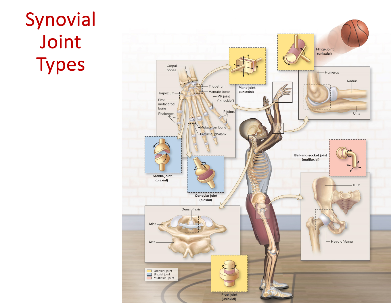
Ball-and-Socket Joint
Spherical head of one bone fitting into cuplike socket
Multiaxial, permitting movement in three planes
The most freely mobile type of joint
Extension + Flexion
Flexion (“Fetal Position”)
Decreases the angle between bones: brings bones closer together
EX: Bending a finger
Extension
Increases angle between articulating bones
EX: Straightening your fingers after making a fist
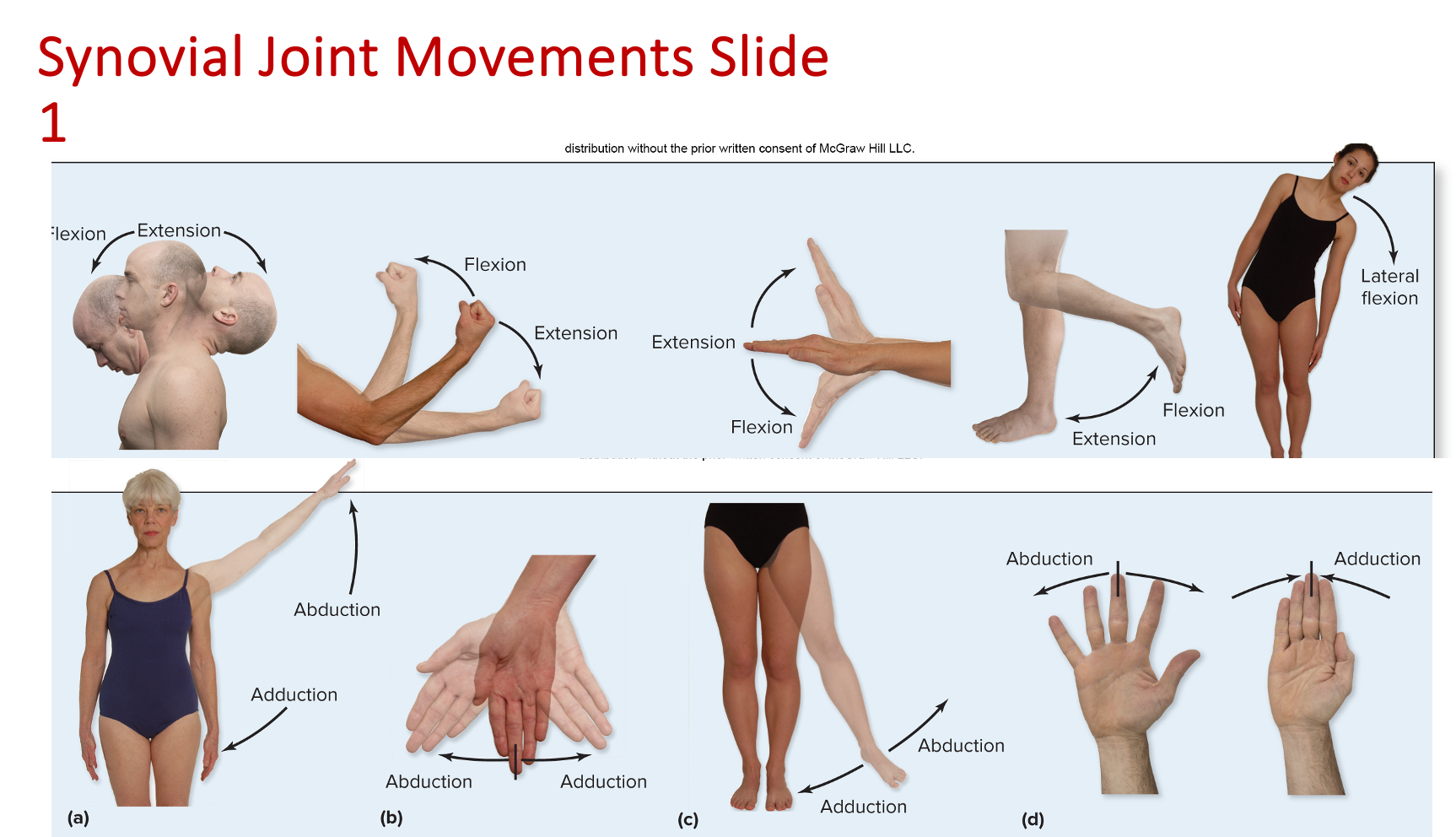
Lateral Flexion
Trunk of body moving in coronal plane laterally
Occurs between vertebrae in the cervical + lumber region
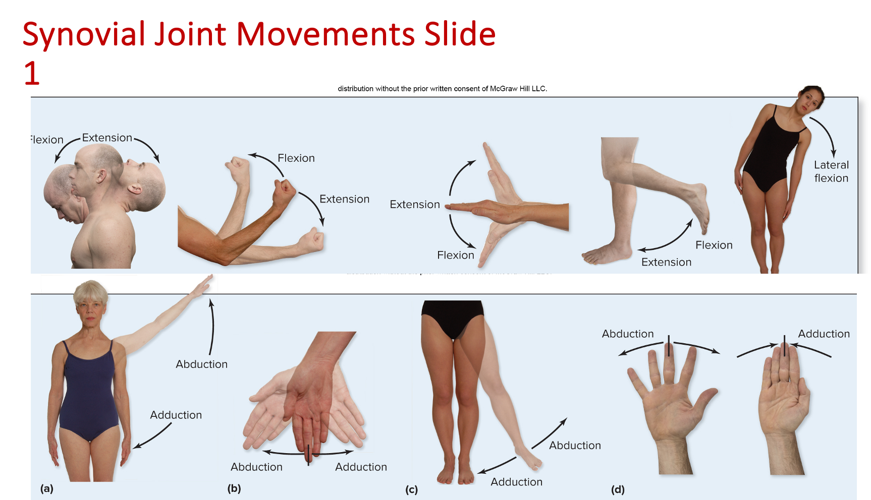
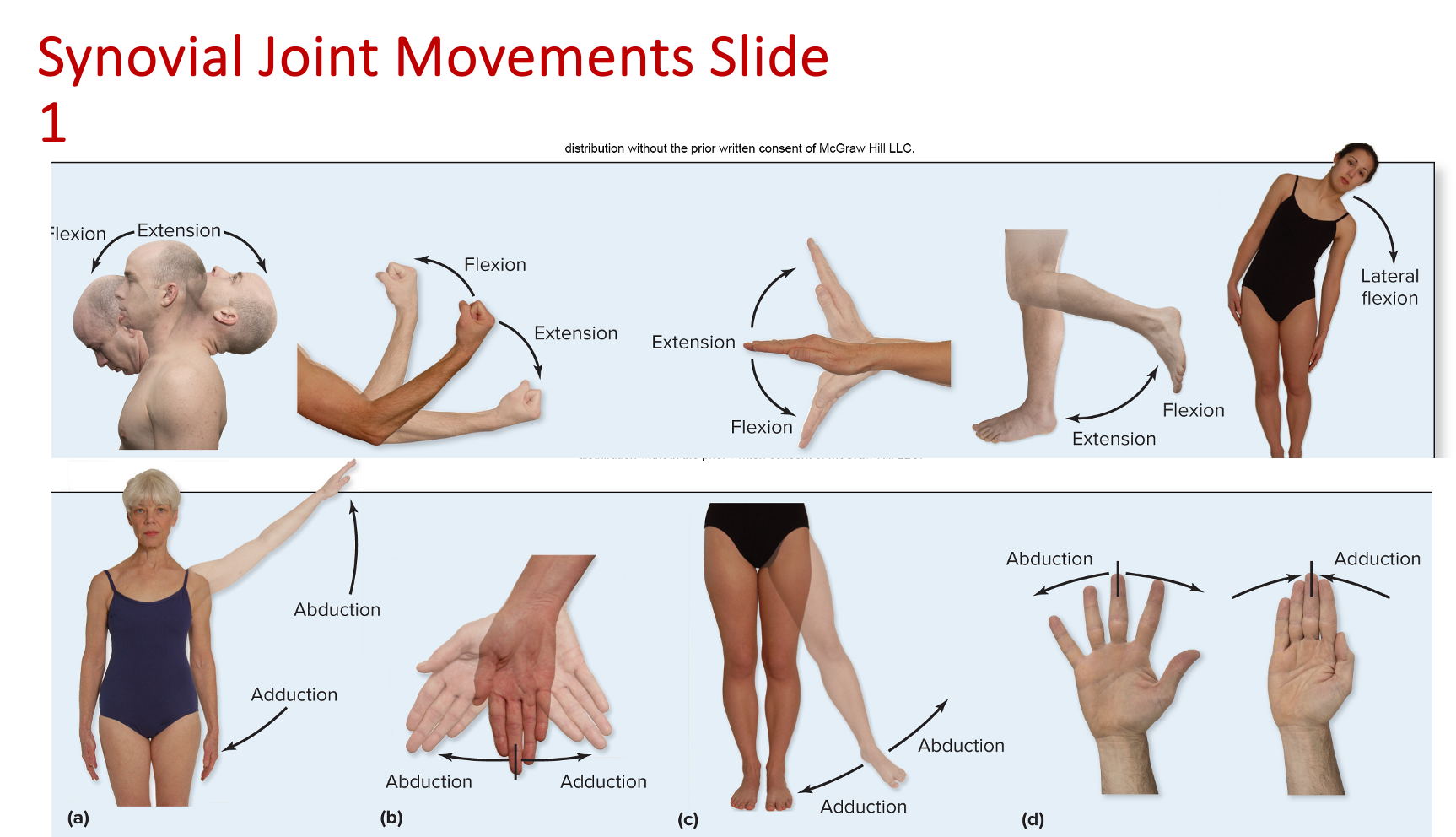
Abduction + Adduction
Abduction (“Alien taking body part away”)
Lateral movement of body part away from midline
EX: Arm or thigh moved laterally from body midline
Adduction (“Adding size to the body”)
Medial movement of body part toward the midline
EX: Arm or thigh brought back toward midline
Circumduction
Proximal end of appendage relatively stationary
Distal end makes a circular motion
Movement creates an imaginary cone-shape
EX: Drawing a circle on a chalkboard
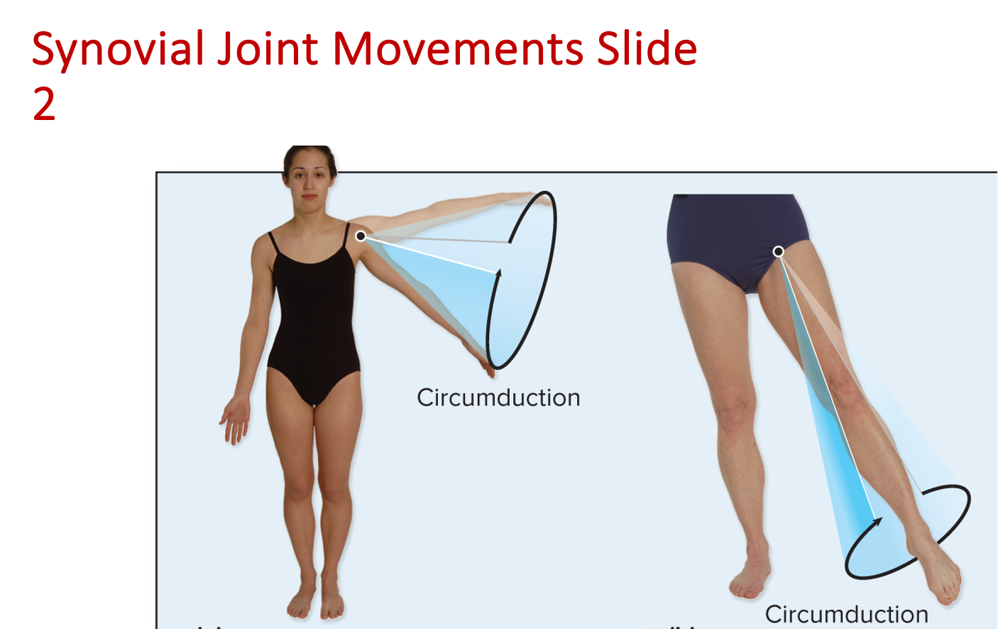
Rotation
Bone pivots on its longitudinal axis
Lateral Rotation + Medial Rotation
Lateral Rotation - turns anterior surface of bone laterally
Medial Rotation - turns anterior surface of bone medially
Pronation + Supination
Pronation - medial rotation of forearm so palm of hand posterior
Supination - lateral rotation of forearm so palm of hand anterior (“Asking for soup”)
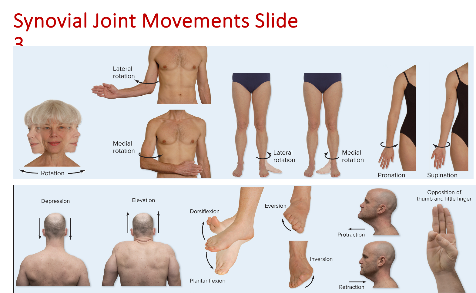
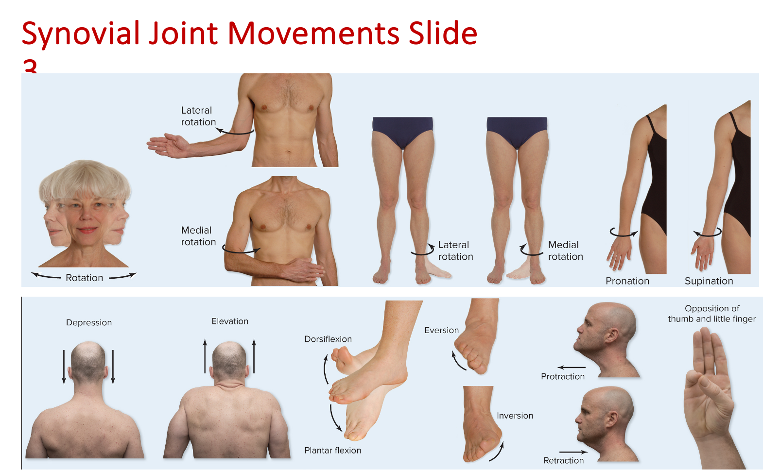
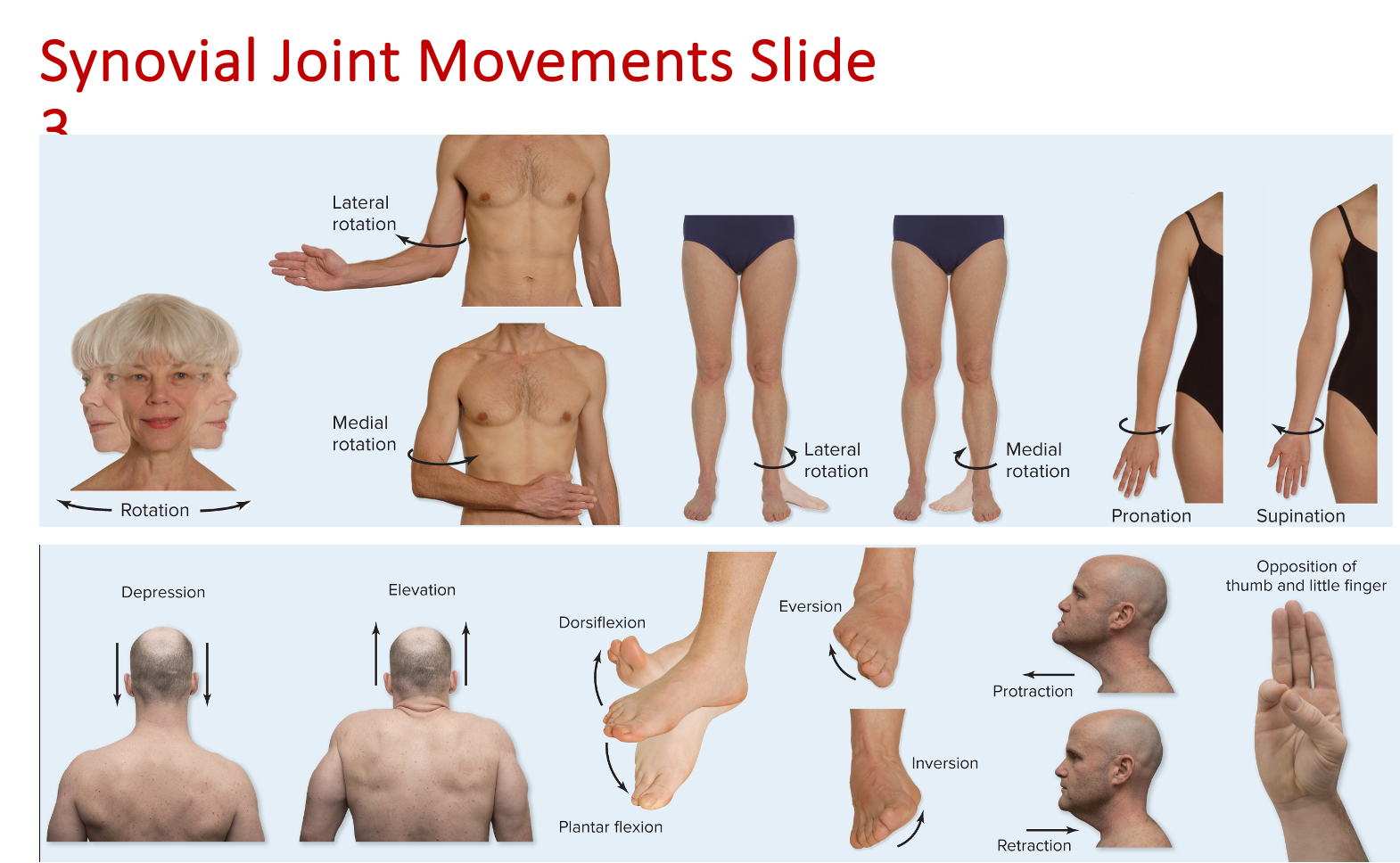
Depression + Elevation
Depression
Inferior movement of a body part
EX: Movement of mandible when opening mouth
Elevation
Superior movement of a body part
EX: Movement of mandible when closing mouth
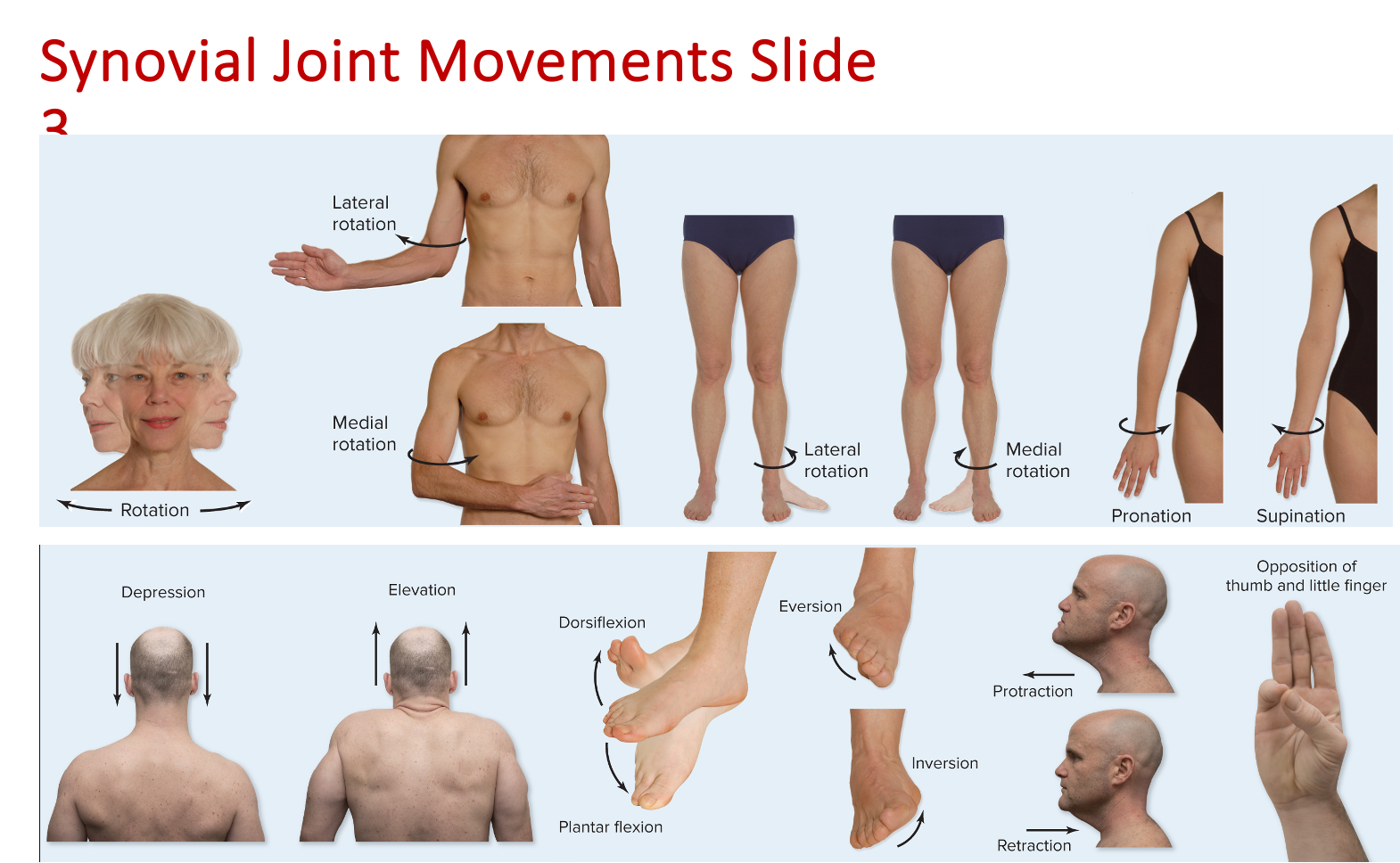
Dorsiflexion + Plater Flexion
Dorsiflexion (flexing foot)
EX: When digging in your hells
Planter Flexion (point foot)
EX: Ballerina on tiptoes
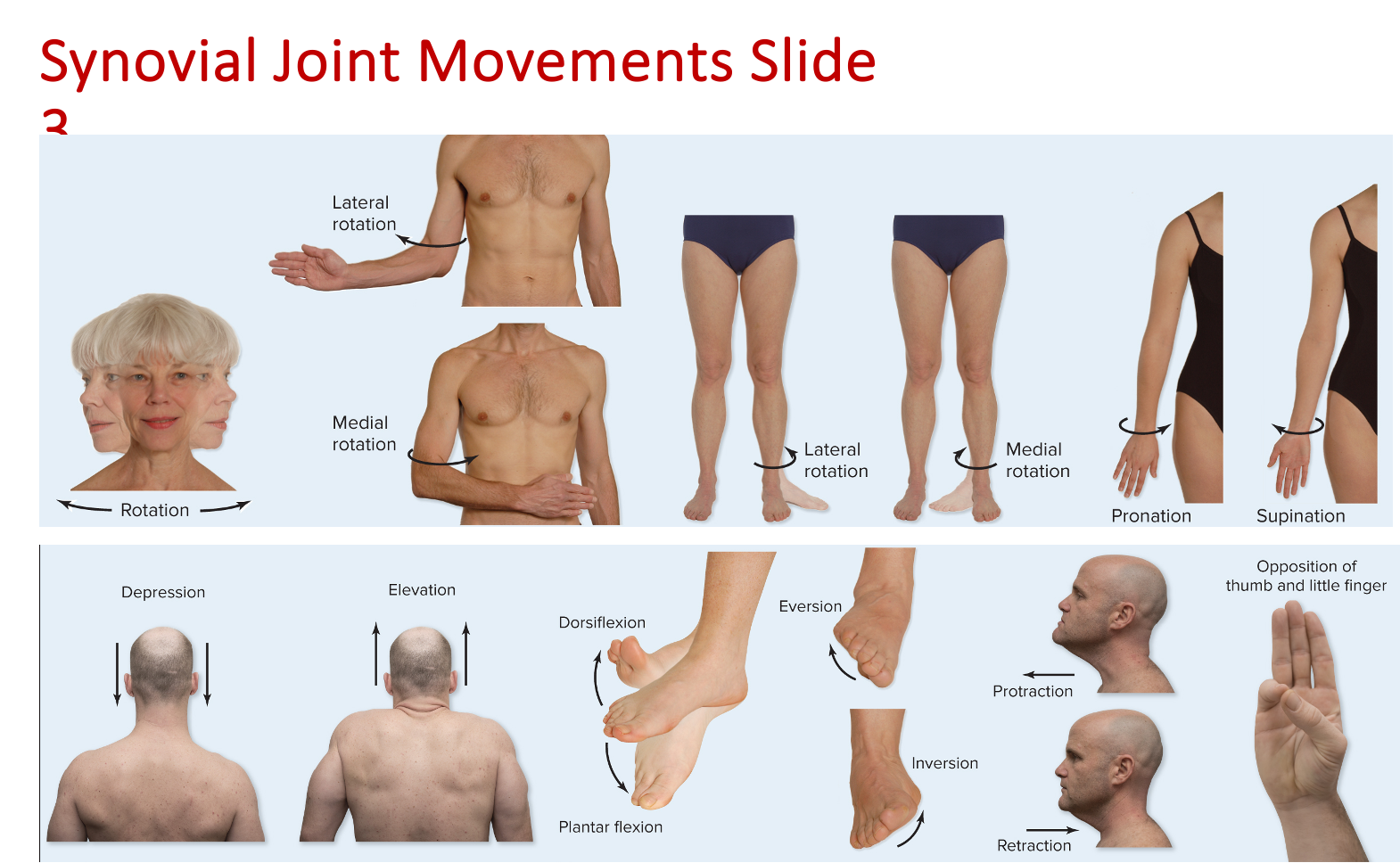
Eversion + Inversion
Eversion (“Eversion forever)
Sole turns laterally
Inversion (“Inversion leaves you inverted)
Sole turns medially
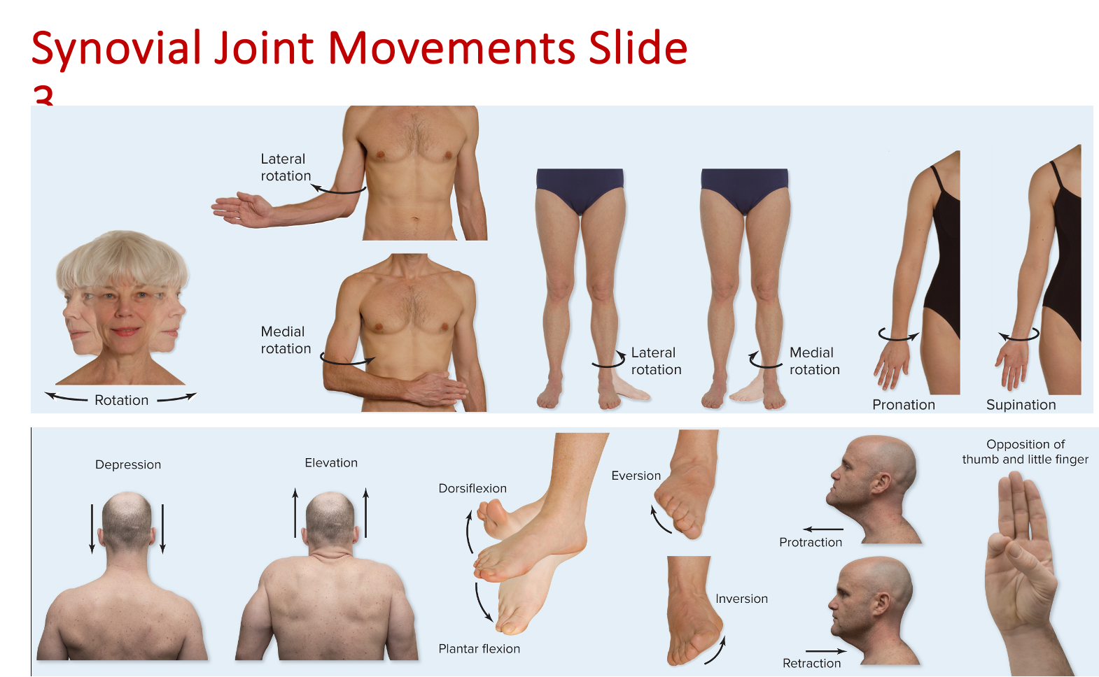
Protraction + Retraction
Protraction
Anterior movement from anatomic position
EX: Jutting jaw anteriorly at temporomandibular joint
Retraction
Posterior movement from anatomic position
EX: Pulling in jaw posteriorly at temporomandibular joint
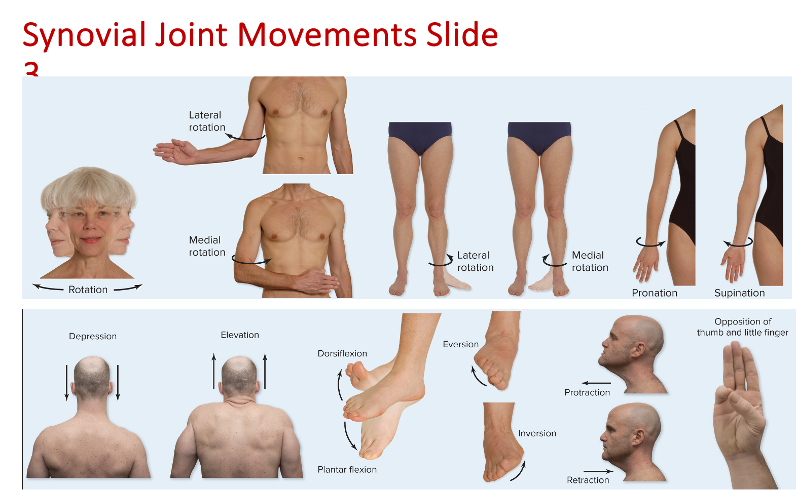
Opposition
Movement of thumb toward tips of fingers at carpometacarpal joint
Enables the thumb to grasp objects
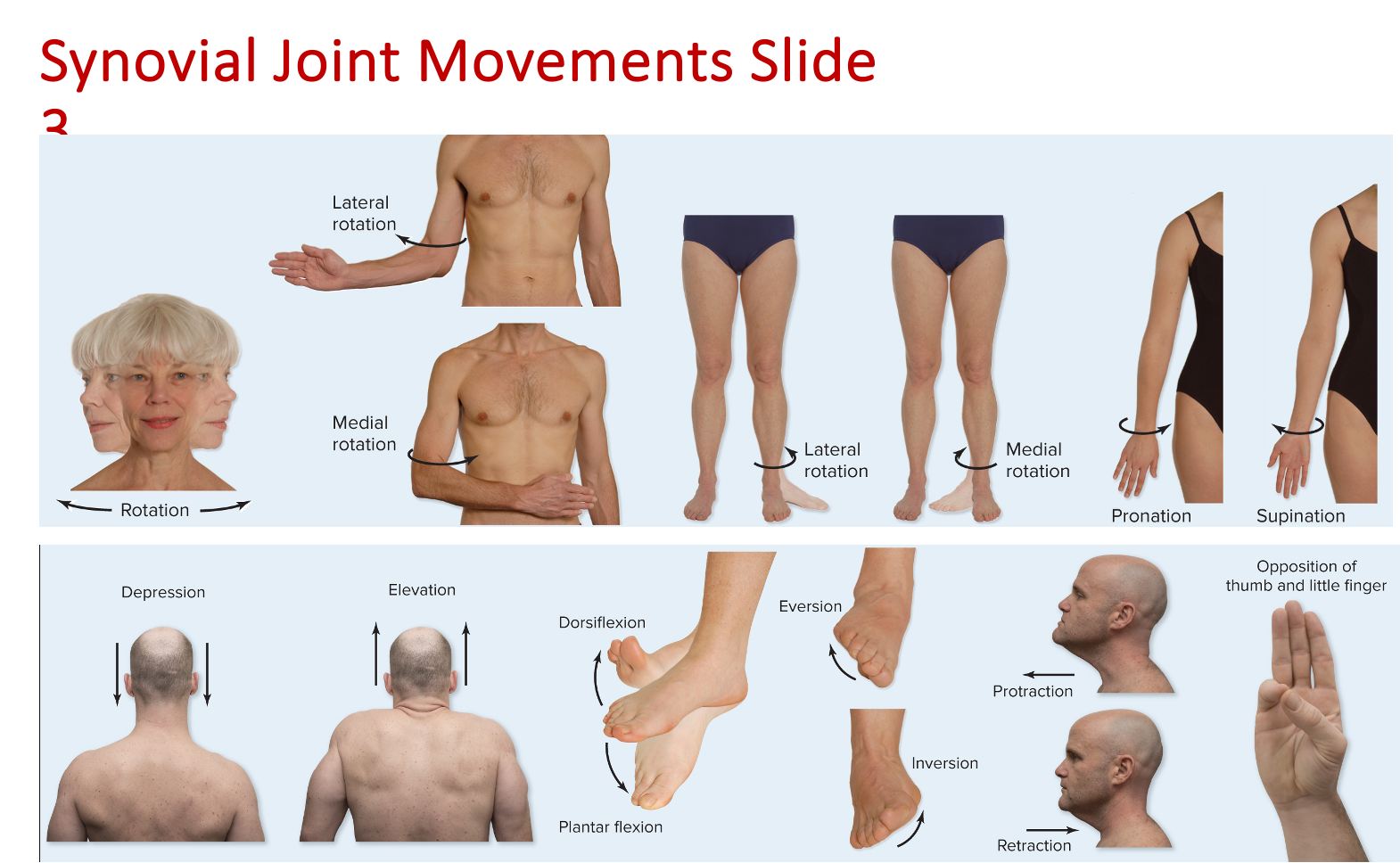
Glenohumeral joint
“Shoulder” Joint
Diarthrodial/Synovial
Ball + Socket Joint
Components:
Glenoid fossa of scapula
Head of humerus
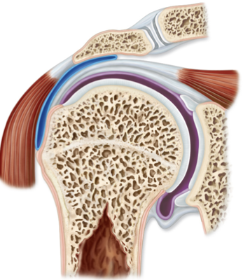
Humeroulnar joint
“Elbow” Joint
Diarthrodial/Synovial
Hinge Joint
Components:
Trochlea of humerus
Trochlear notch of ulna
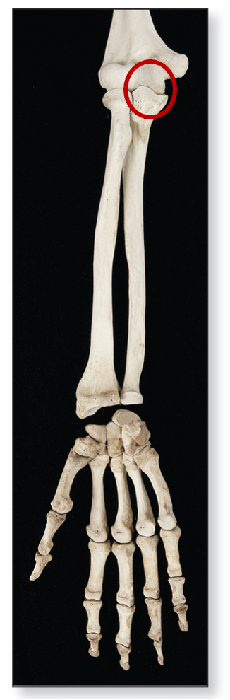
Humeroradial joint
“Elbow” Joint
Diarthrodial/Synovial
Hinge Joint
Components:
Capitulum of humerus
Head of radius
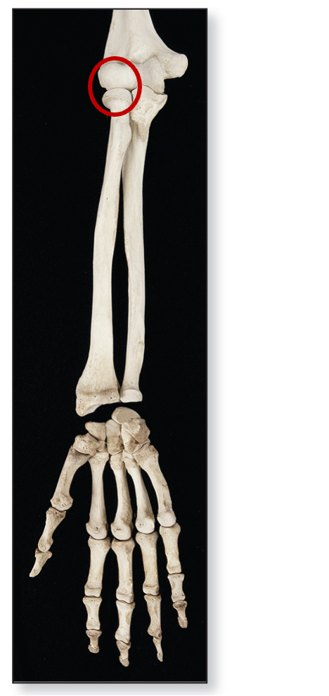
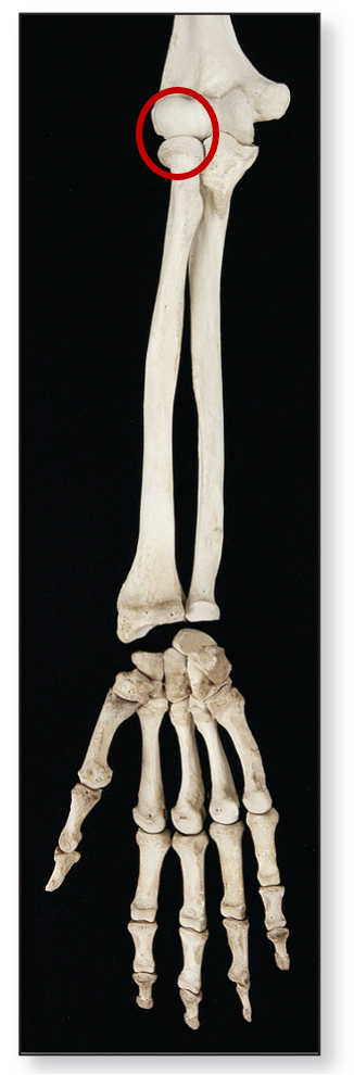
Proximal radioulnar joint
“Elbow” Joint
Diarthrodial/Synovial
Pivot Joint
Components:
Head of radius
Radial notch of ulna
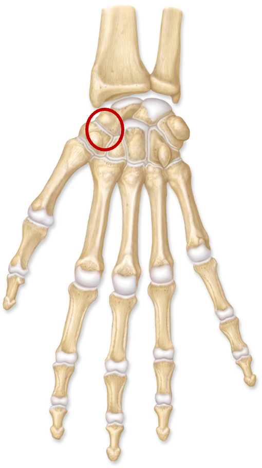
Intercarpal & intertarsal joints
Diarthrodial/Synovial
Plane Joint
Components:
Articular surfaces of carpal/tarsal bones
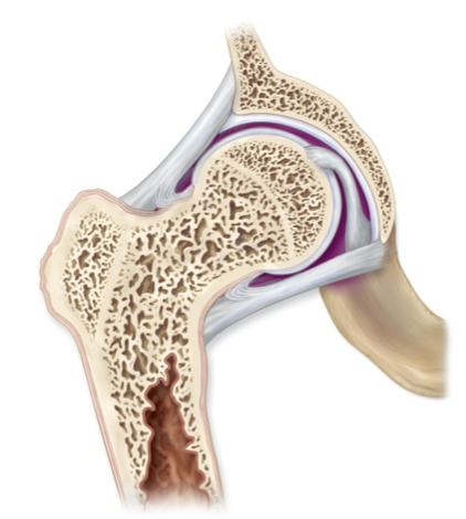
Coxal joint
“Hip” Joint
Diarthrodial/Synovial
Ball + Socket Joint
Components:
Acetabulum of coxa
Head of femur
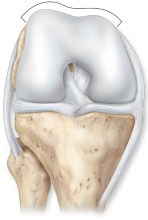
Tibiofemoral joint
“Knee” Joint
Diarthrodial/Synovial
Hinge Joint
Components:
Condyles of femur
Condyles of tibia
Medial + lateral menisci
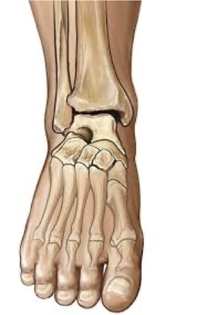
Talocrural joint
Diarthrodial/Synovial
Hinge Joint
Components:
Medial malleolus of tibia
Lateral malleolus of fibula
Articular surfaces of talus
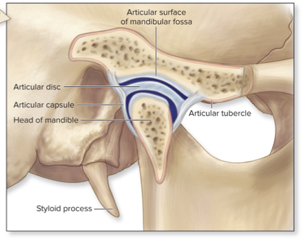
Temporomandibular joint
“Jaw” Joint
Diarthrodial/Synovial
Hinge and plane Joint
Components:
Mandibular fossa of temporal bone
Anterior tubercle of temporal bones
Articular disc
Head of mandible
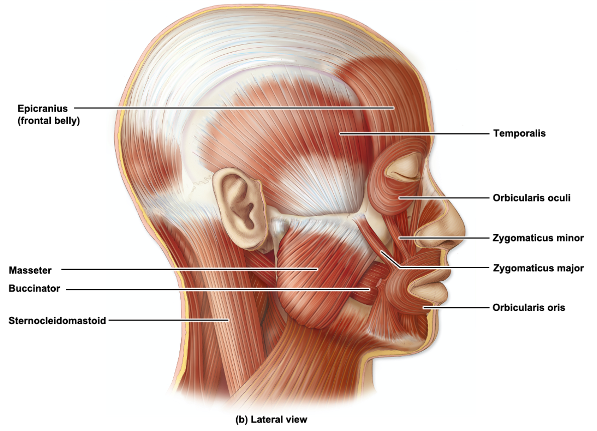
What is the origin, insertion, and action of the sternocleidomastoid muscle?
Origin - manubrium of sternum + clavicle
Insertion - mastoid process
Action - rotation, slight lateral flexion
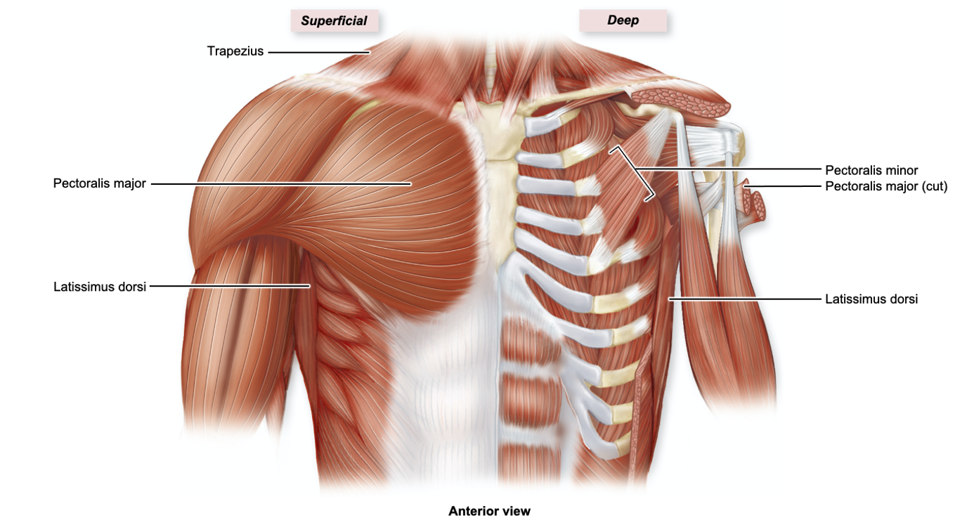
What is the origin, insertion, and action of the pectoralis major muscle?
Origin - ribs + body of sternum
Insertion - humerus
Action - flexes, adducts, + medially rotates arm
What is the origin, insertion, and action of the trapezius muscle?
Origin - clavicle + acromion process + spine of scapula
Insertion - occipital bone + lower thoracic vertebrae
Action - elevates + depresses scapula, rotates scapula superiorly, retracts scapula
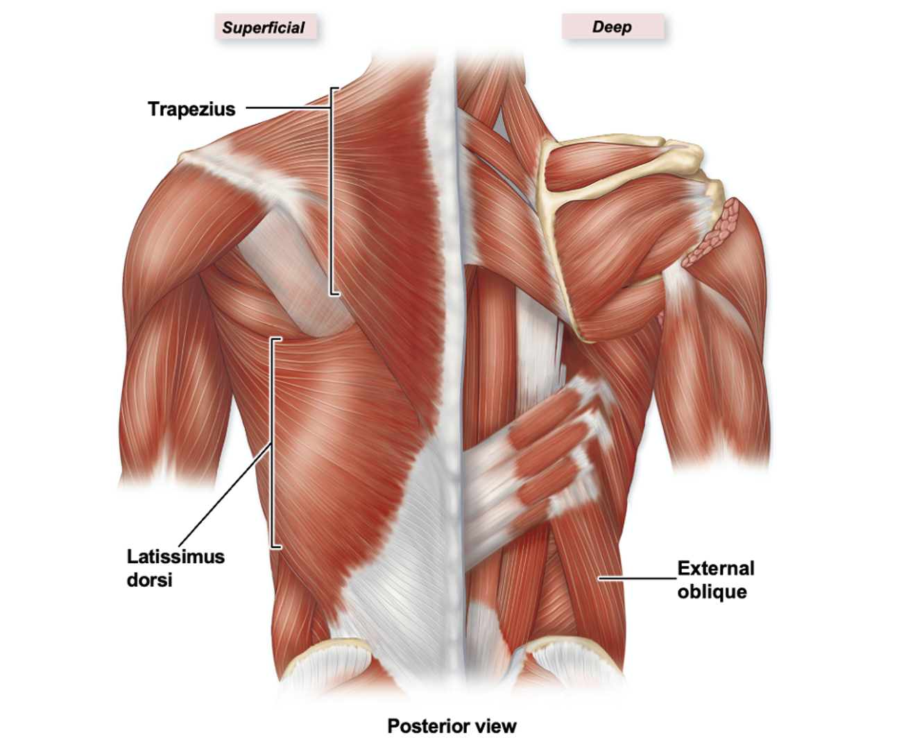
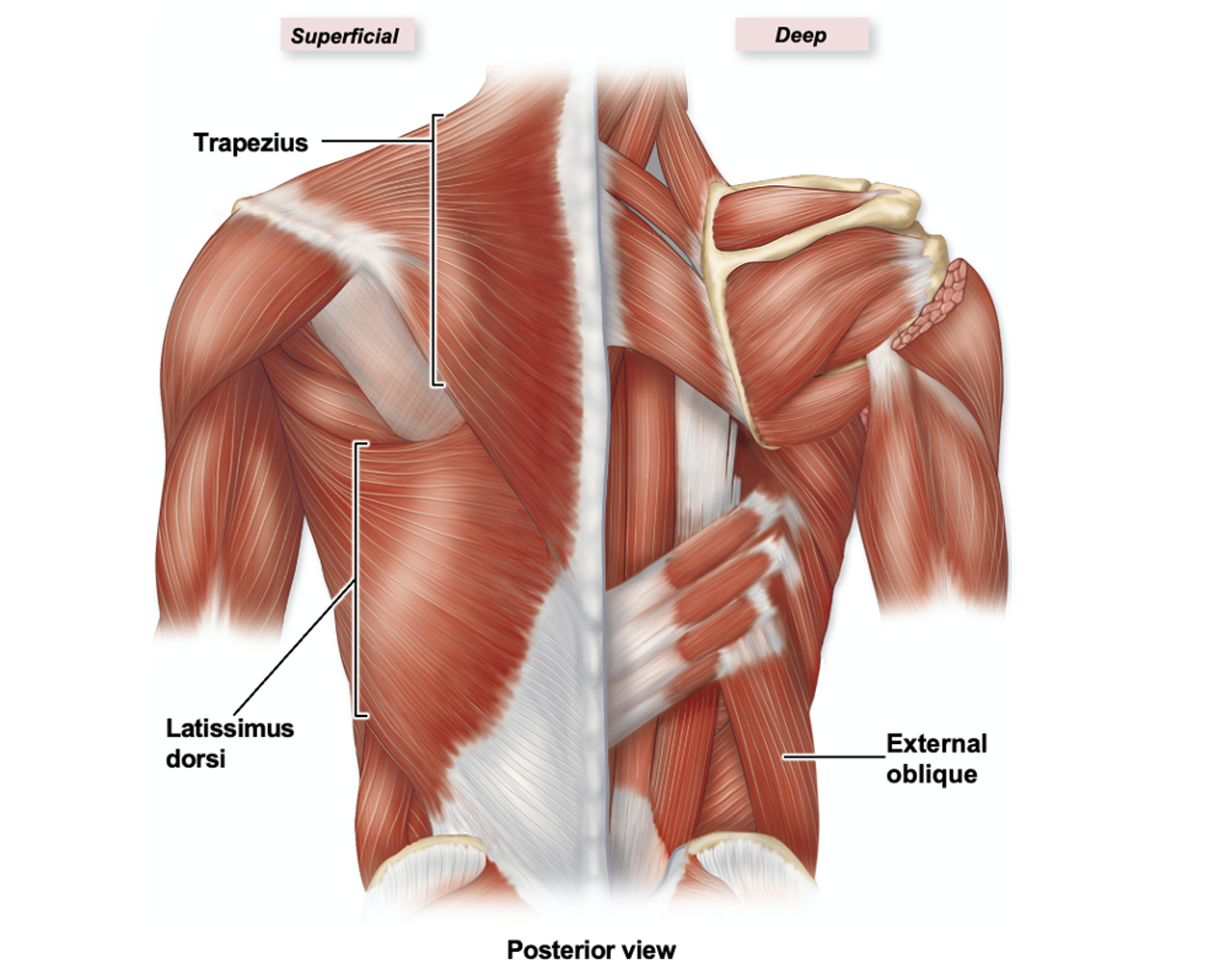
What is the origin, insertion, and action of the latissimus dorsi muscle?
Origin - thoracic spine, ribs, + iliac crest
Insertion - humerus
Action - extends, adducts, + medially rotates arm
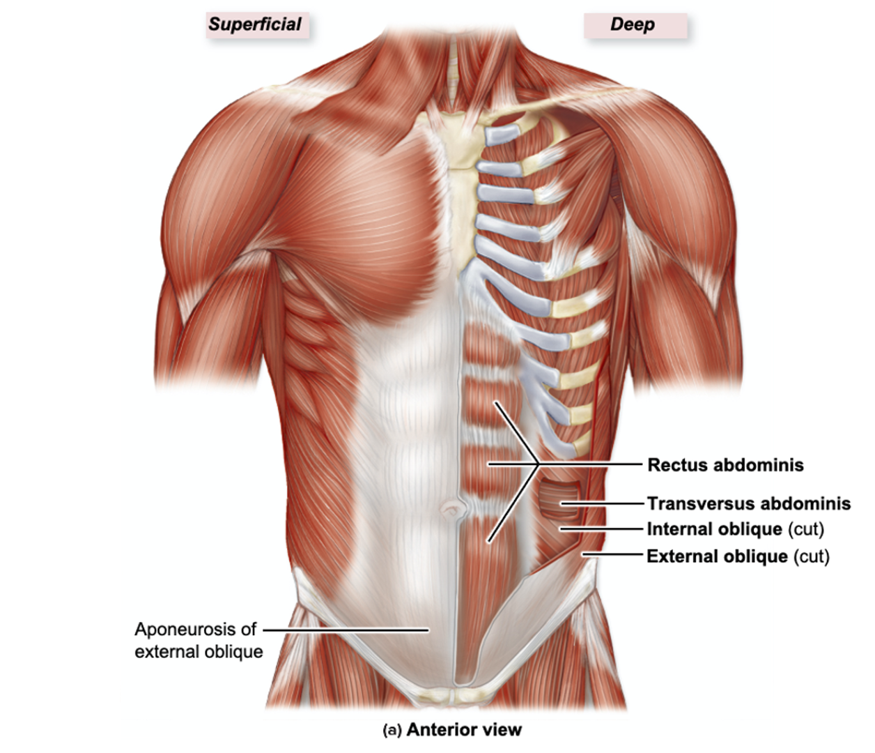
What is the origin, insertion, and action of the rectus abdominis muscle?
Origin - pubic symphysis + crest
Insertion - xiphoid process + ribs
Action - flexes trunk, compresses abdominal viscera, used in forced expiration
What are some of the main functions of head and neck muscles?
Head movement, mastication (chewing), facial expressions, and speech
What are some of the main functions of chest and back muscles?
Torso movement, but some help with arm movement
Back - vertebral column movement
What are some of the main functions of respiration muscles?
Breathing in and out by changing pressure in the thorax
What are some of the main functions of abdominal wall muscles?
Form several layers of muscle that contribute to abdominal movement + structure
What are the abdominal wall muscles from superficial → deep?
External obliques, rectus abdominis (medial) and internal obliques (lateral), transversus abdominis
Compact Bone Slide - Osteocytes in Lacunae
Structure: mature bone cells in spaces between concentric lamellae (lacunae)
Function: osteocytes function to maintain the bone matrix, lacunae house osteocytes
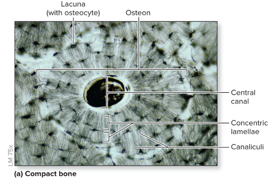
Compact Bone Slide - Canaliculi
“Telephone poles”
Structure: interconnecting channels that project from the osteocyte and lacuna to connect to the central canal
Function: allows osteocytes to exchange nutrients + other substances with one another
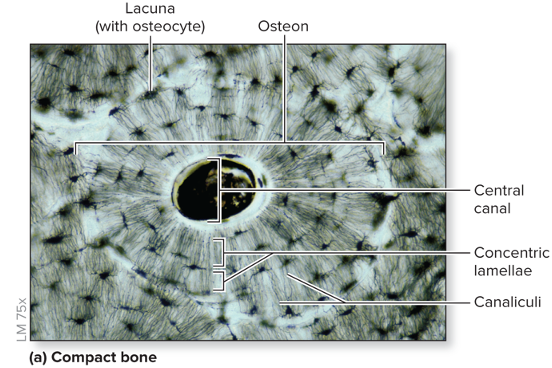
Compact Bone Slide - Osteon
“Bull’s-Eye Target”
Structure: small cylindrical structures, oriented parallel to bone diaphysis
Function: basic functional + structural unit of mature compact bone
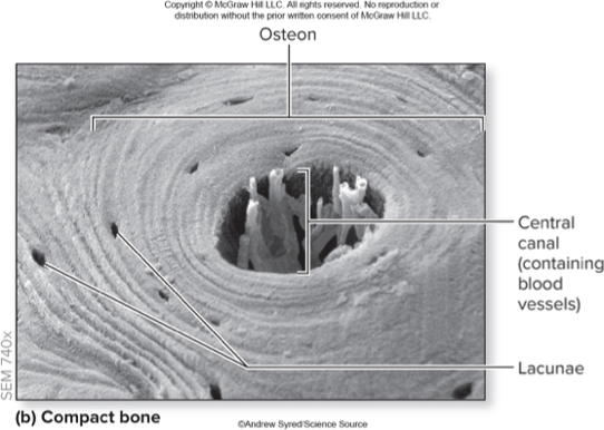
Compact Bone Slide - Central Canal
Structure: cylindrical channel at center of osteon + parallel to it, blood vessels + nerves extend through the channel
Function: provides a tunnel for the supply of blood, oxygen, and nutrients to bone
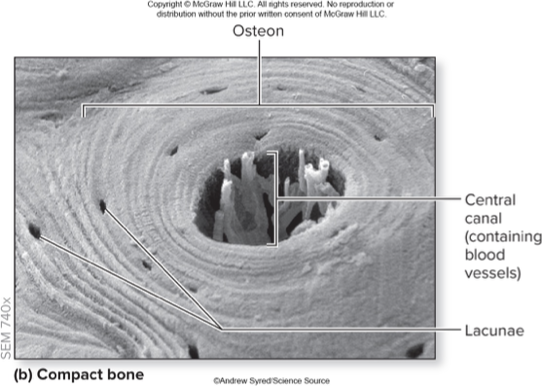
Compact Bone Slide - Concentric Lamellae
Structure: layers of bone matrix arranged concentrically around central canals
Function: gives bone strength + resilience
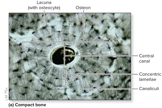
Bone Tissue - Spongy Bone
Toward the medullary cavity of the bone, contains trabeculae and red/yellow marrow
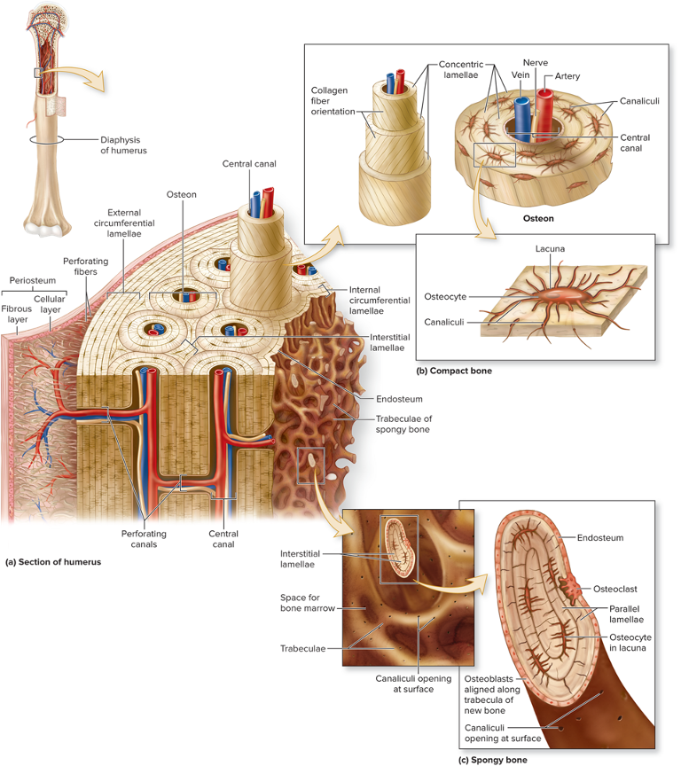
Spongy Bone - Trabeculae
Open lattice of narrow rods + plates of bones
Meshwork of crisscrossing bars
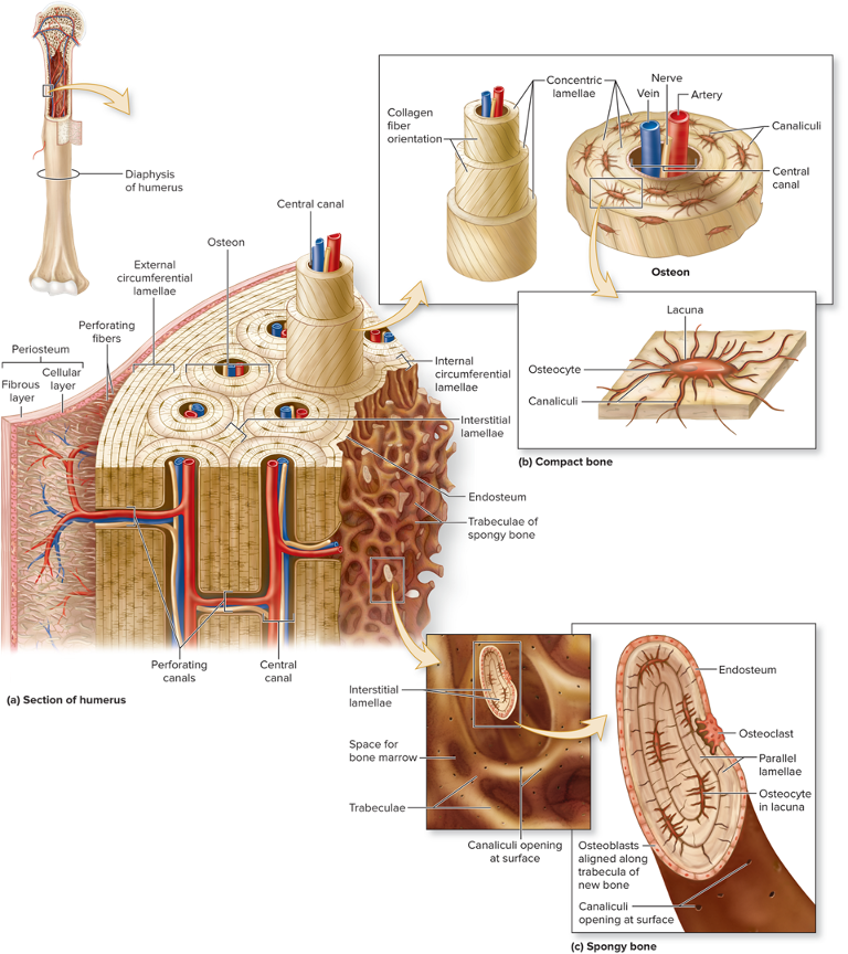
Spongy Bone - Red or Yellow Marrow
Fills spaces between trabeculae
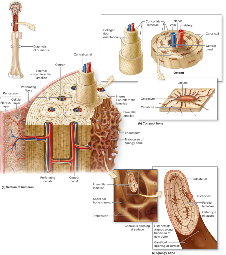
Compact Bone - Circumferential Lamellae
External - rings of bone run immediately internal to the periosteum
Internal - rings of bone run internal to the endosteum
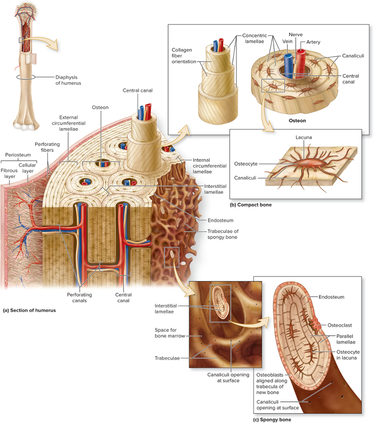
Compact Bone - Interstitial Lamellae
Components of compact bone between osteons or partially resorbed osteons
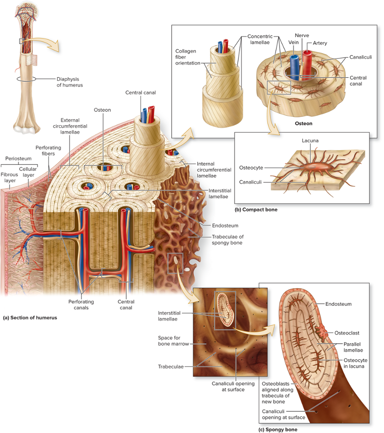
Compact Bone - Perforating Canals
Run perpendicular to central canals, contains blood vessels + nerves
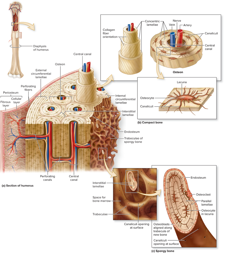
Bone Tissue - Periosteum
Outside of bone
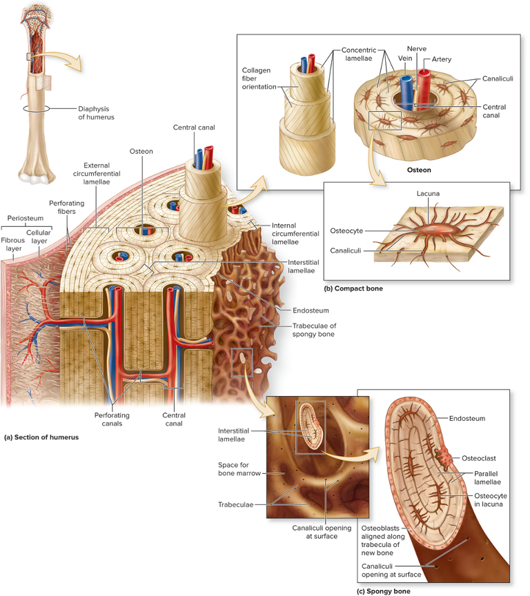
Bone Tissue - Endosteum
Covers all internal surfaces of bone within medullary cavity
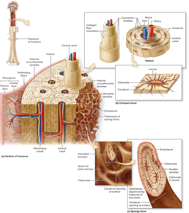
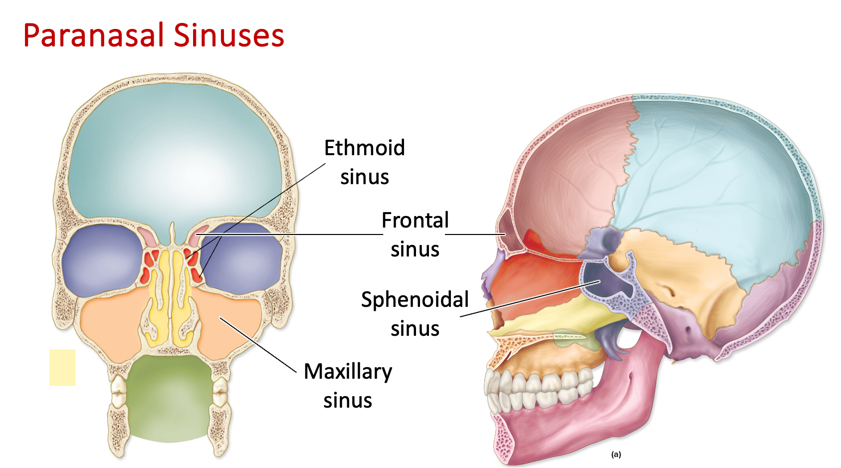
Paranasal Sinus
One of many small hollow spaces in bones around the nose
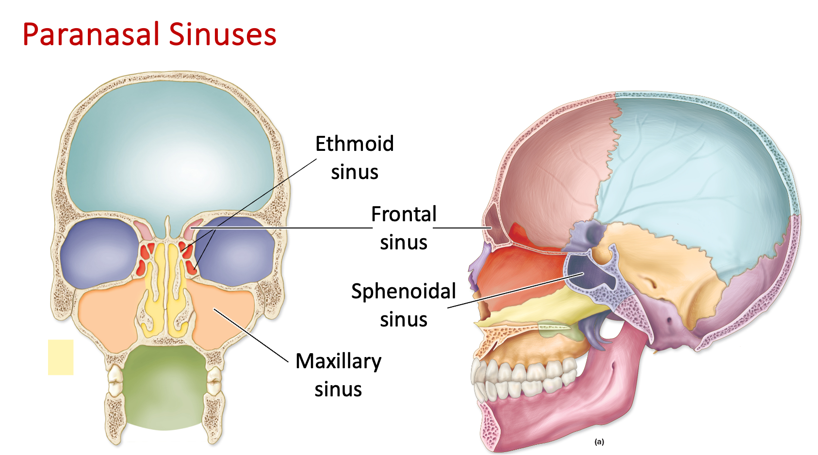
Frontal Sinus
Cavity/opening in the frontal region
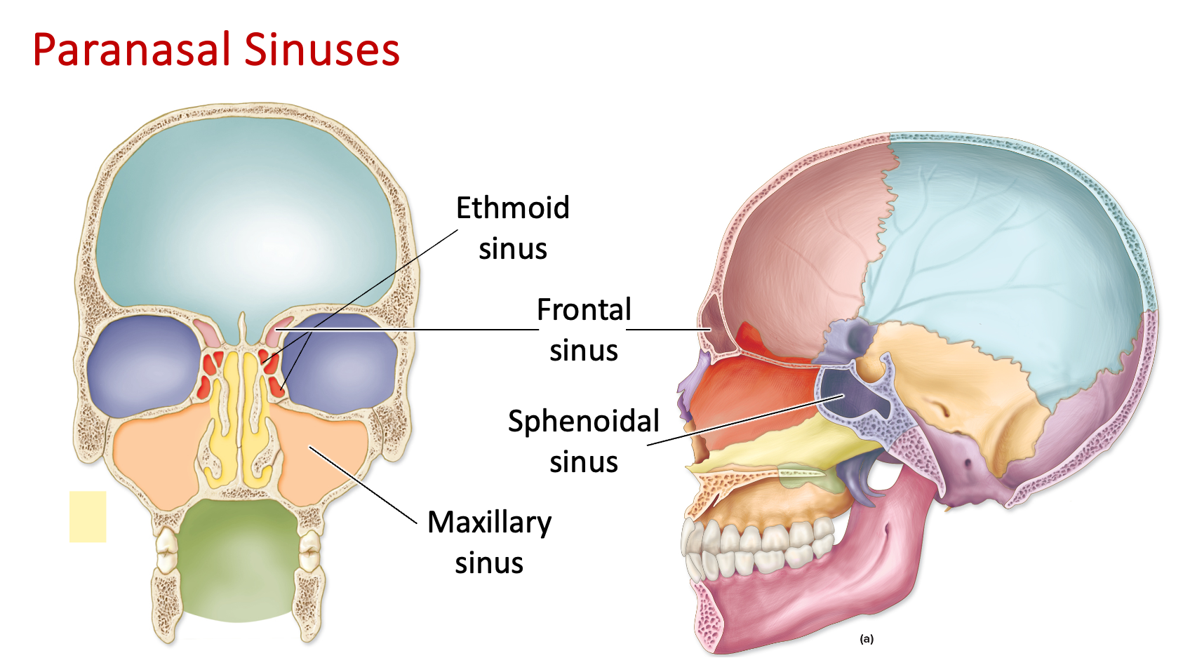
Sphenoid Sinus
Cavity/opening in the sphenoid bone (inferior to ethmoid sinus)
“E comes before S”
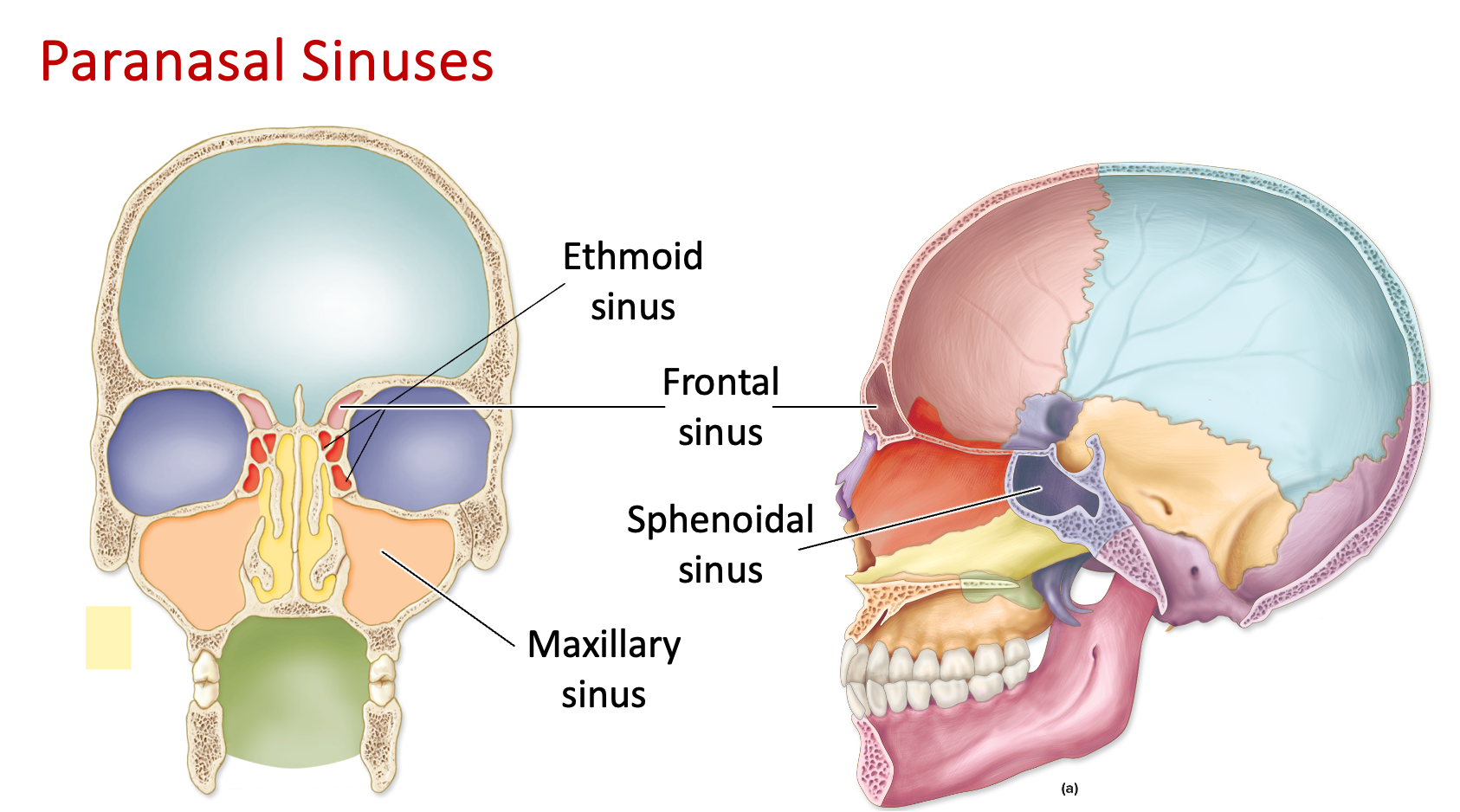
Ethmoid Sinus
Cavity/opening in the ethmoid bone, between the eyes (superior to sphenoid sinus)
“E comes before S”
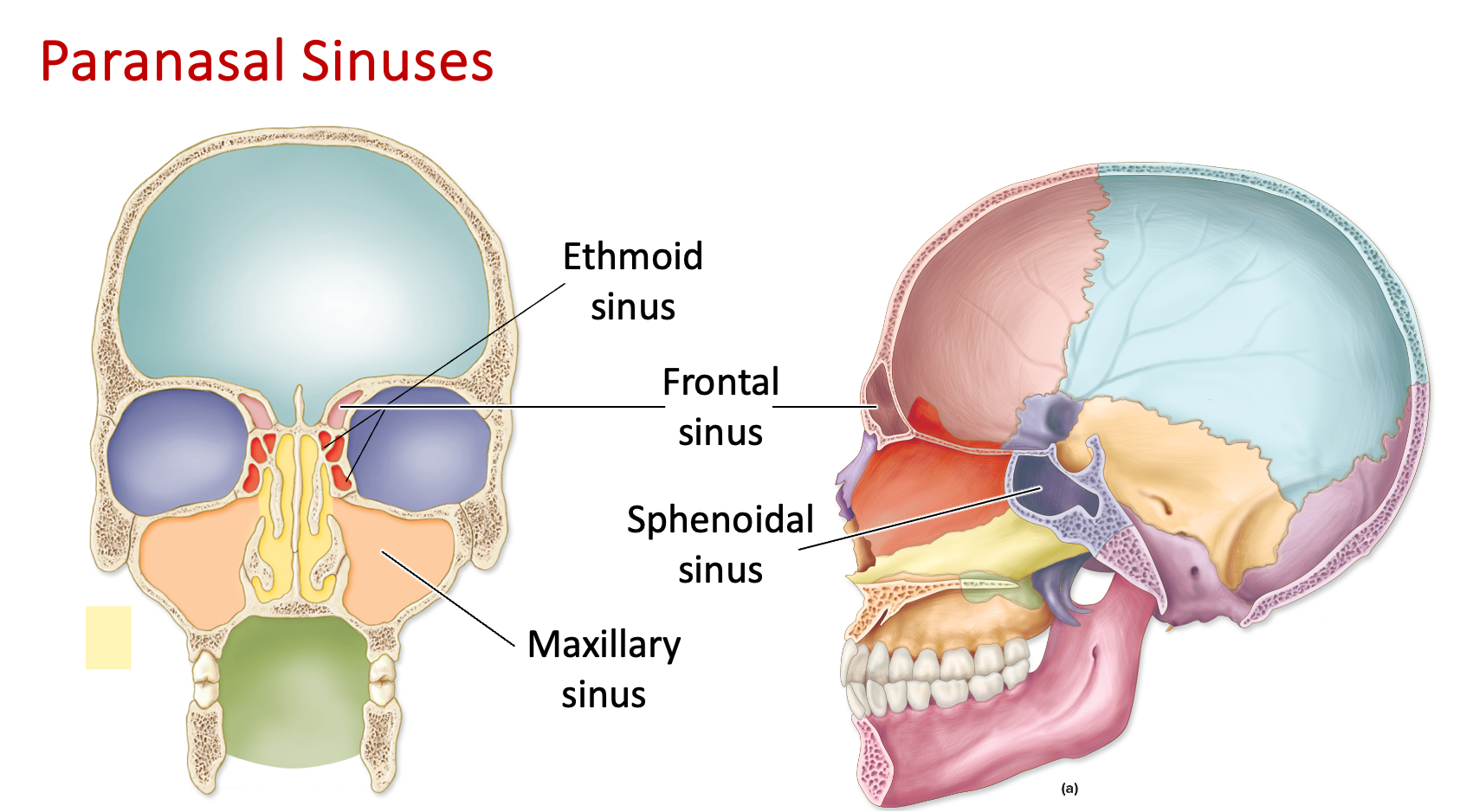
Maxillary Sinus
Cavity/opening in the maxillary bone/cheekbone regions
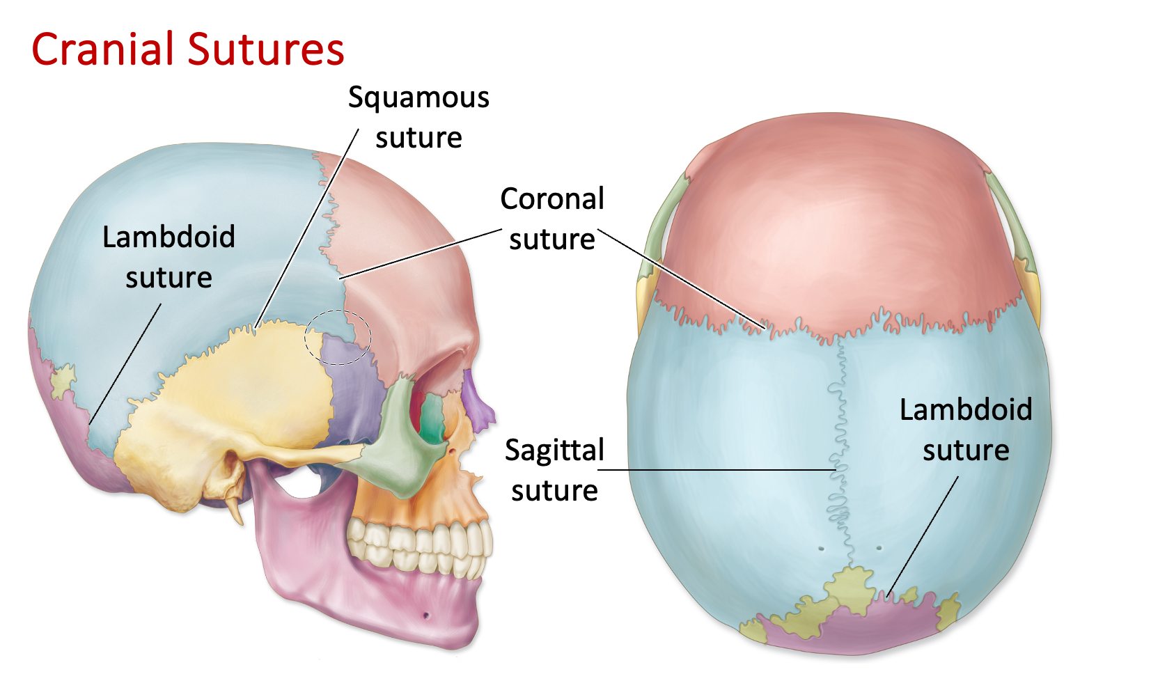
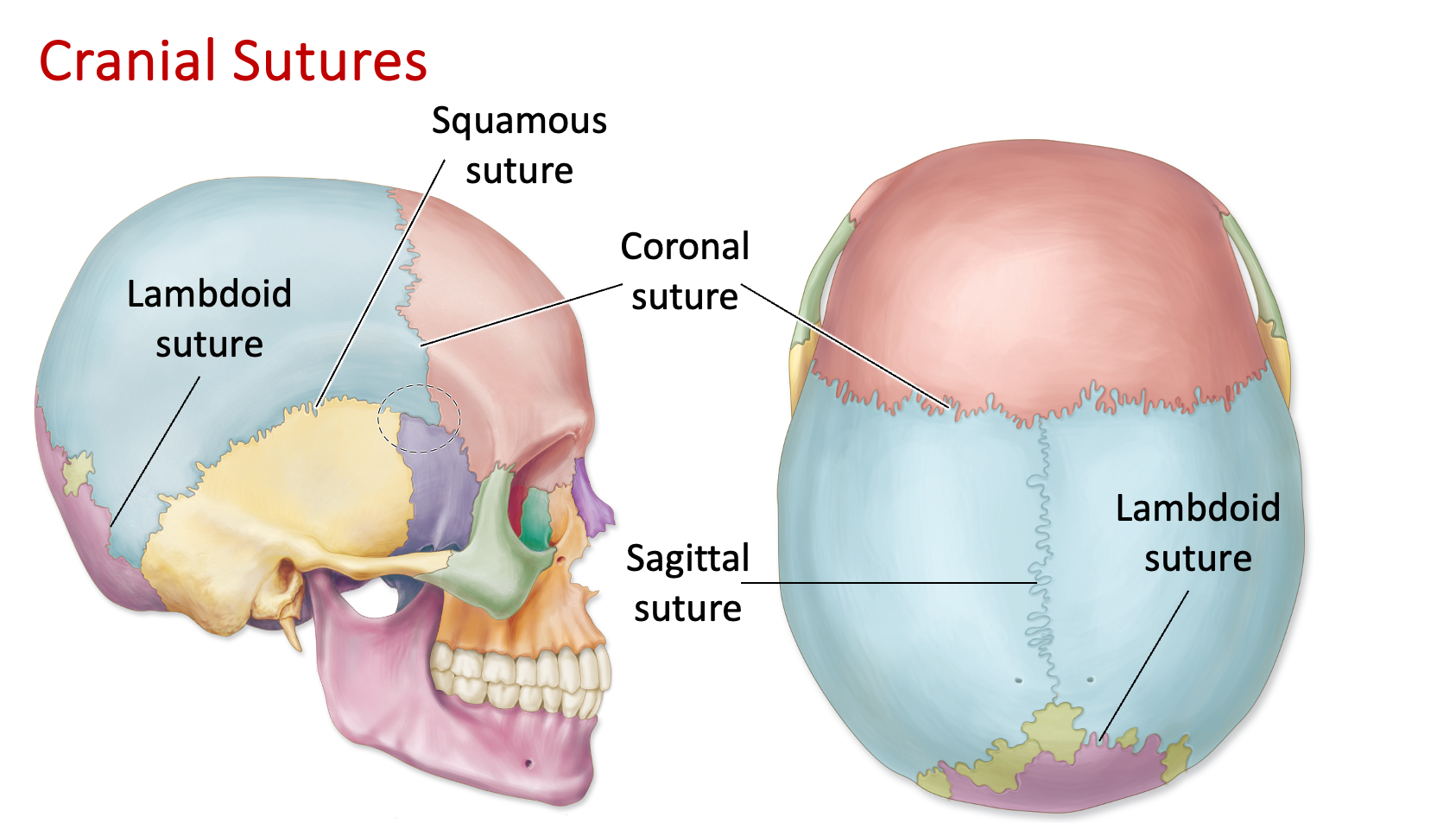
Coronal Suture
Suture in the skull that runs across the coronal plane
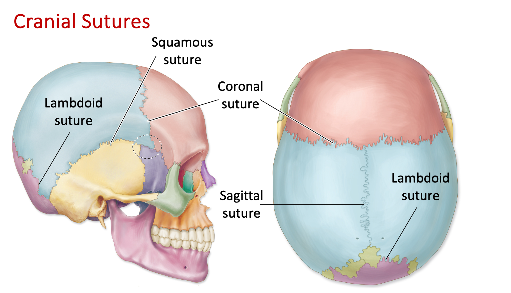
Sagittal Suture
Suture in the skull that runs across the sagittal plane
Lambdoidal/Lambdoid Suture
Suture found at the back of the skull that connects the parietal bones with the occipital bone
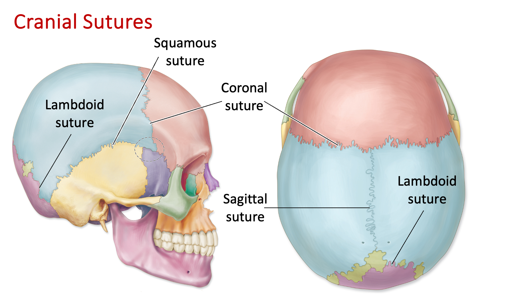
Squamosal/Squamous Suture
Suture between the parietal and temporal bones on each side of the skull
Where is the frontal bone located?
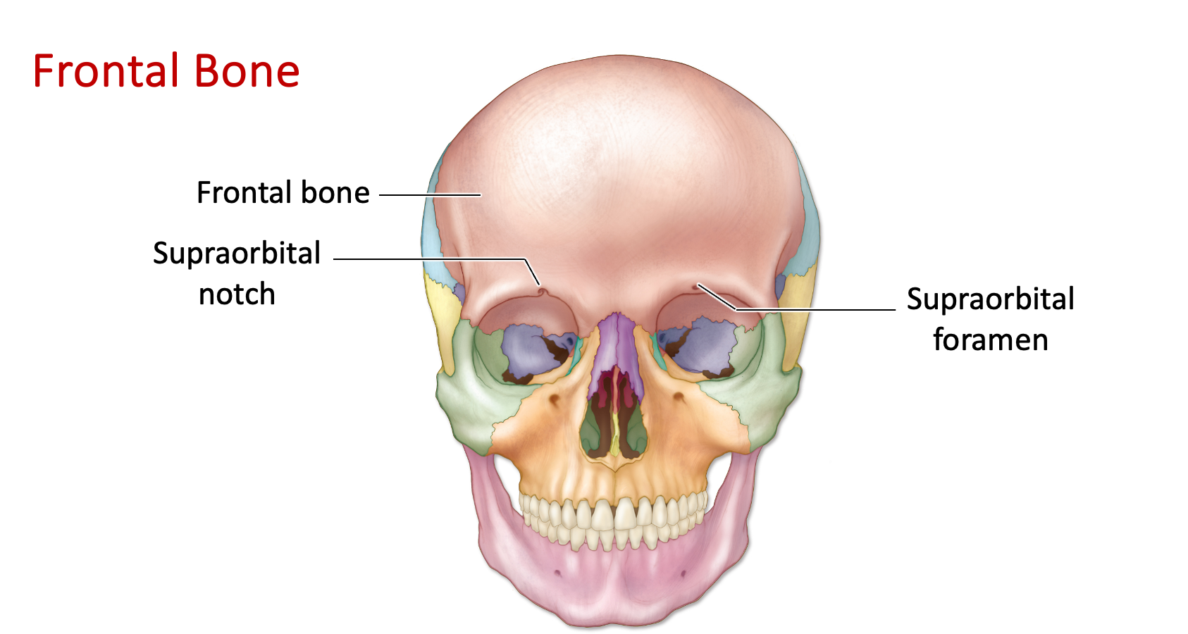
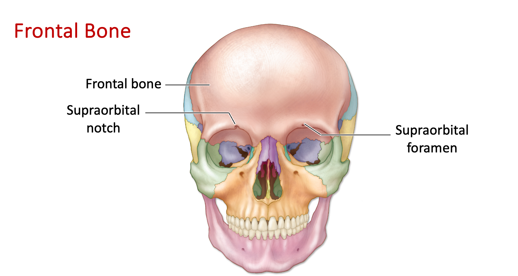
Frontal Bone - Supraorbital Notch/Foramen
“Above orbital notch”
Located above the eye socket (orbit)
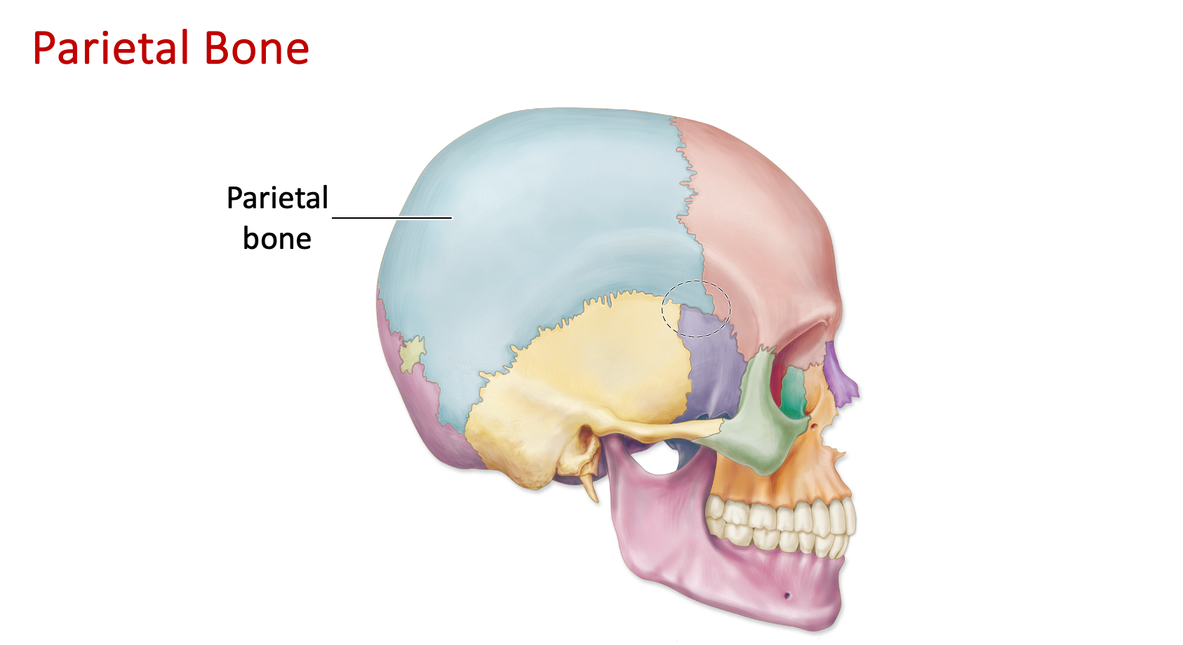
Where is the parietal bone located?
“Paired bones”
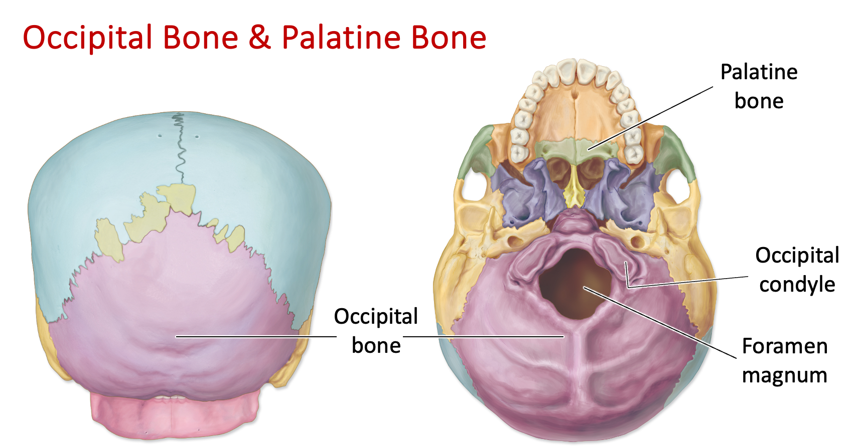
Where is the occipital bone located?
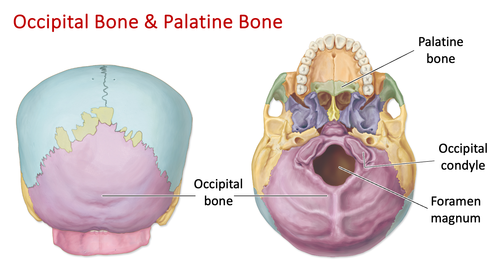
Occipital Bone - Foramen Magnum
“Magnum = huge”
A large hole, foramen, in the occipital bone
Where the brain stem exits, and the spinal cord is connected
Allows for nodding
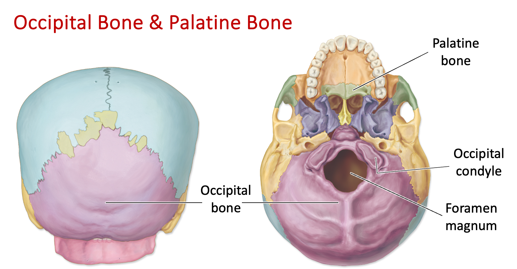
Occipital Bone - Occipital Condyles
Rounded projections under the occipital bone
Articulates with vertebral column
Next to foramen magnum
Allows for nodding movement
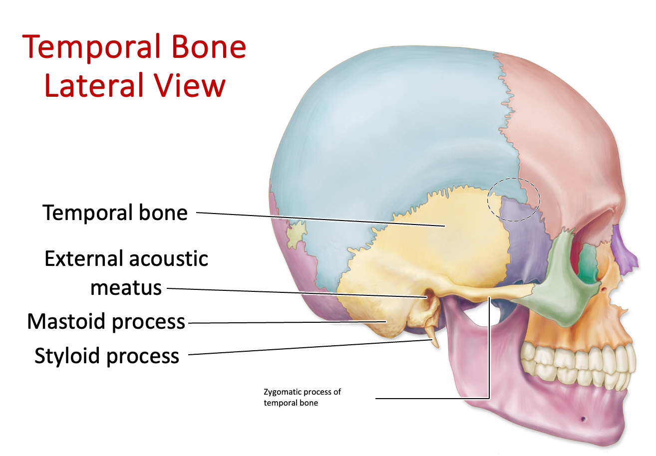
Where is the temporal bone located?
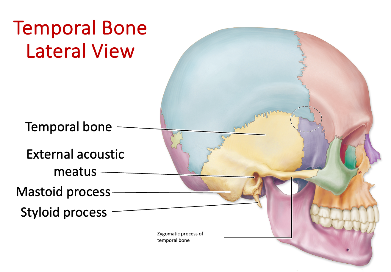
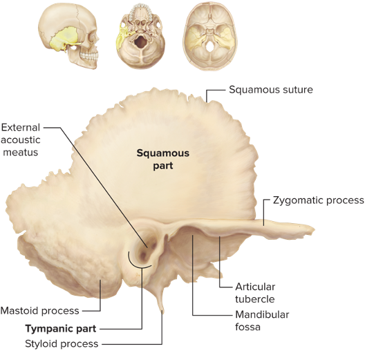
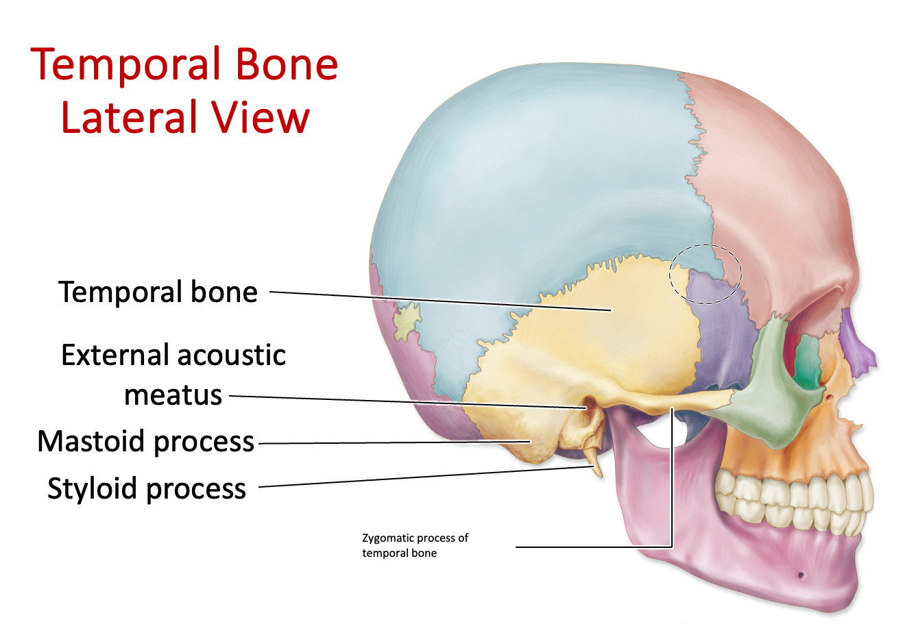
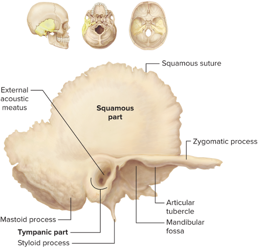
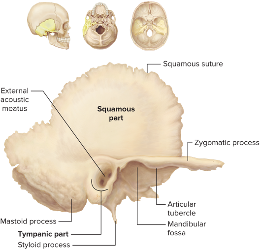
Temporal Bone - External Auditory/Acoustic Meatus
Hole in the temporal bone that permeates, where we hear things
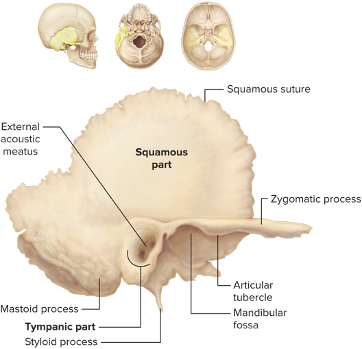
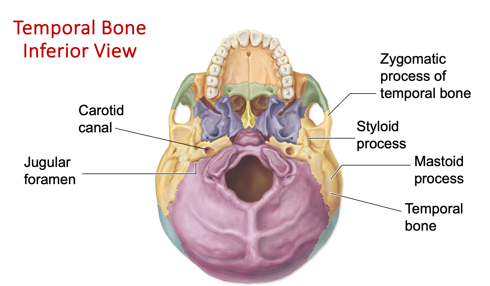
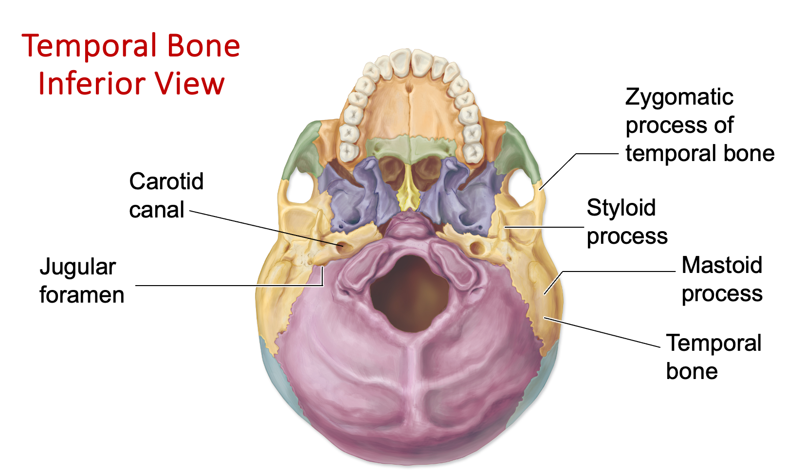
Temporal Bone - Styloid Process
Penlike, sharp projection of the temporal bone
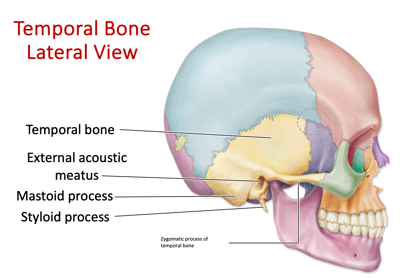
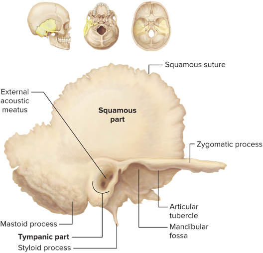
Temporal Bone - Zygomatic Process
Bony projection on the temporal bone that articulates with the zygomatic bone
Cheekbones - think the Kardashians
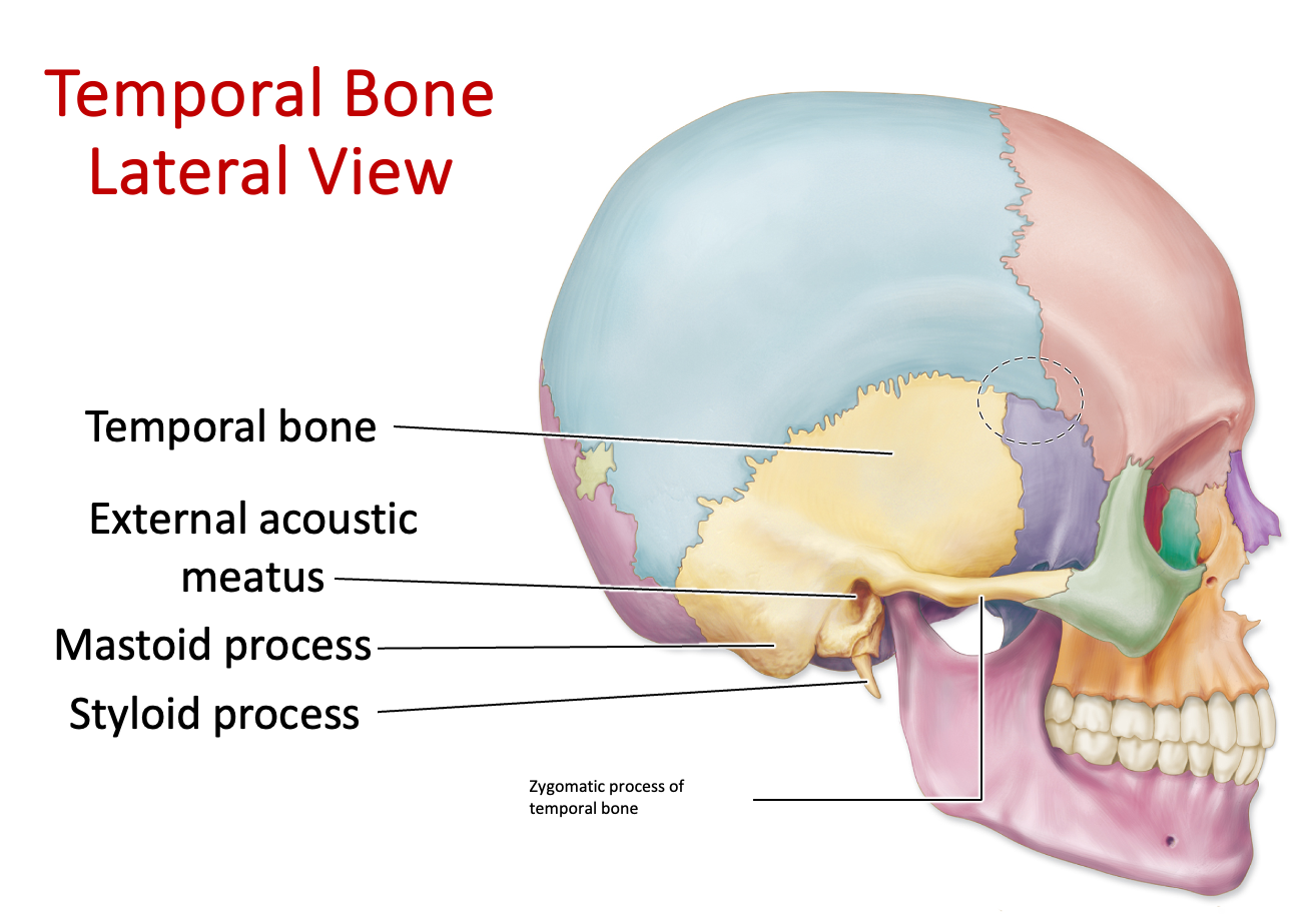
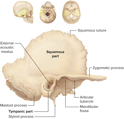
Temporal Bone - Mastoid Process
A bony projection of the temporal bone located behind the ear, looks like a big chunk
Temporal Bone - Carotid Canal
Passage in the temporal bone that allows the internal carotid artery to enter the skull and supply blood to the brain
Temporal Bone - Jugular Foramen
Jugular vein runs through this foramen, lateral to foramen magnum
Where is the sphenoid bone located?
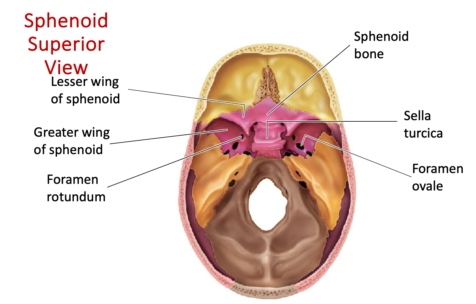 ”Bat-shaped bone”
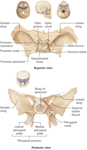
Sphenoid Bone - Sella Turcica
Houses the pituitary gland
Named for its resemblance to a saddle
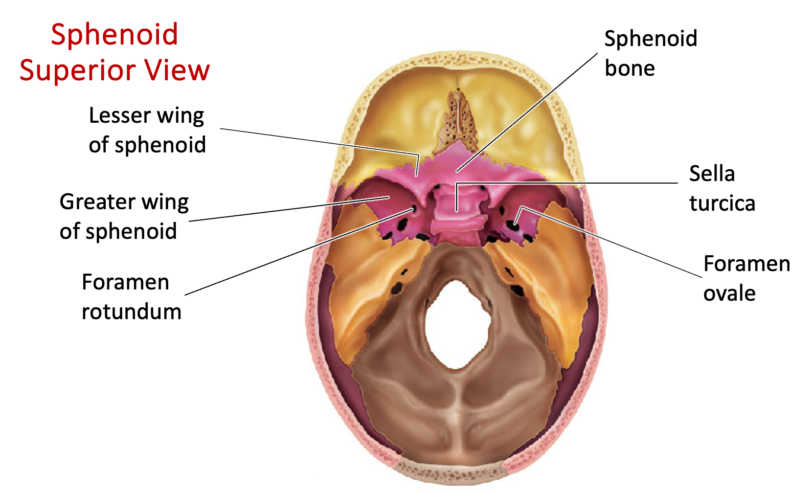
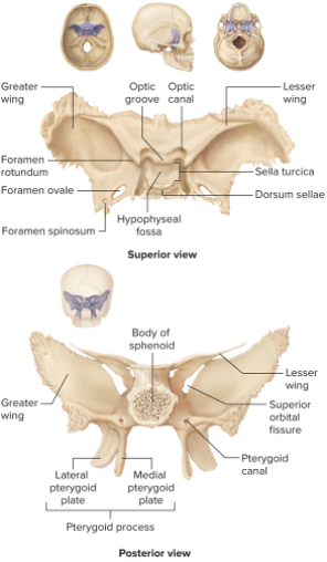
Sphenoid Bone - Greater Wings
Larger, lower portion of the sphenoid bone in the skull
Sits inferiorly to lesser wings