Lab 2 Exam Review
Lab 4 (Skeletal System - Bony Tissue + Axial Skeleton)
EXAM Objectives:
ID structures and state functions for all histology slides ✅
Compact bone CT slide and models ✅
Identify all bones on lab manual lists ✅
Identify all assigned bone parts, markings, structures ✅
Articulated & Disarticulated ✅
Know functions where appropriate ✅
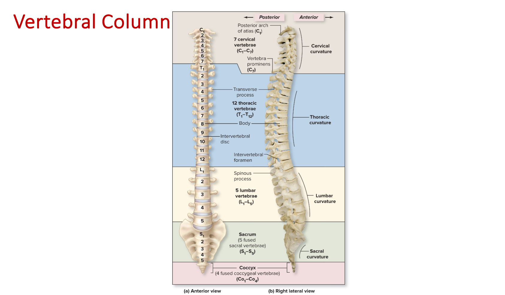
Normal spinal curvature: names, locations, member vertebrae in each ✅
Histology: You should be able to identify the following structures on an image or microscope slide.
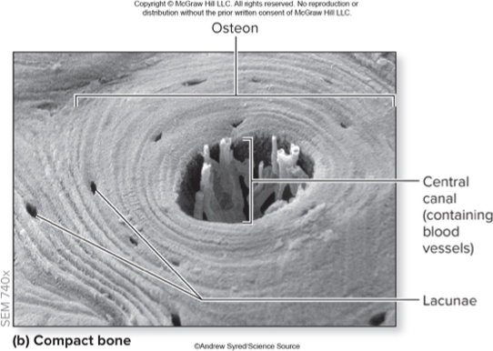
Compact bone slide
Osteocytes in lacunae
Structure: mature bone cells in spaces between concentric lamellae (lacunae)
Function: osteocytes function to maintain the bone matrix, lacunae house osteocytes
Canaliculi (“Telephone poles”)
Structure: interconnecting channels that project from the osteocyte and lacuna to connect to the central canal
Function: allows osteocytes to exchange nutrients + other substances with one another
Osteon (“Bull’s-Eye Target”)
Structure: small cylindrical structures, oriented parallel to bone diaphysis
Function: basic functional + structural unit of mature compact bone
Central canal
Structure: cylindrical channel at center of osteon + parallel to it, blood vessels + nerves extend through the channel
Function: provides a tunnel for the supply of blood, oxygen, and nutrients to bone
Concentric lamellae
Structure: layers of bone matrix arranged concentrically around central canals
Function: gives bone strength + resilience
Models: You should be able to identify the following structures on various lab models.
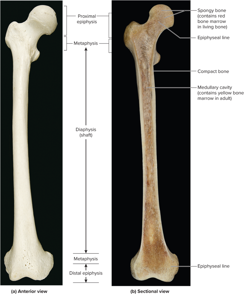
Bone tissue
Spongy bone - towards the medullary cavity of the bone
Trabeculae - open lattice of narrow rods + plates of bones, meshwork of crisscrossing bars
Red or yellow marrow - fills in the spaces between trabeculae
Compact bone
Circumferential lamellae
External - rings of bone run immediately internal to the periosteum
Internal - rings of bone run internal to the endosteum
Interstitial lamellae - components of compact bone between osteons or partially resorbed osteons
Perforating canals - run perpendicular to central canals, contains blood vessels + nerves
Periosteum - outside of bone
Endosteum - covers all internal surfaces of bone within the medullary cavity

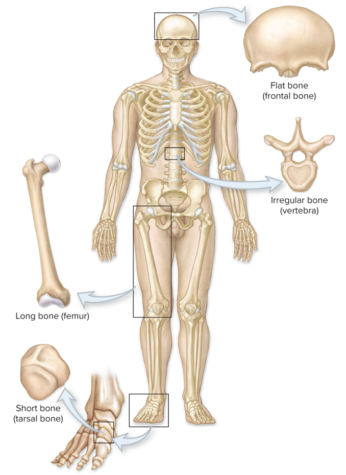
Types of bones
Long - greater in length than width (femur, humerus)
Short - length nearly equal to width (carpals + tarsals)
Flat - flat, thin surfaces, may be slightly curved (cranial bones)
Irregular - elaborate, sometimes complex shapes (vertebrae)
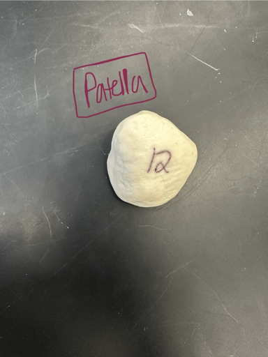
Sesamoid - small, sesame seed-shaped bones along the tendons of some muscles, classified as short bones. (Patella is the largest sesamoid bone)
Sutural - small bones located within sutures of the skull, vary in number and shape among individuals
Long bone structures
Diaphysis/shaft - elongated, usually cylindrical shaft
Medullary cavity - hollow, cylindrical space within the diaphysis. Contains red bone marrow in children and yellow bone marrow in adults.
Metaphysis - region between the diaphysis and epiphysis
Epiphysis - knobby region at each end of the long bone
Epiphyseal plate - thin layer of hyaline cartilage located within metaphysis. In adults, the epiphyseal line, is the remnant of the epiphyseal plate.
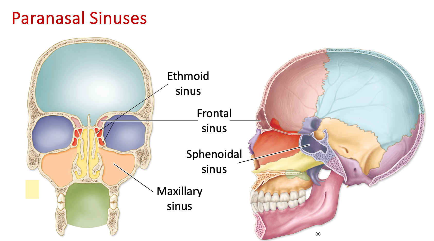
Paranasal sinuses (opening/cavity)
Frontal - cavity in the frontal region
Sphenoid - cavity in the sphenoid bone (inferior to ethmoid sinus)
Ethmoid - cavity in the ethmoid bone, between the eyes (superior to sphenoid sinus)
Maxillary - cavity in the maxillary bone/cheekbone regions
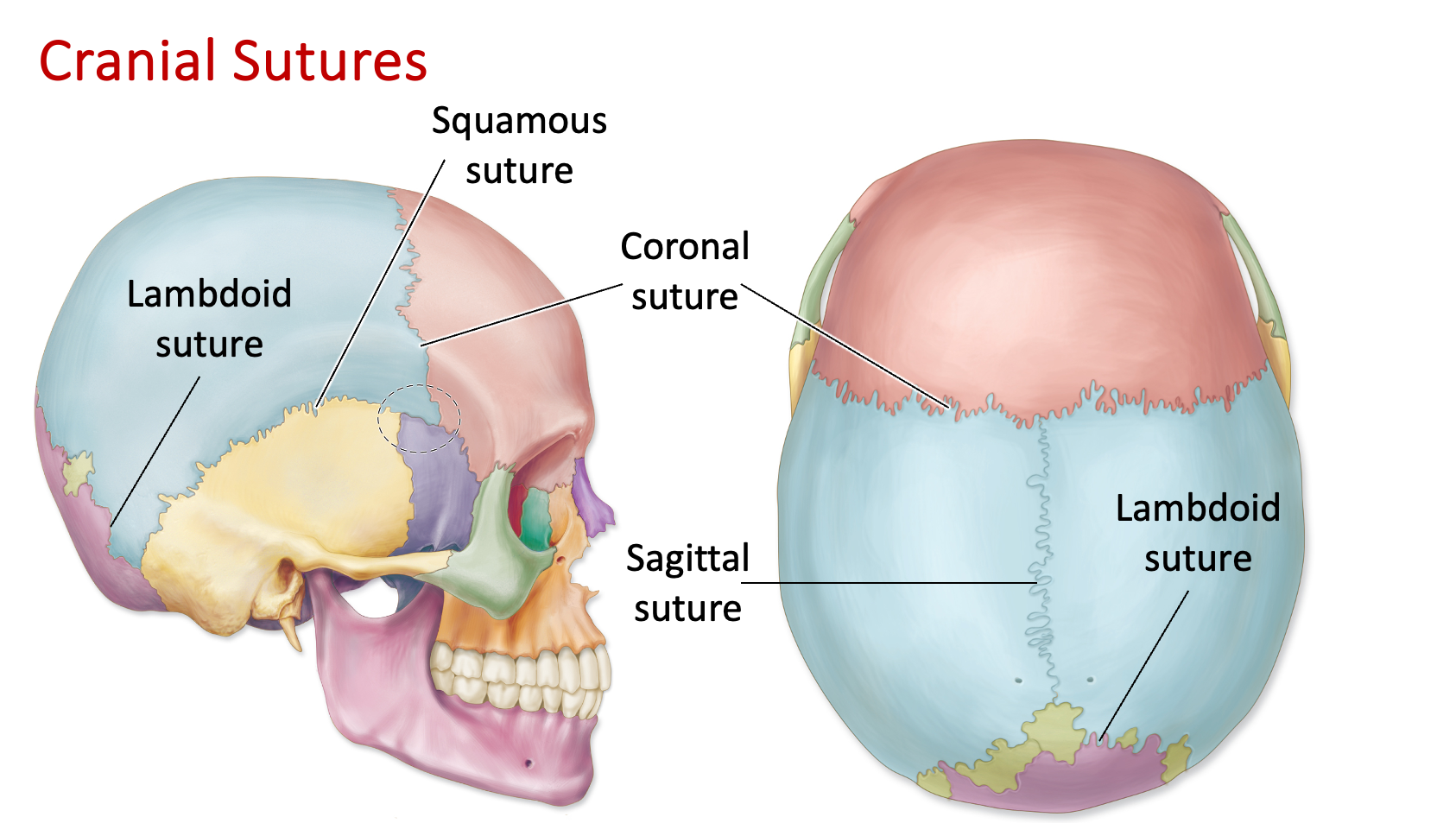
Sutures
Coronal - a suture in the skull that runs across the coronal plane
Sagittal - a suture in the skull that runs across the sagittal plane
Lambdoidal/lambdoid - a suture found at the back of the skull that connects the parietal bones with the occipital bone
Squamosal/squamous - a suture between the parietal and temporal bones on each side of the skull
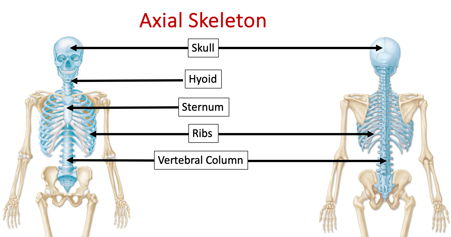
Axial Skeleton
Skull & facial bones (articulated & disarticulated models)
Sphenoid, ethmoid, and temporal bones will be the only disarticulated bones on the test
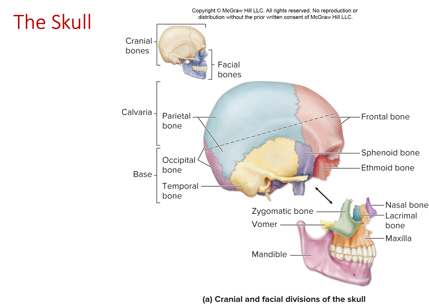
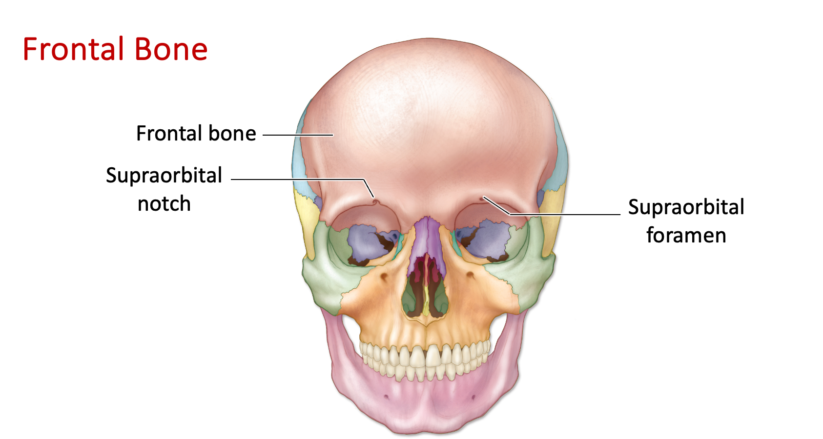
Frontal bone
Supraorbital notch/foramen (“above orbital notch”) - located above the eye socket (orbit)
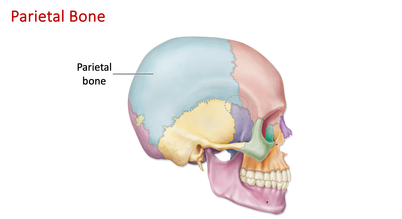
Parietal bone (“paired bones”)
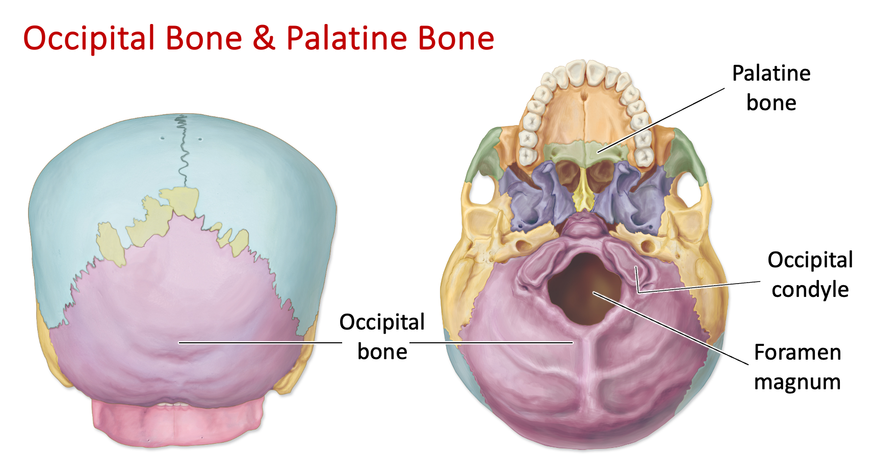
Occipital bone
Foramen magnum (magnum = huge)
A large hole, foramen, in the occipital bone
Where the brain stem exits, and the spinal cord is connected
Allows for nodding
Occipital condyles
Rounded projections under the occipital bone
Articulates with vertebral column
Next to foramen magnum
Allows for nodding movement
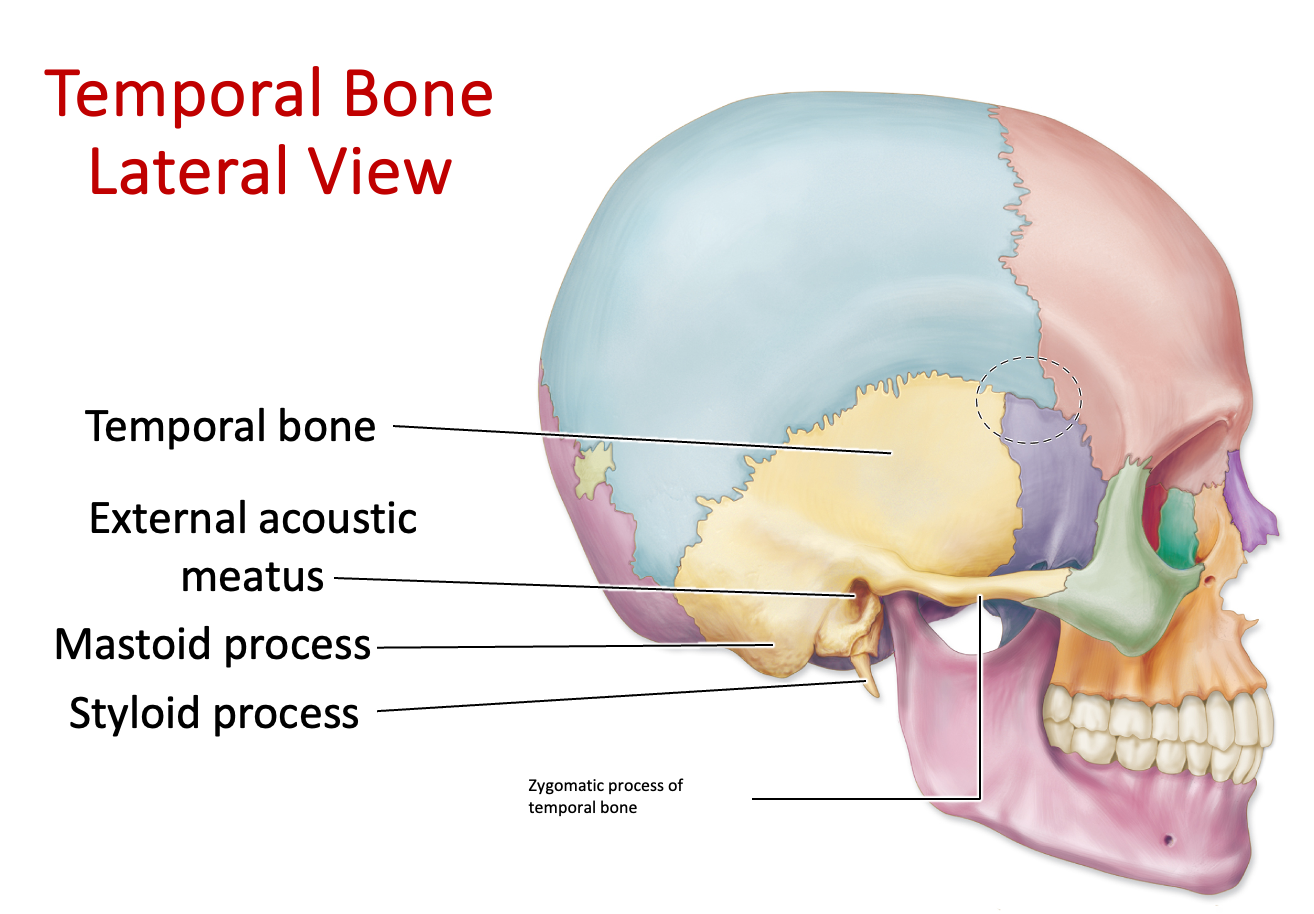
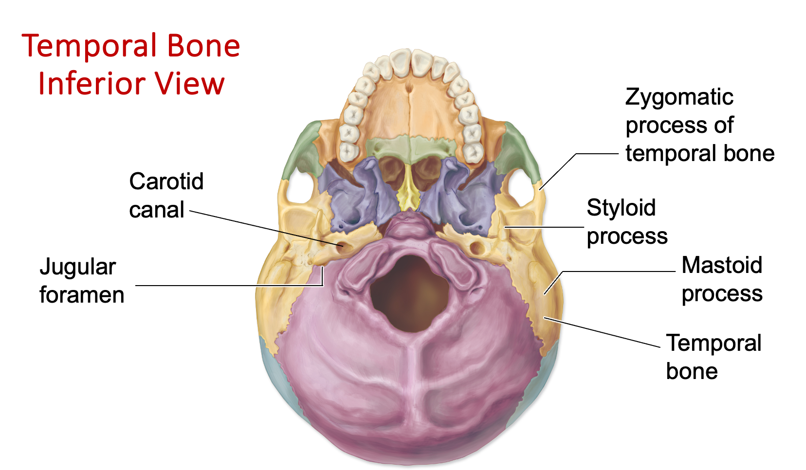
Temporal bone
External auditory/acoustic meatus - hole in the temporal bone that permeates, where we hear things
Styloid process - penlike, sharp projection of the temporal bone
Zygomatic process - a bony projection on the temporal bone that articulates with the zygomatic bone (cheekbones - think the Kardashians)
Mastoid process - a bony projection of the temporal bone located behind the ear, looks like a big chunk
Carotid canal - passage in the temporal bone that allows the internal carotid artery to enter the skull and supply blood to the brain
Jugular foramen - jugular vein runs through this foramen, lateral to foramen magnum
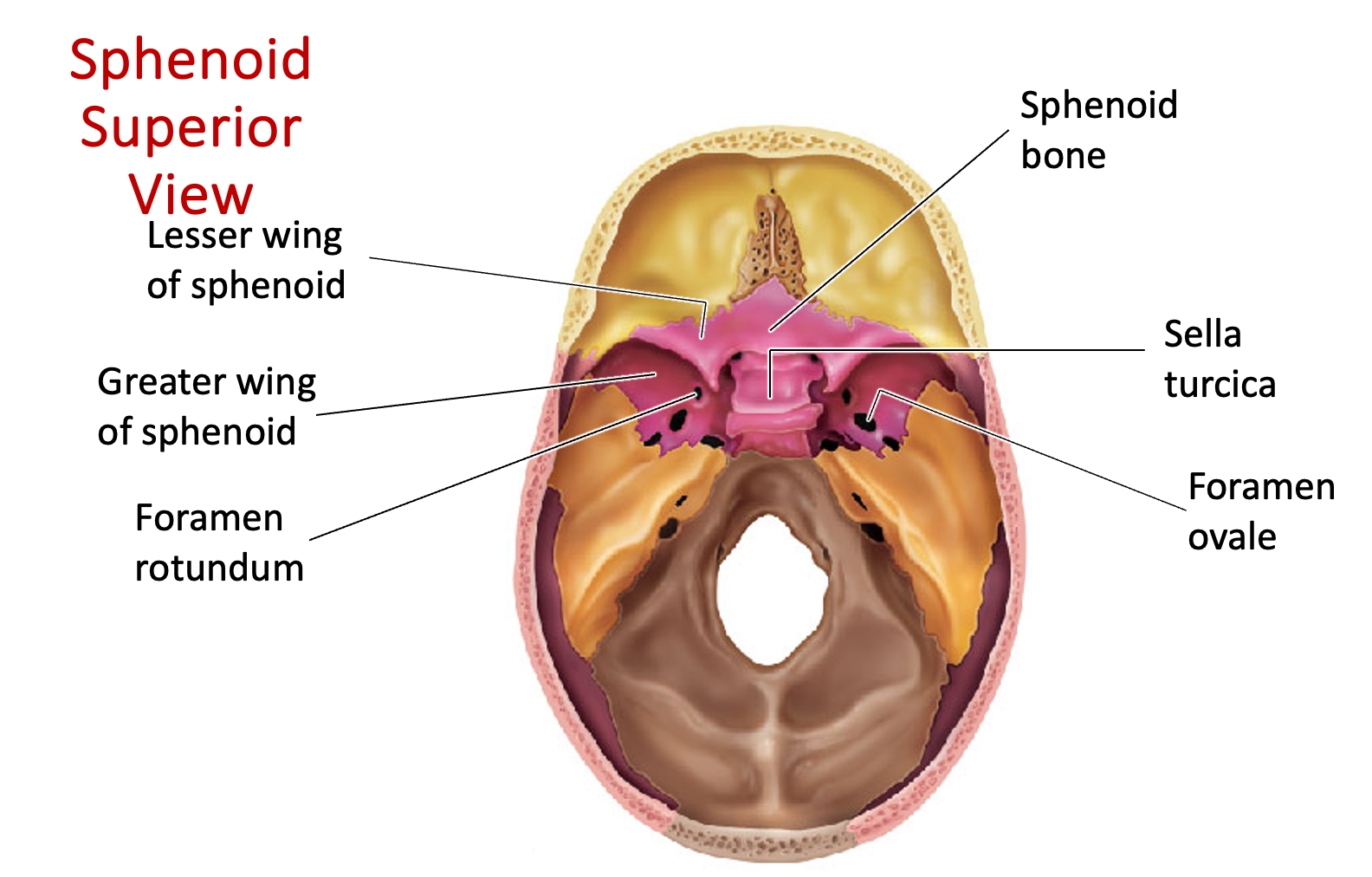
Sphenoid bone - located at the base of the skull, bat-shaped bone
Sella turcica - structure in the sphenoid bone that houses the pituitary gland; named for its resemblance to a saddle
Greater wings - larger, lower portion of the sphenoid bone in the skull, sits inferiorly to lesser wings
Lesser wings - smaller, upper portion of the sphenoid bone in the skull, sits superiorly to greater wings
Pterygoid processes - bony projections on the sphenoid bone near the base of the skull (“feet of the bat”)
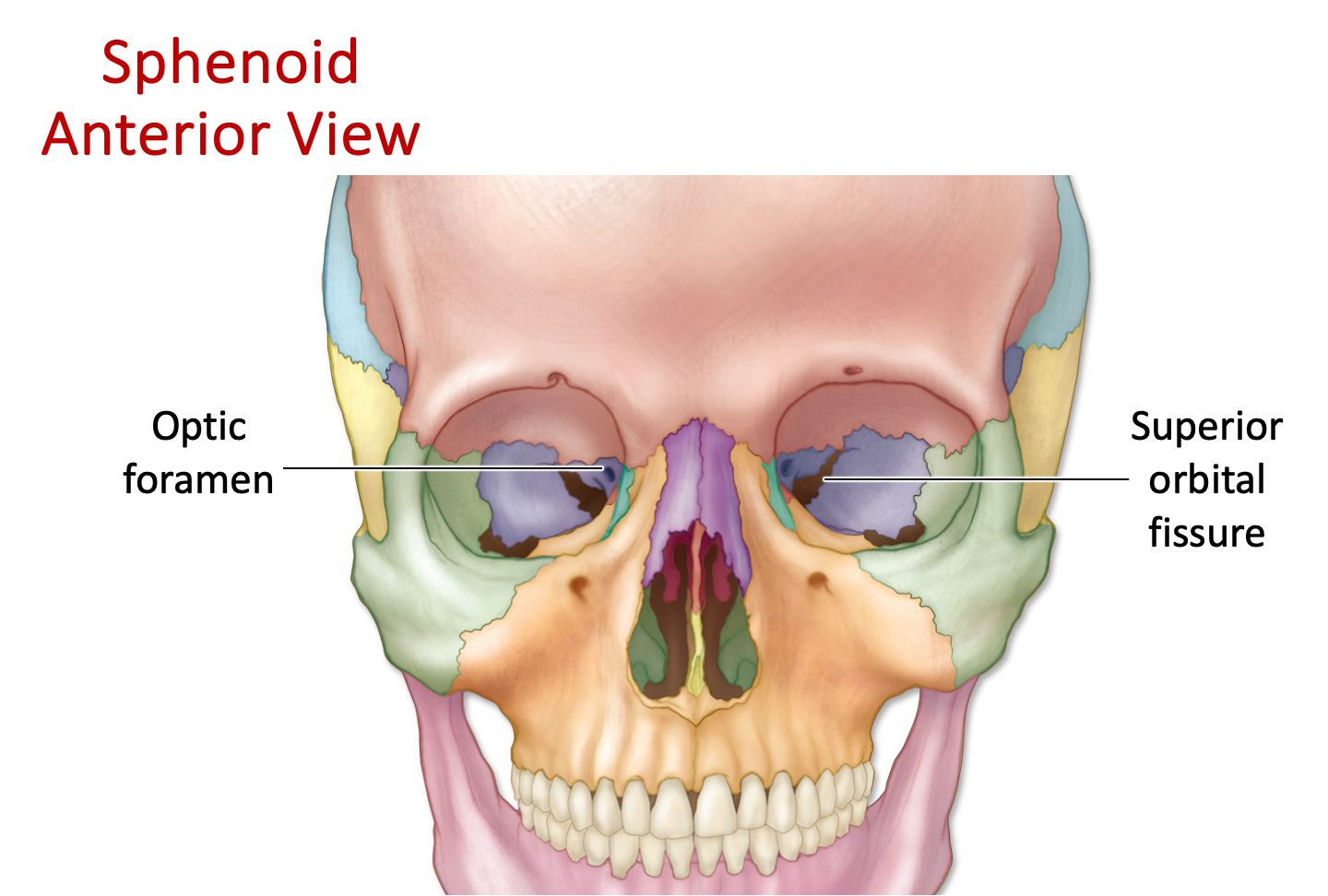
Optic foramen/canal - sits below the lesser wings, where the optic nerve runs
Foramen ovale - lateral to rotundum, ovale = oval
Foramen rotundum - lateral and posterior to optic foramen, rotundum = rounded

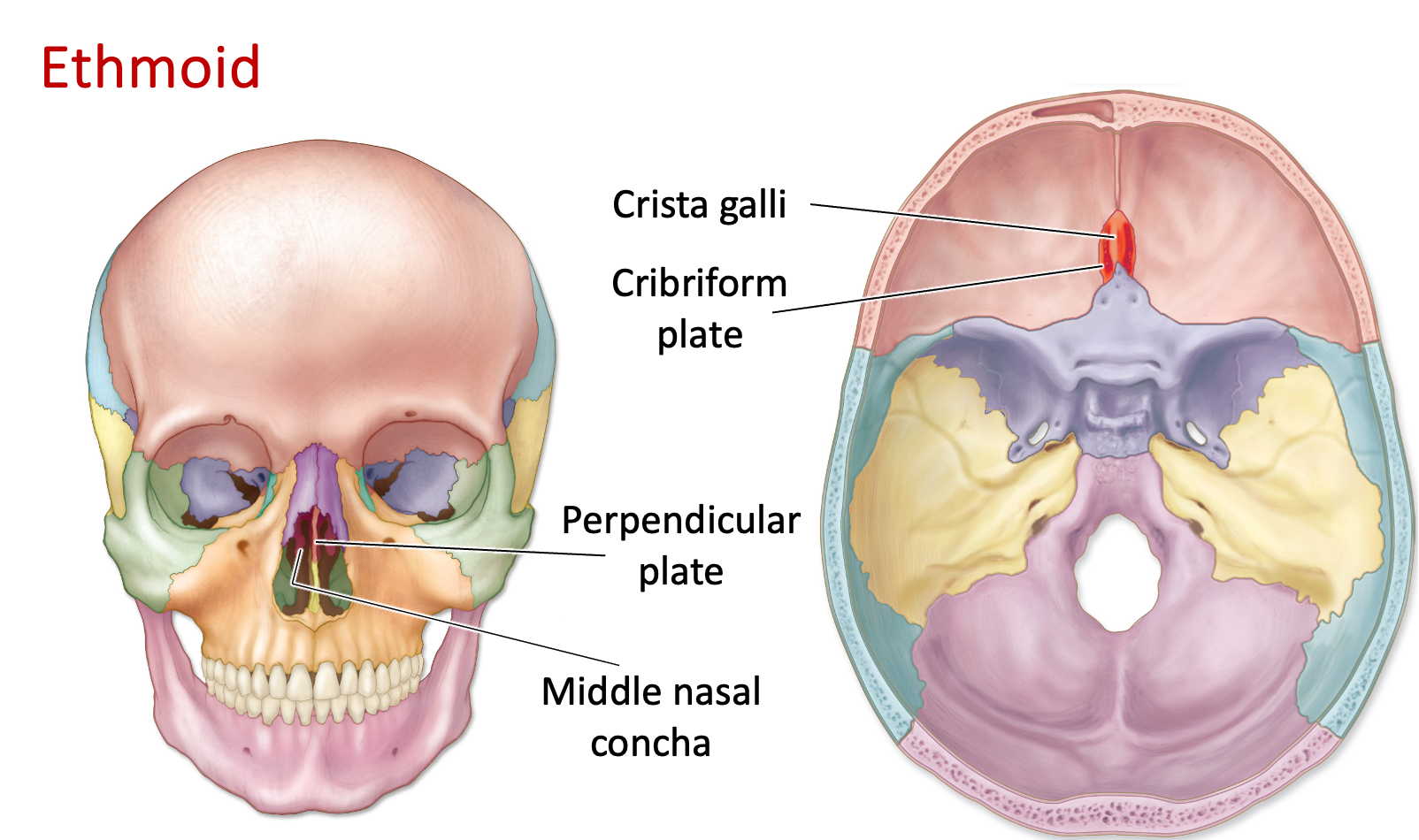
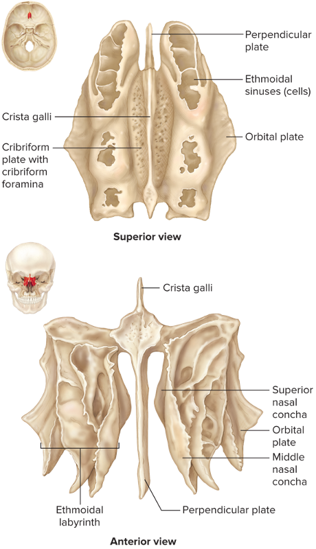
Ethmoid bone - located at the roof of the nose and between the eyes
Crista galli - bony ridge in the ethmoid bone that projects upwards into the cranial cavity
Hold onto the “rhino horn” - indicates it is anterior
Perpendicular plate - part of the ethmoid bone that forms the superior portion of the nasal septum, separating the two nasal cavities
Superior nasal conchae - bony shelves on the lateral walls of the nasal cavity, sits superiorly to the middle nasal conchae
Middle nasal conchae - located in the nasal cavity and the ethmoid bone, sits inferiorly to the superior nasal conchae
Cribriform plate (“the crib”) - part of the ethmoid bone in the skull, the crista galli sits on top of this
Olfactory foramina - holes in the cribriform plate of the ethmoid bone where the olfactory nerves come through
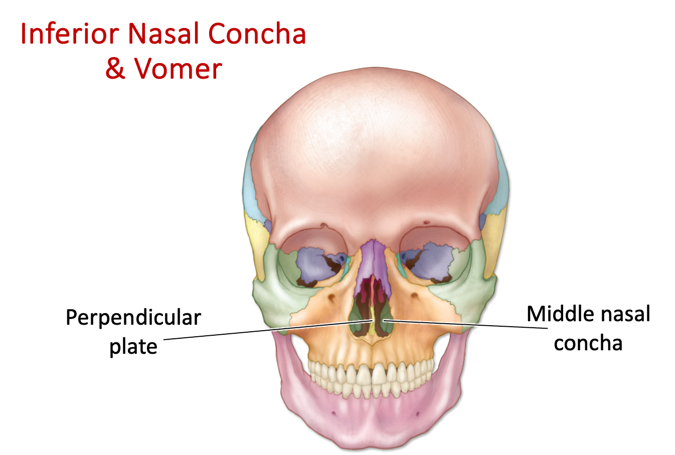
Inferior nasal concha - thin bone in the nasal cavity of the ethmoid bone that sits below the middle nasal concha
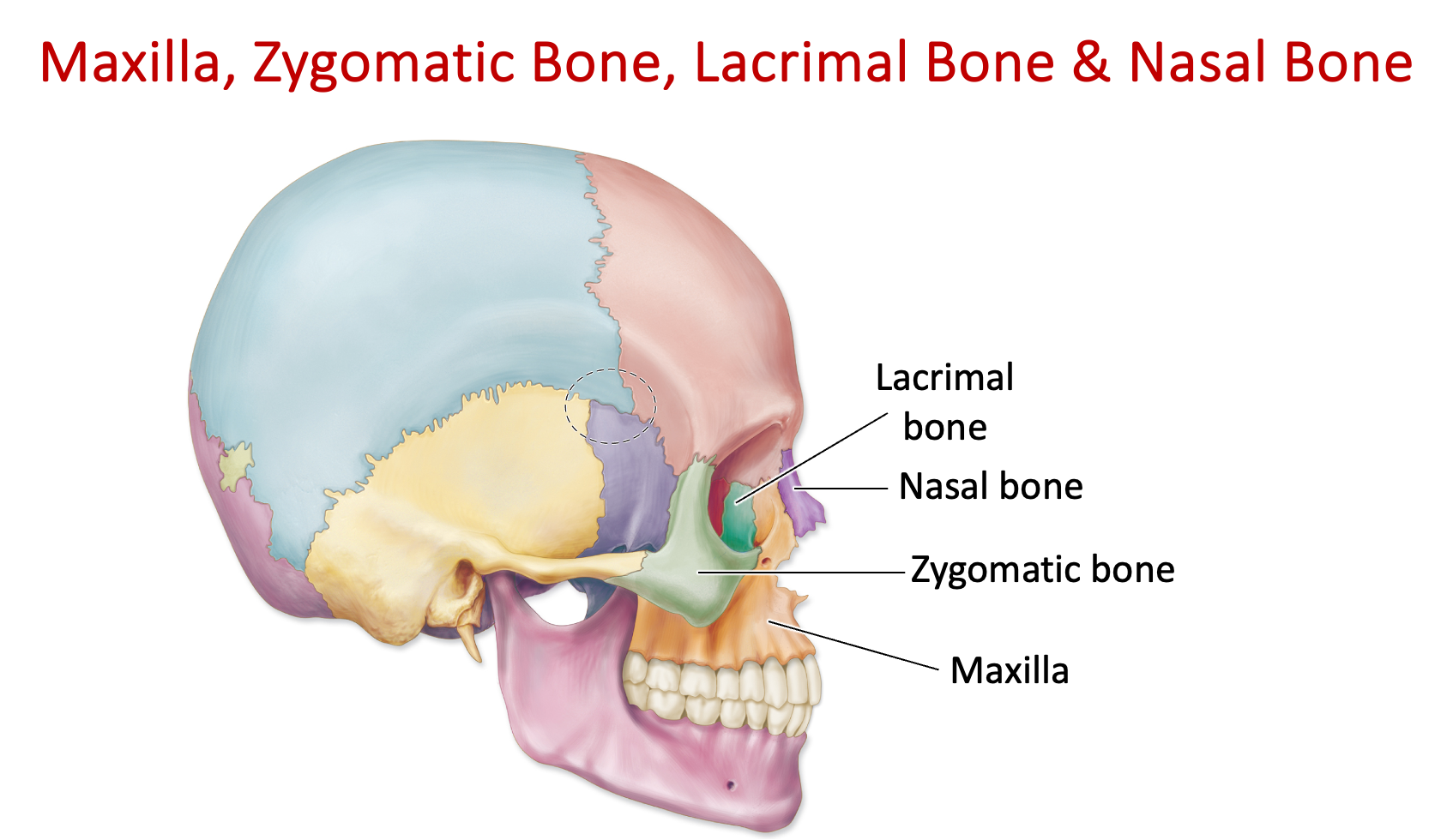
Maxillary bone - forms the upper jaw and part of the eye socket. Contains the maxillary sinus and houses the teeth.
Palatine bone (“Tasting - pallet”) - located behind the maxilla bone
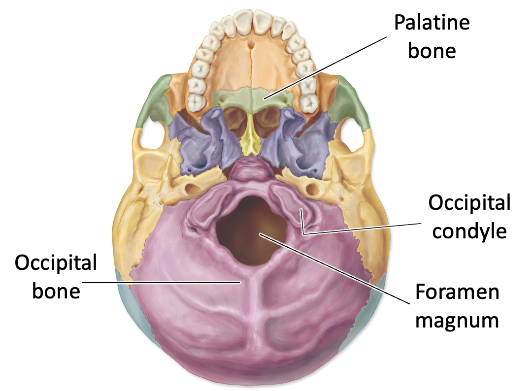
Vomer - inferior portion of the perpendicular plate

Zygomatic bone - located in the cheekbone region
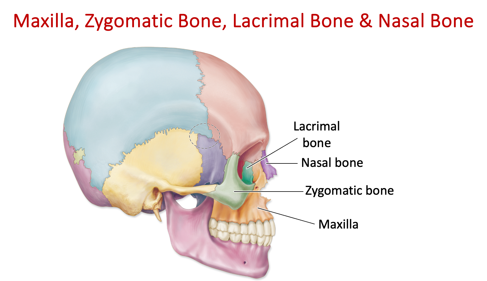
Lacrimal bone - bone forming part of the eye socket; houses the tear (lacrimal) ducts and surrounds tear duct pore
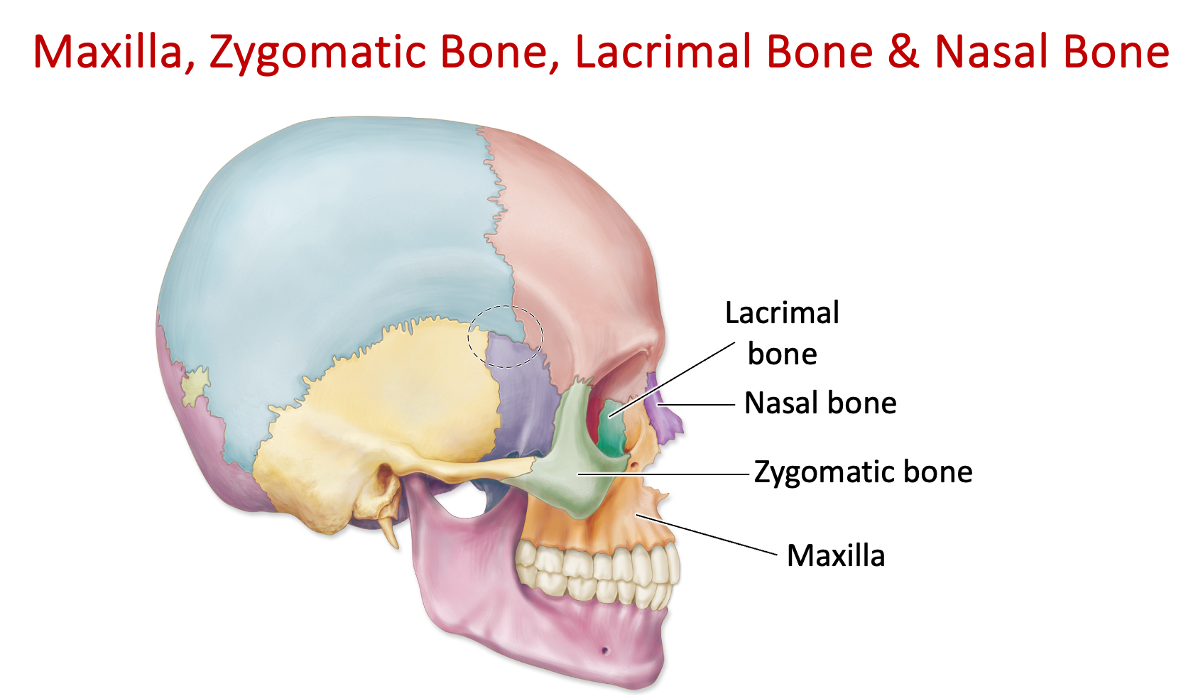
Nasal bone - located in the upper part of the nose
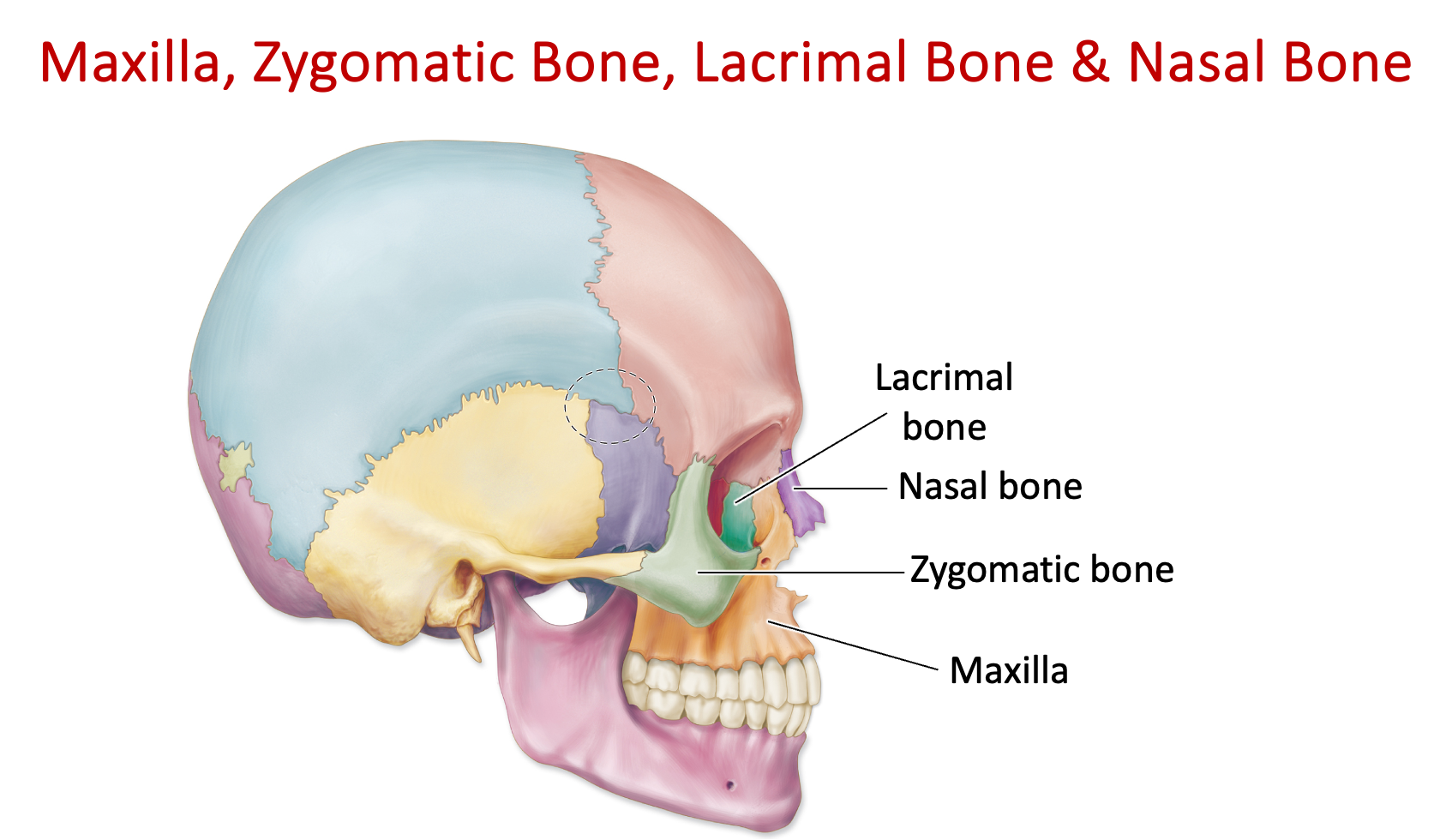
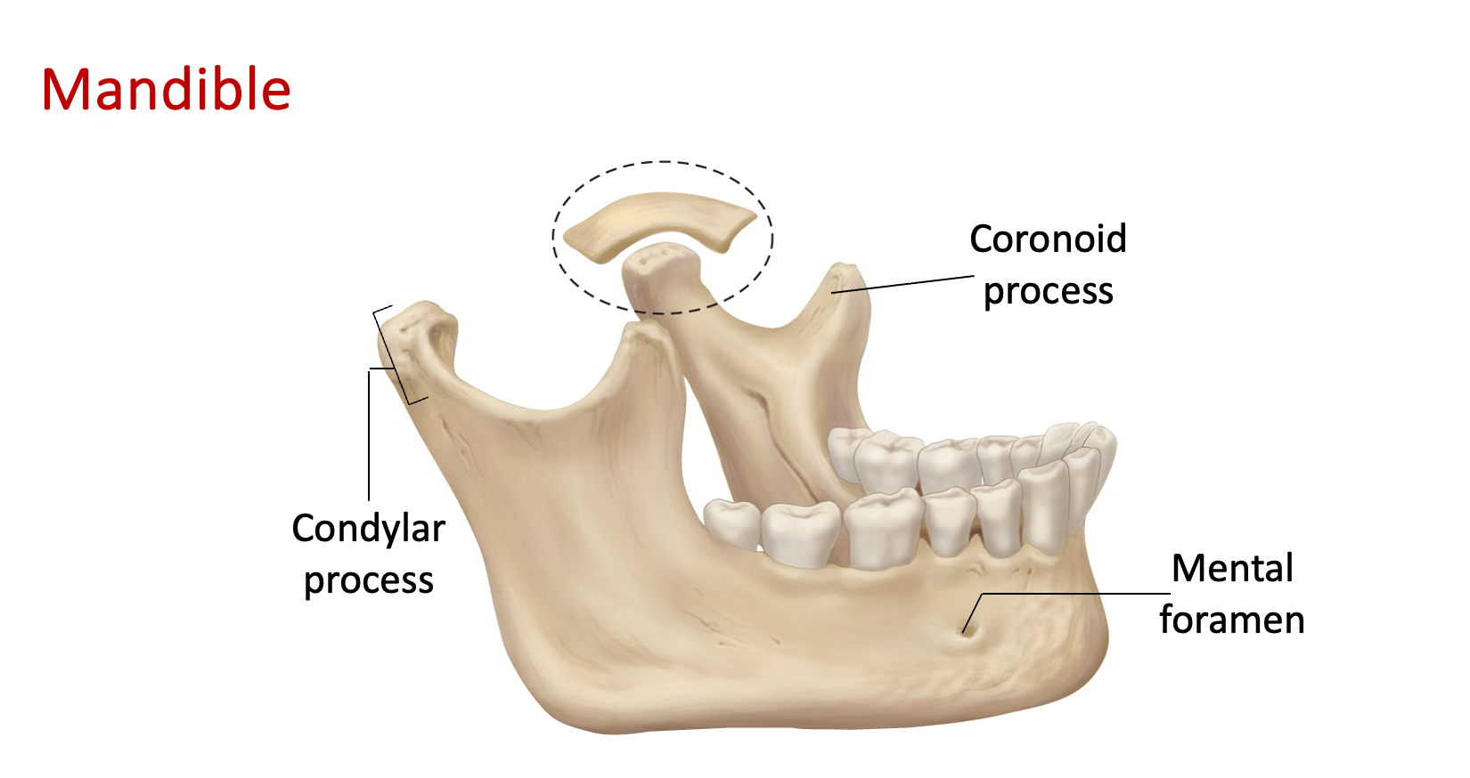
Mandible - structure in the skull forming the lower jaw, contains the lower teeth. The only bone that moves.
Mental foramen - foramen in the mandible where vessels and nerves run through
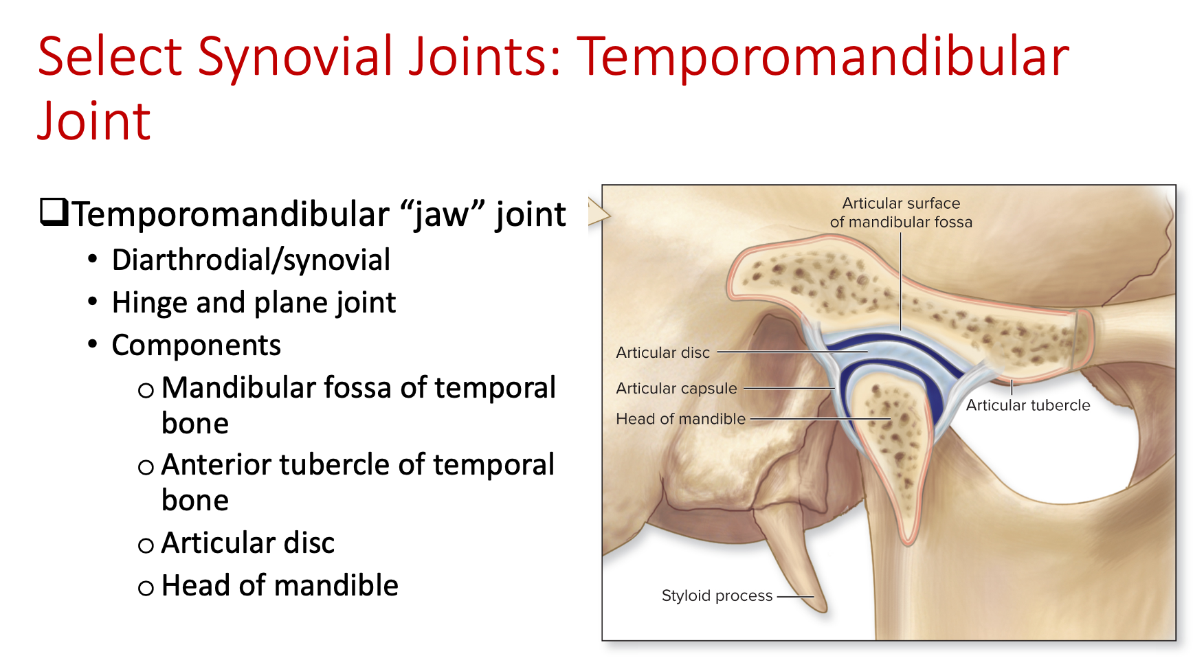
Mandibular condyles - rounded condyle bones in the mandible that articulates with another bone. Creates synovial joints - temporomandibular joints (TMJ).
Coronoid process - flat-ended bones located in the mandible
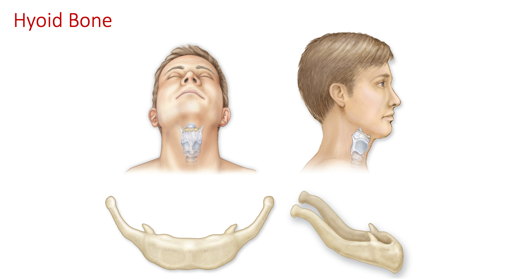
Hyoid bone - small U-shaped bone in the neck, not connected to any other bone, supports muscles that allow movement of lower jaw
Spine
“Breakfast at 7 (7 cervical vertebrae), lunch at 12 (12 thoracic vertebrae), dinner at 5 (5 lumber vertebrae)”
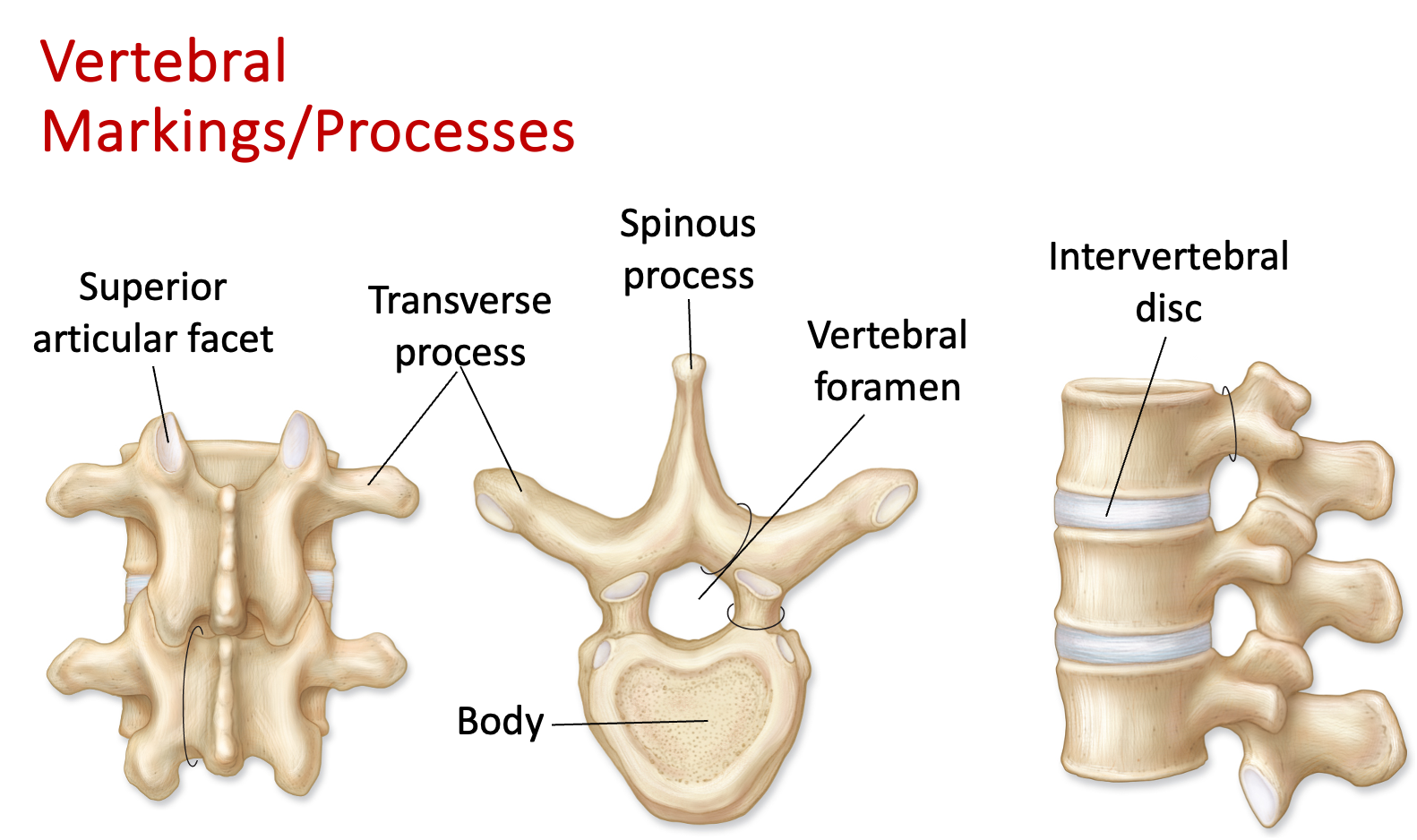
Typical vertebral structures
Spinous process - a bony projection off the posterior aspect of a vertebra. The bony part of the vertebra that you feel as you run your hand down your back.
Transverse processes - bony projections on each side of a vertebra that stick out, located on the transverse plane
Vertebral body - The thick, cylindrical anterior portion of a vertebra providing structural support and protection for the spinal cord
Vertebral canal - A bony canal formed by vertebral foramina, housing the spinal cord and its protective coverings
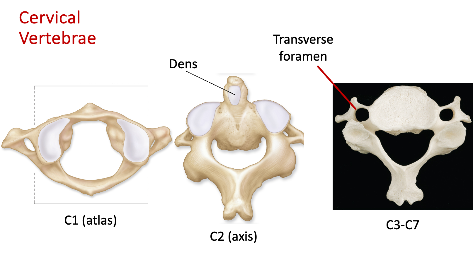
Cervical spine - the spinous process is split
Transverse foramina - little holes in the cervical vertebrae that vertebral arteries run through to supply the brain
C1 – atlas
C2 – axis
Dens - keeps the atlas from sliding into spinal cord/brainstem and allows for rotation
C3-7
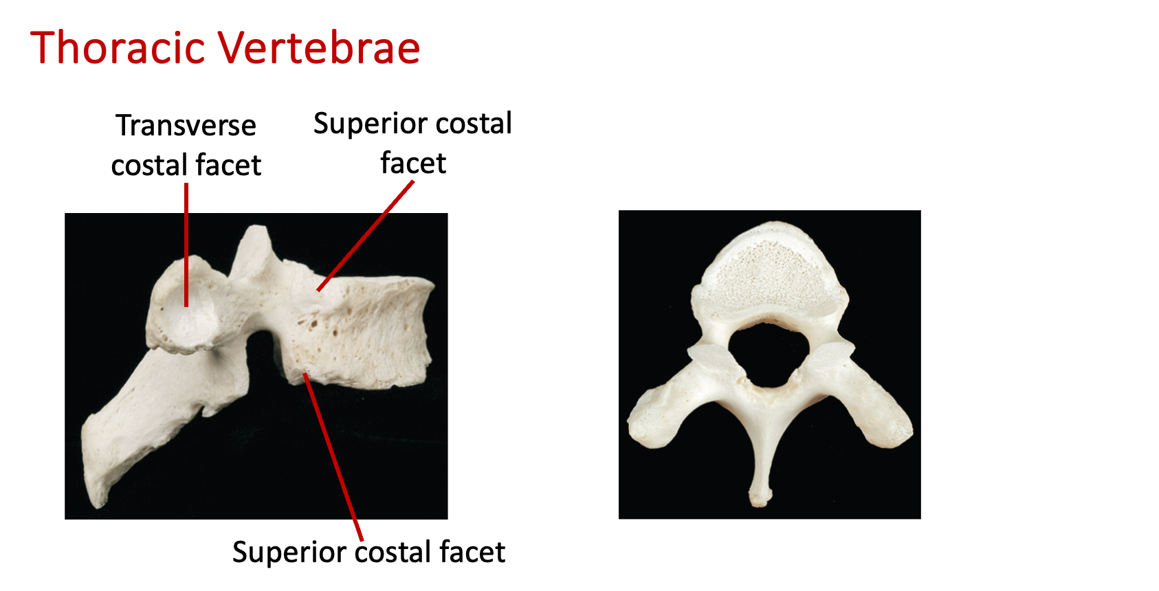
Thoracic spine (looks like a giraffe)
T1-12
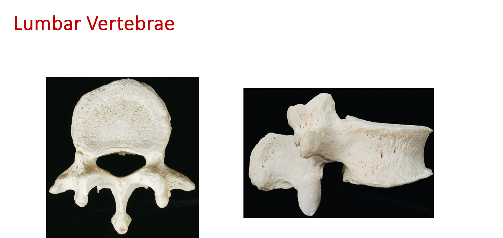
Lumbar spine (looks like a moose)
L1-5
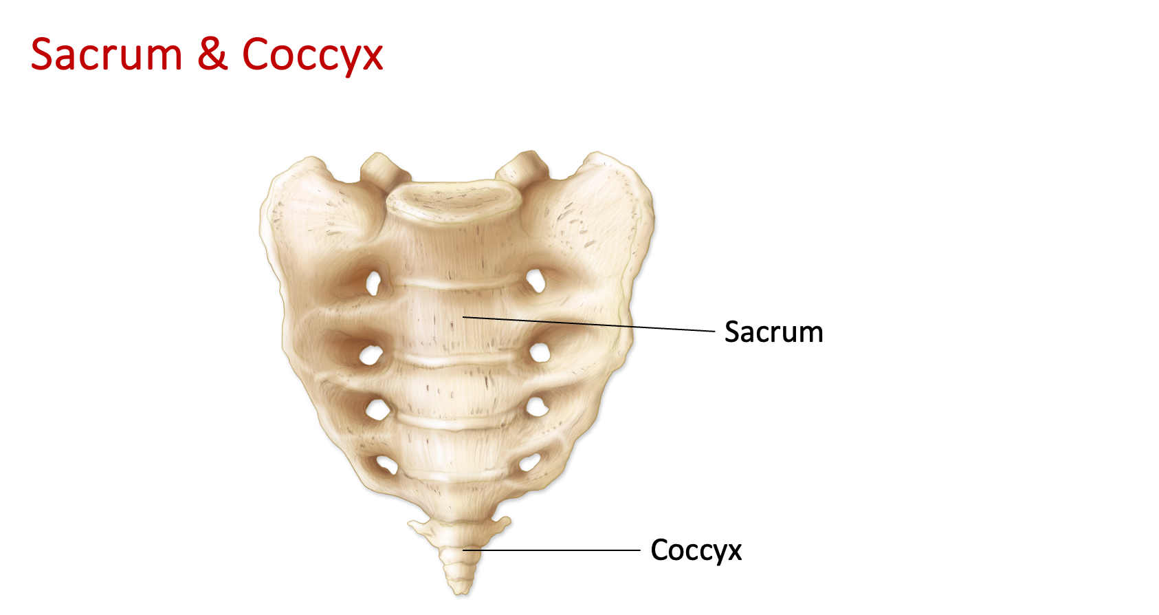
Sacral spine - lowest part of the vertebral column, composed of five fused vertebrae
Coccyx - the small, triangular bone at the base of the spine, composed of fused vertebrae (curved = male, straightened = female)
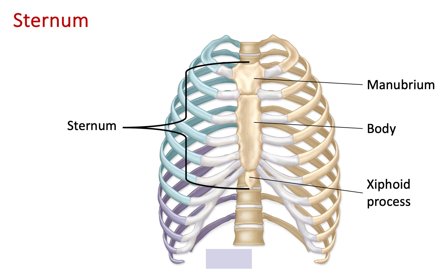
Thoracic cage - bony structure formed by the ribs, sternum, and thoracic vertebrae, protecting the heart and lungs. Allows for breathing and supports the upper body.
Sternum - bone located in the center of the chest, connecting the ribs and protecting vital organs
Manubrium - part of the sternum located at the top, articulating with the clavicles and first rib
Body - the long, flat bone in the center of the chest, connects to the ribs through cartilage
Xiphoid process - small, cartilaginous extension at the bottom of the sternum
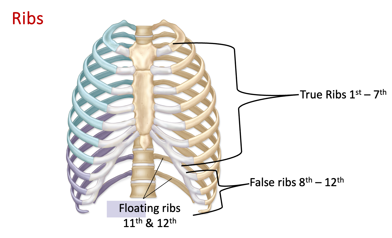
Ribs – 12 pairs
True (7 pairs) - directly attached to the sternum through costal cartilage, known as ribs 1-7
False (5 pairs, including 2 pairs floating ribs) - Ribs 8-12, 5 pairs of ribs including 2 pairs of floating ribs, attached to the sternum indirectly via costal cartilage
Floating (2 pairs) - ribs 11-12 that are not attached to the sternum, only to the vertebrae
Costal cartilages
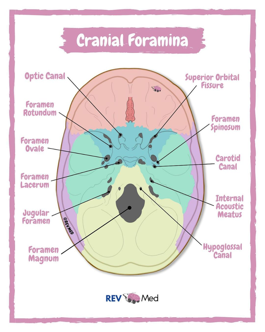
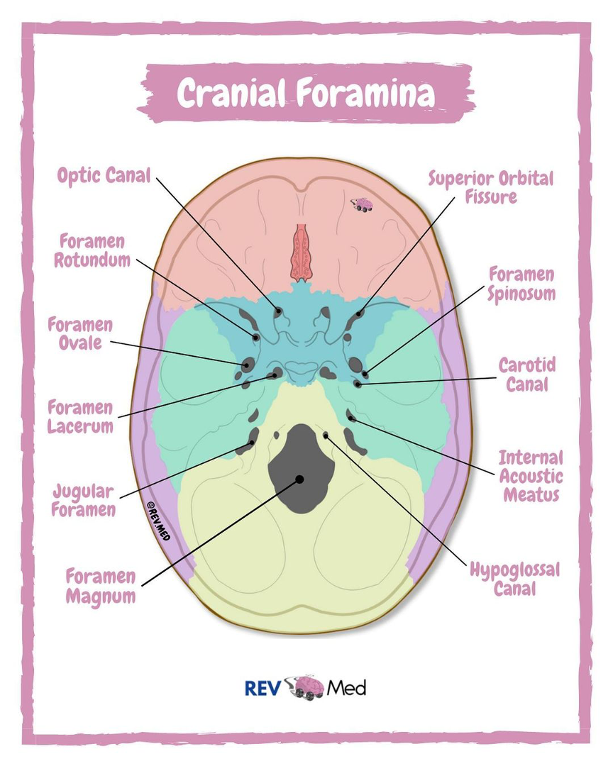
*Foramina Mnemonic - OROS-LAJ, “Old Rotund Owls Spin Lazily Across Jugs”
Optic
Rotundum
Ovale
Spinosum
Lacerum
Acoustic
Jugular
Lab 5 (Skeletal System - Appendicular Skeleton + Joints)
EXAM Objectives:
Identify all bones on lab manual lists ✅
Identify all assigned bone parts, markings, structures ✅
Articulated & Disarticulated ✅
Right v. Left ✅
Male v. Female pelvis ✅
Know functions where appropriate ✅
Joints: Identify joints ✅
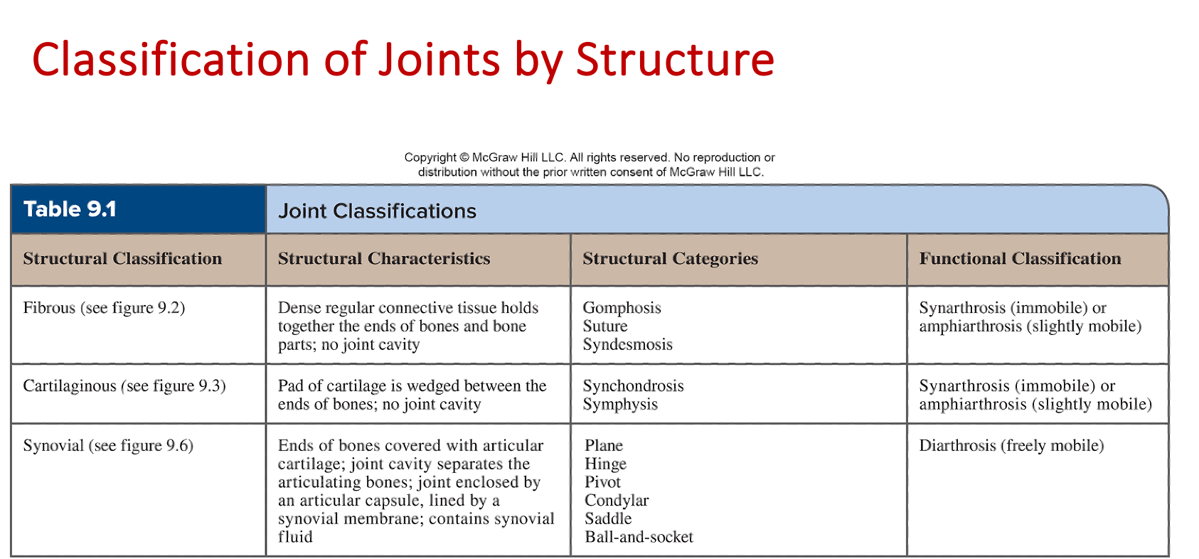
Functional classification of each joint ✅
Structural classification & subclassification of each joint ✅
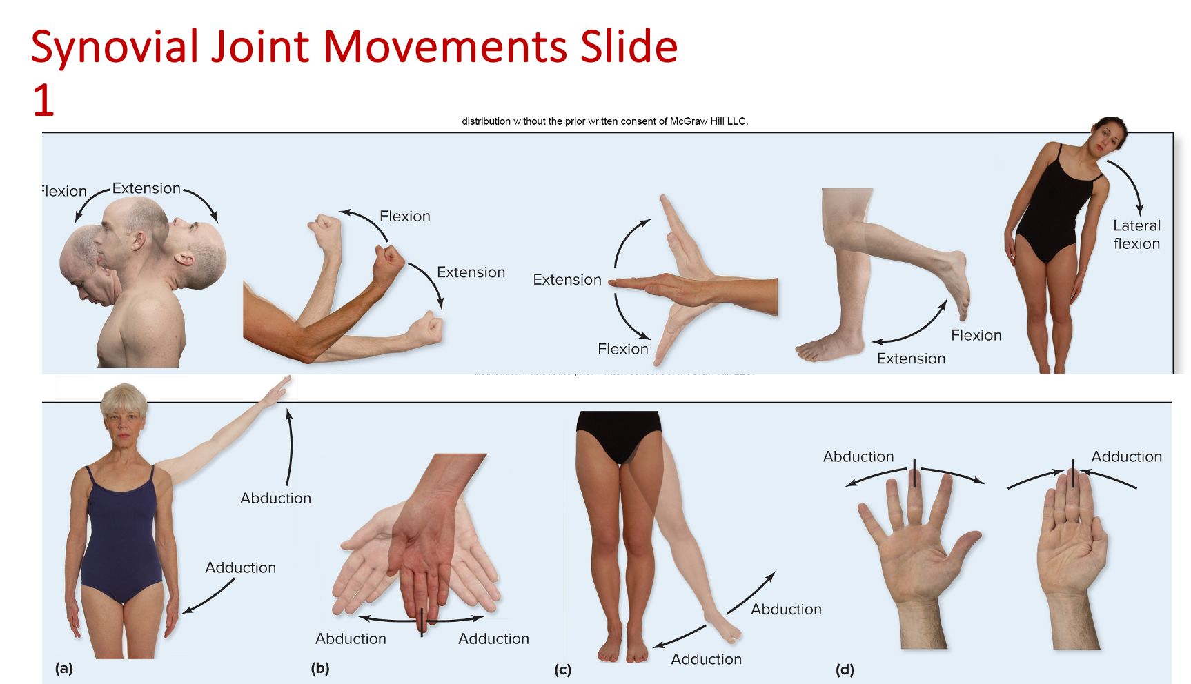
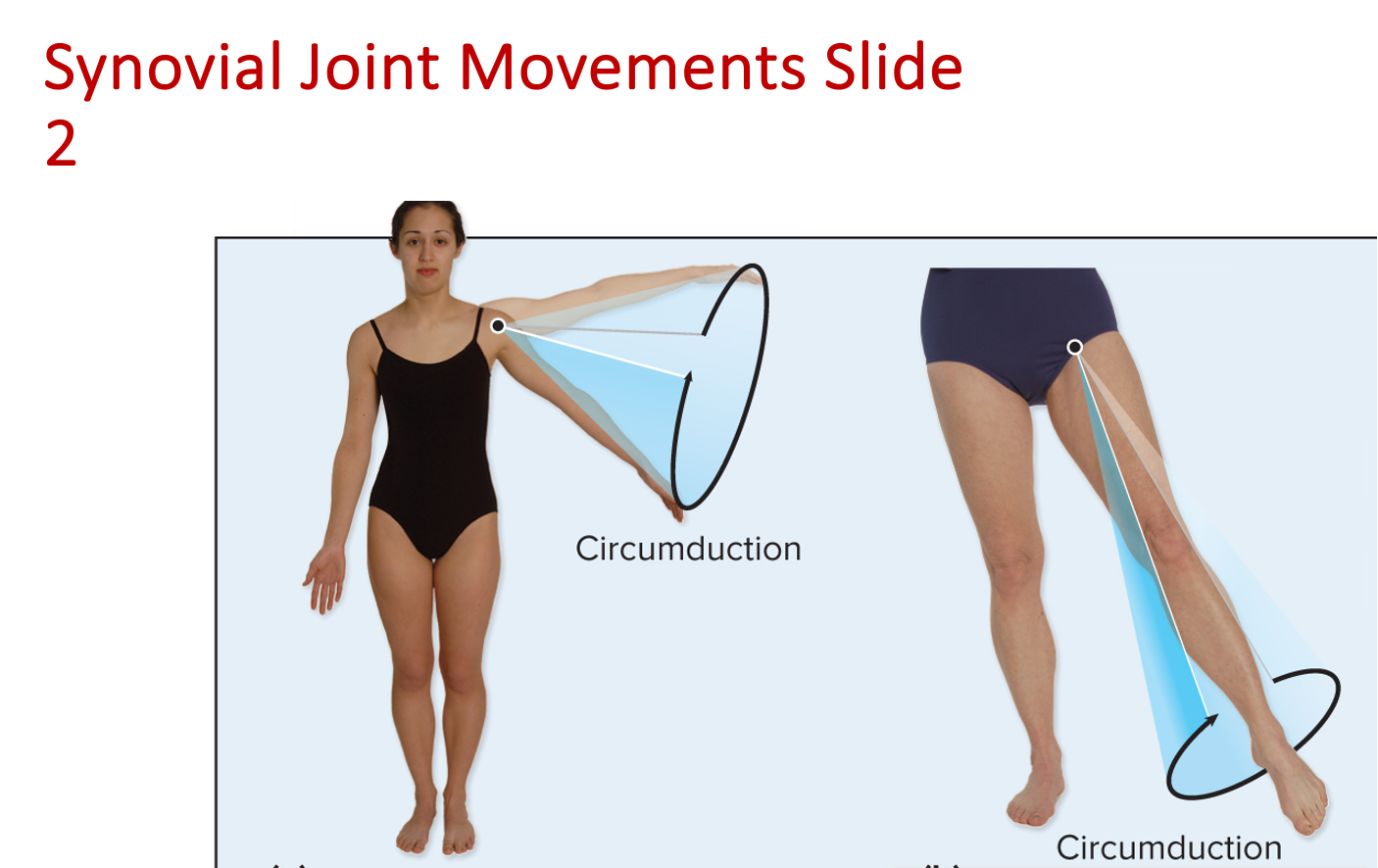
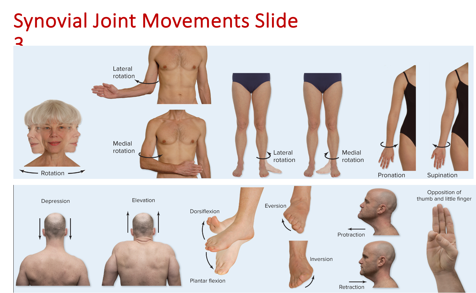
Body movements ✅
Models: You should be able to identify the following structures on various lab models. Additionally, you should be able to distinguish between the male and female pelvis.
Appendicular skeleton
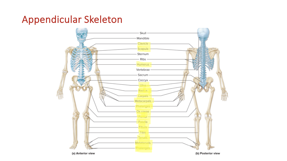
Shoulder girdle
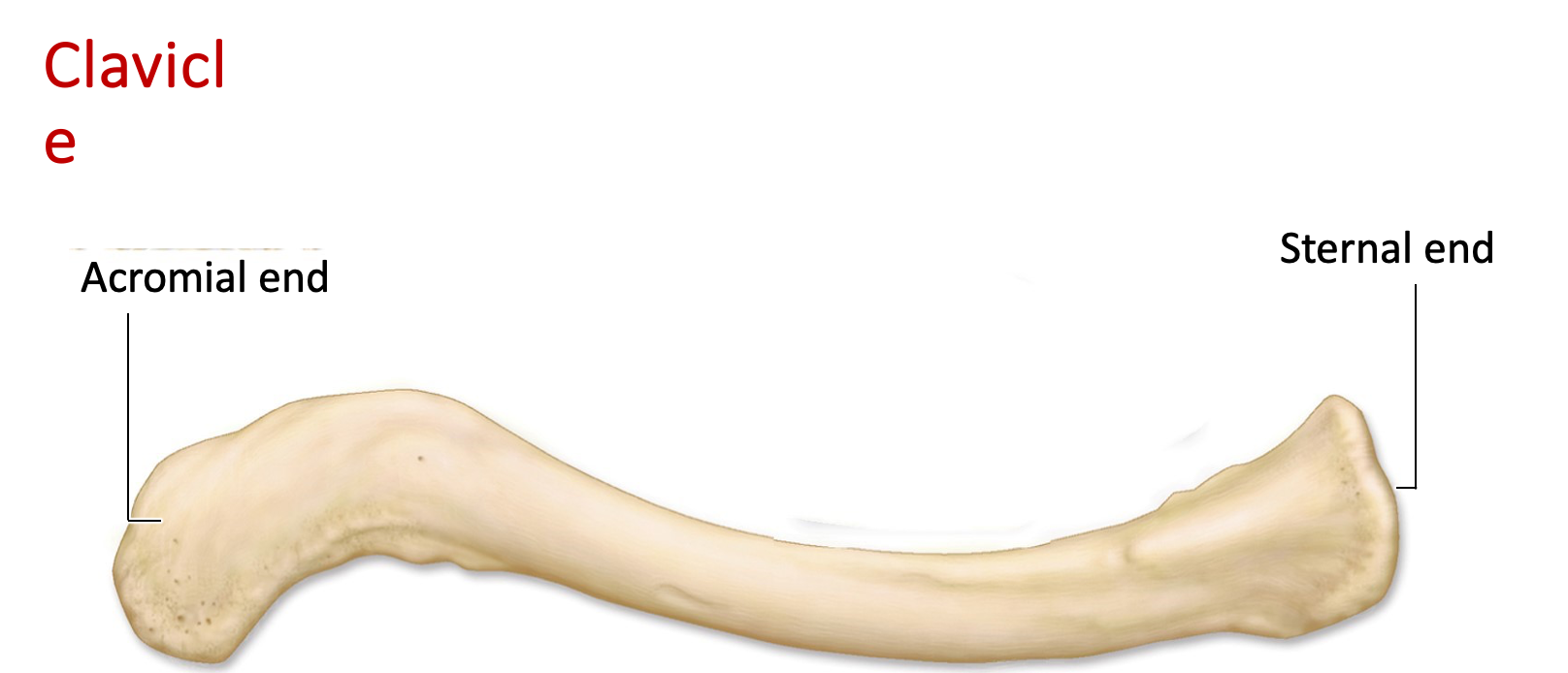
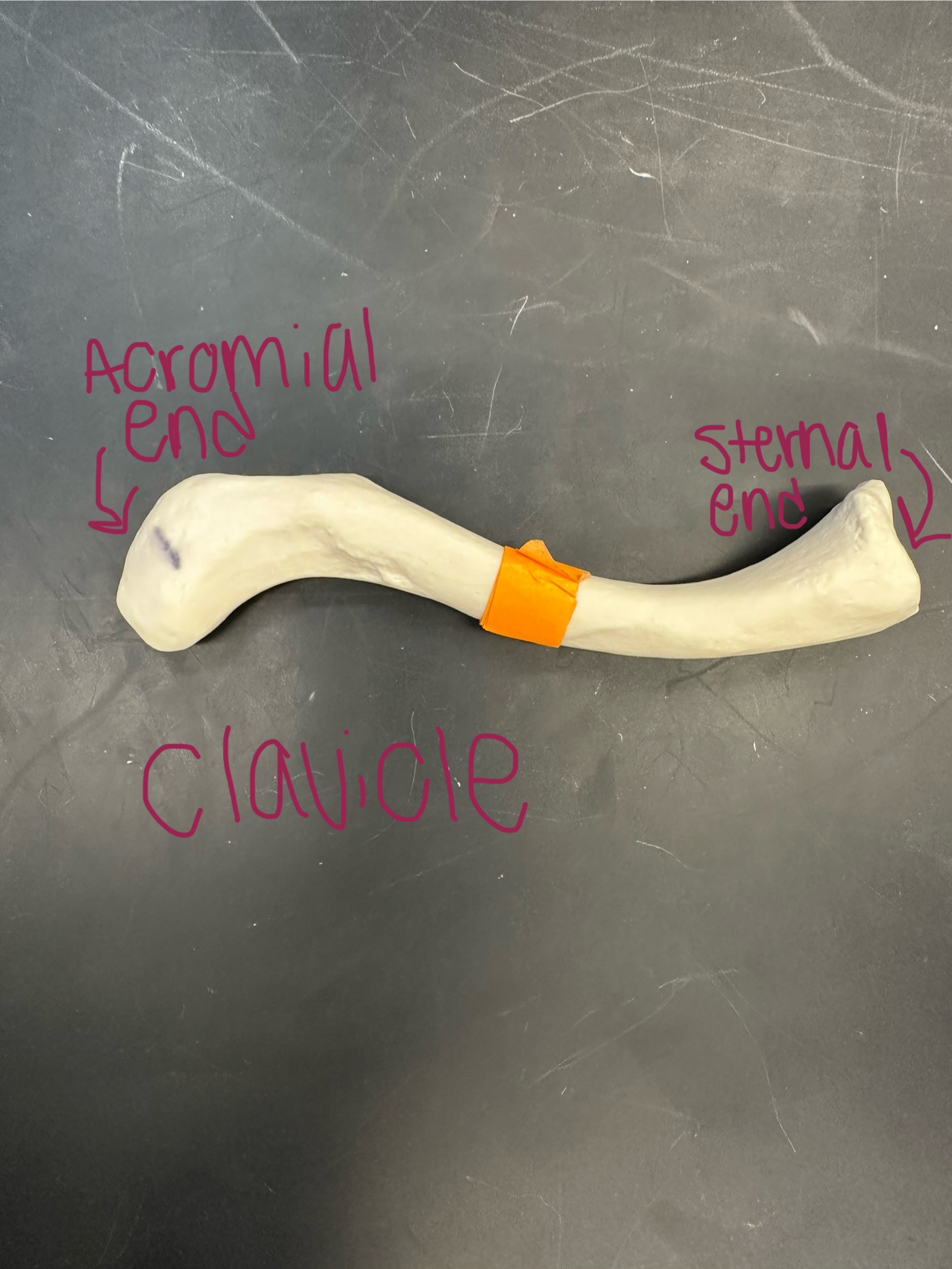
Clavicle - the collarbone, connects the sternum to scapula
Sternal end (“blunt end”) - articulates with the sternum
Acromial end (“flattened end”) - lateral end of the collarbone, articulates with acromion process of the scapula
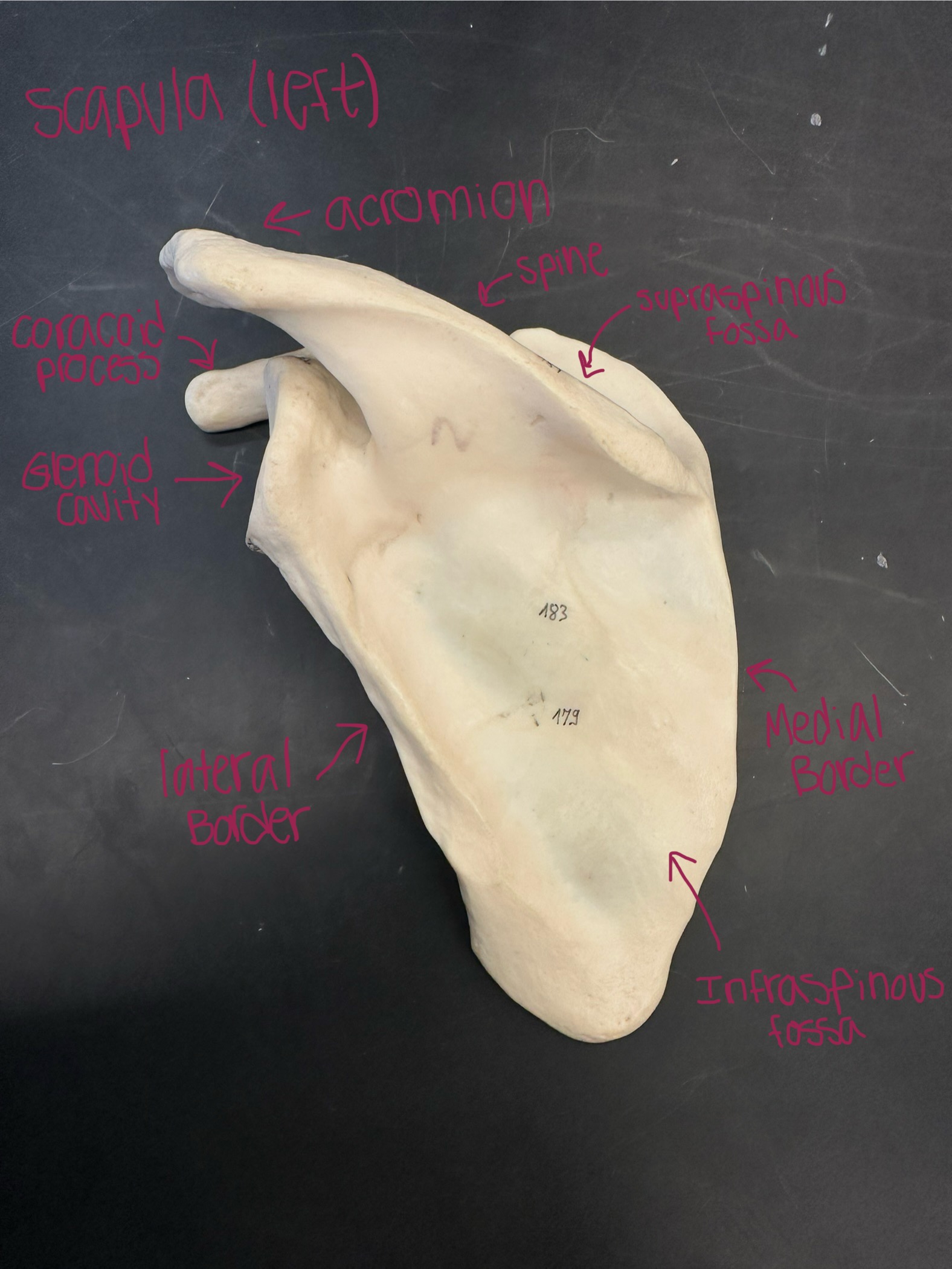
Scapula - shoulder blad, a flat + triangular bone located on the upper back, articulates with the clavicle + humerus
Acromion/acromial process - articulates with the acromial end of scapula, posterior aspect of scapula
Coracoid process - curved projection on the anterior aspect of the scapula
Glenoid fossa/cavity - articulates with head of humerus (lateral border)
Medial border
Lateral border
Spine - ridge on the back of the scapula that runs diagonally across the bone
Supraspinatus fossa (“above the spine”) - located on the posterior aspect of the scapula
Infraspinatus fossa (“below the spine”) - located on the posterior aspect of the scapula
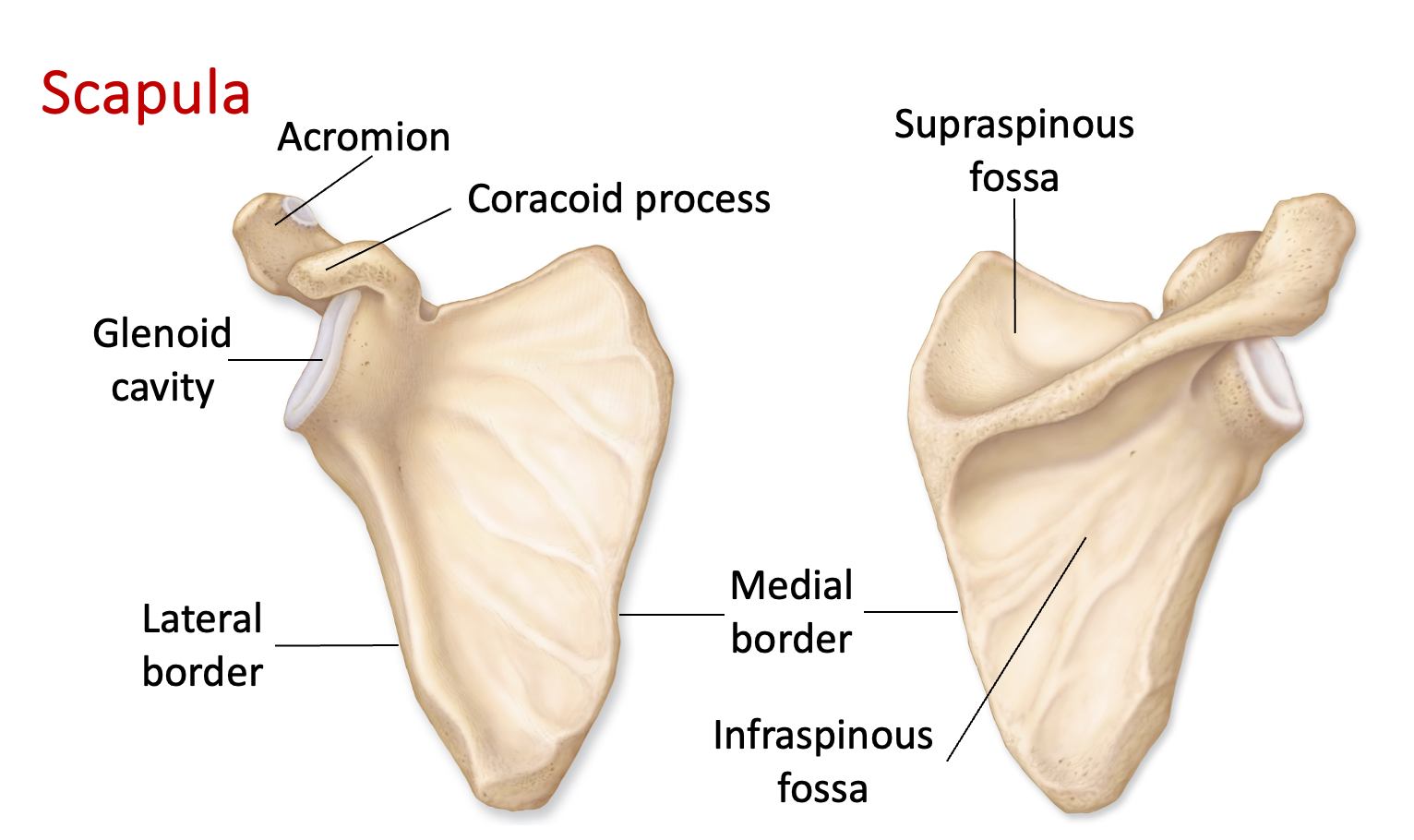
*Determine if the scapula is posterior if you feel the spine
Upper extremity
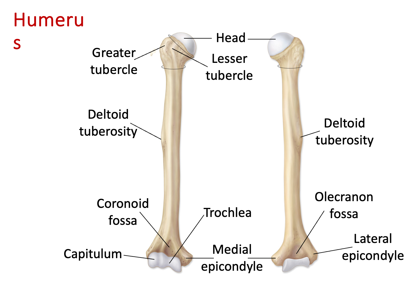
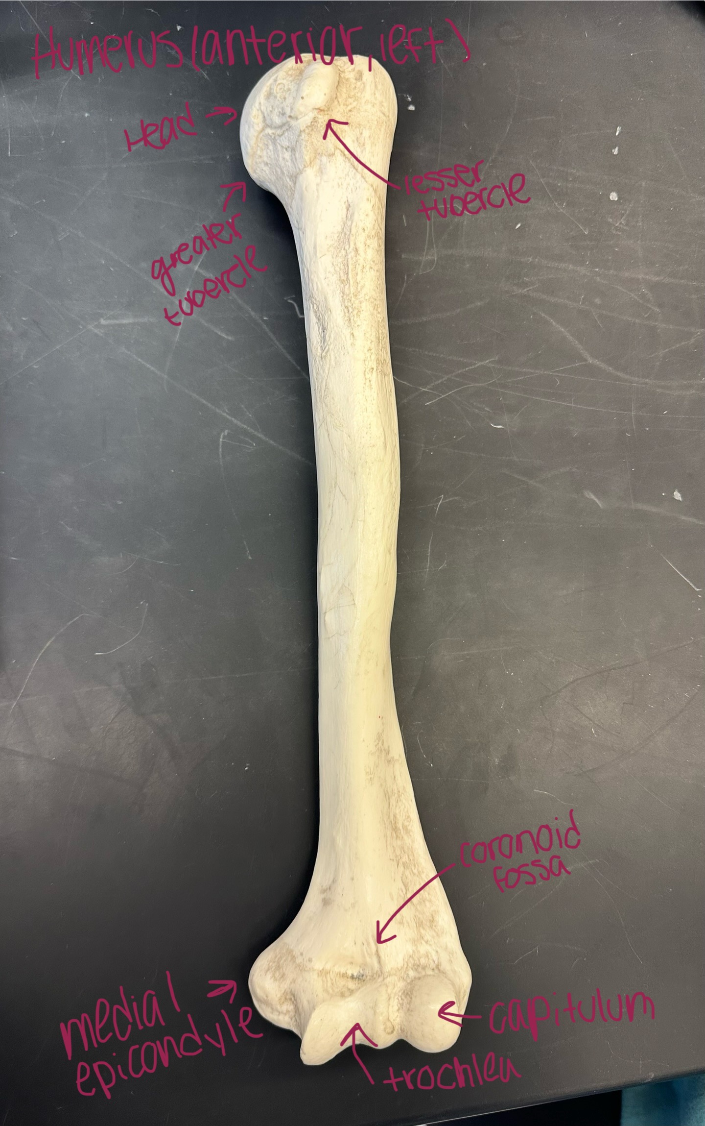
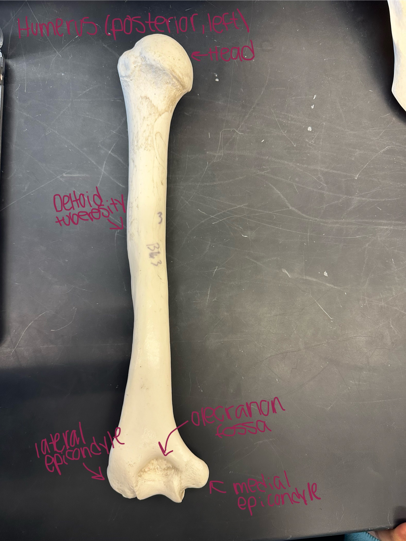
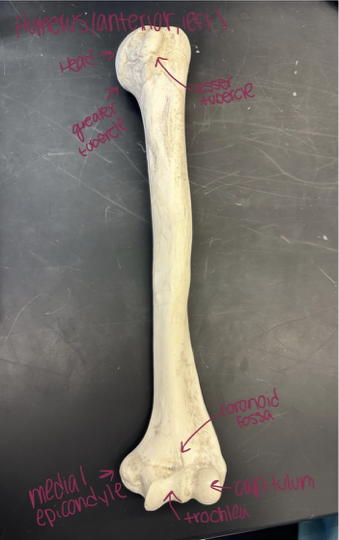
Humerus - long bone of the upper arm that connects the shoulder + elbow joints
Head - rounded proximal end of the humerus bone that articulates with the scapula to form the shoulder joint
Greater tubercle (“bigger grip”) - bony projection on the anterior surface of the humerus on the proximal end
Lesser tubercle (“lesser grip”) - small bony projection on the anterior surface of the humerus on the proximal end
Deltoid tuberosity - bony projection of the humerus about halfway down the shaft, located on the lateral side
Medial epicondyle - bony projection on the medial side of the humerus
Lateral epicondyle - bony projection on the lateral side of the humerus bone
Trochlea - structure located at the distal end of the humerus bone
Capitulum - located on the lateral side of the distal end of the humerus
Coronoid fossa - determines if the humerus is anterior (can’t put your thumb in it)
Olecranon fossa - determines if the humerus is posterior (can put your thumb in it)
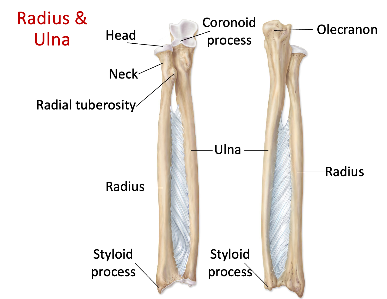
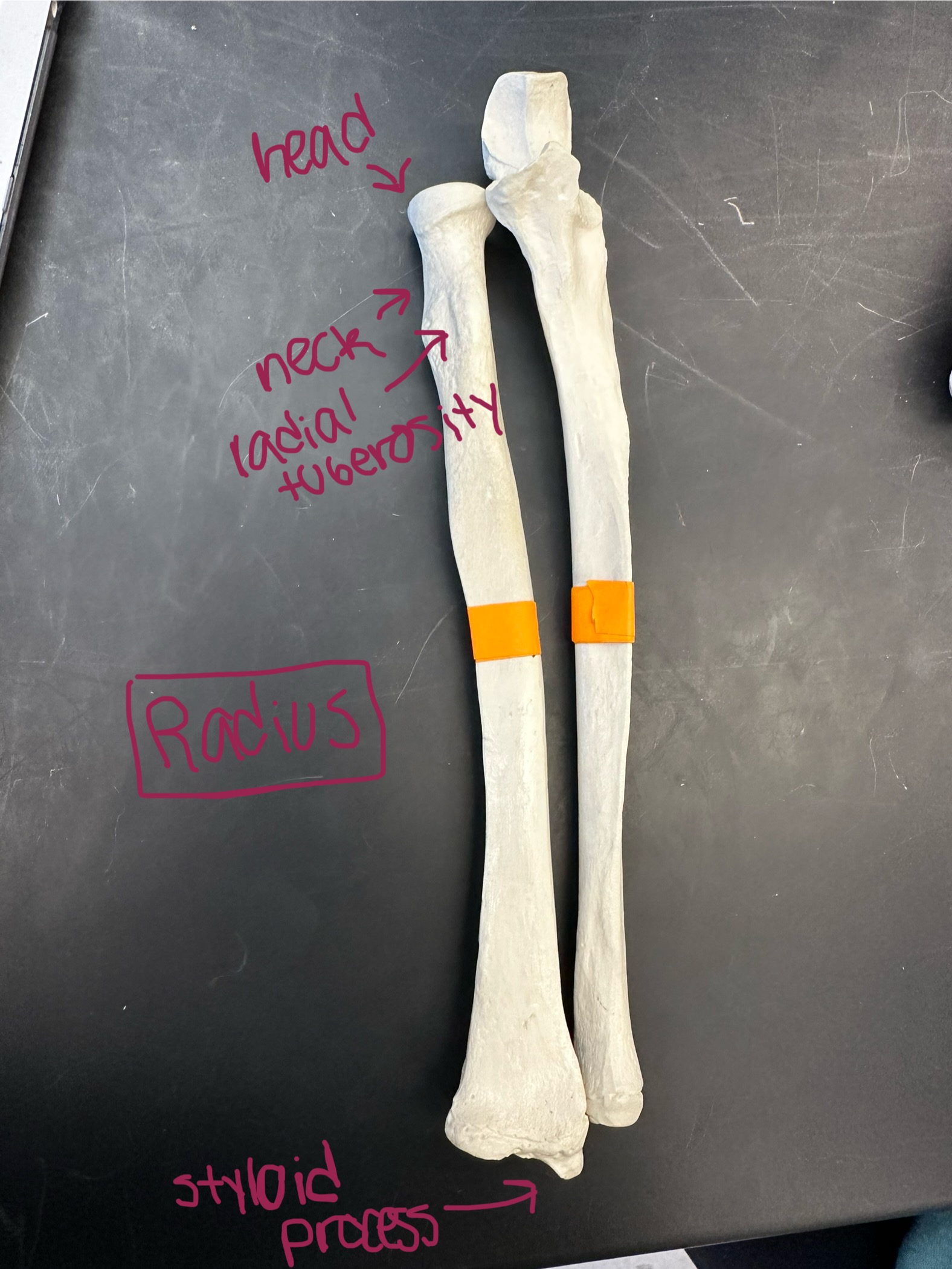
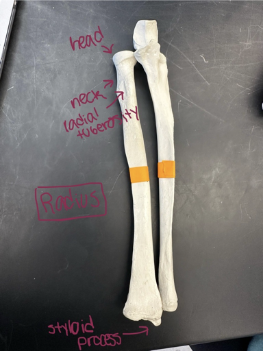
Radius (“lateral, thumb”) - articulates with ulna
Head - located on the proximal end of the radius bone near the elbow joint, articulates with humerus + ulna
Neck - part of the radius bone connecting the head to the shaft
Radial tuberosity - bony projection on the medial aspect of the radius
Styloid process - structure located at the distal end of the radius bone, pointed end indicates lateral border
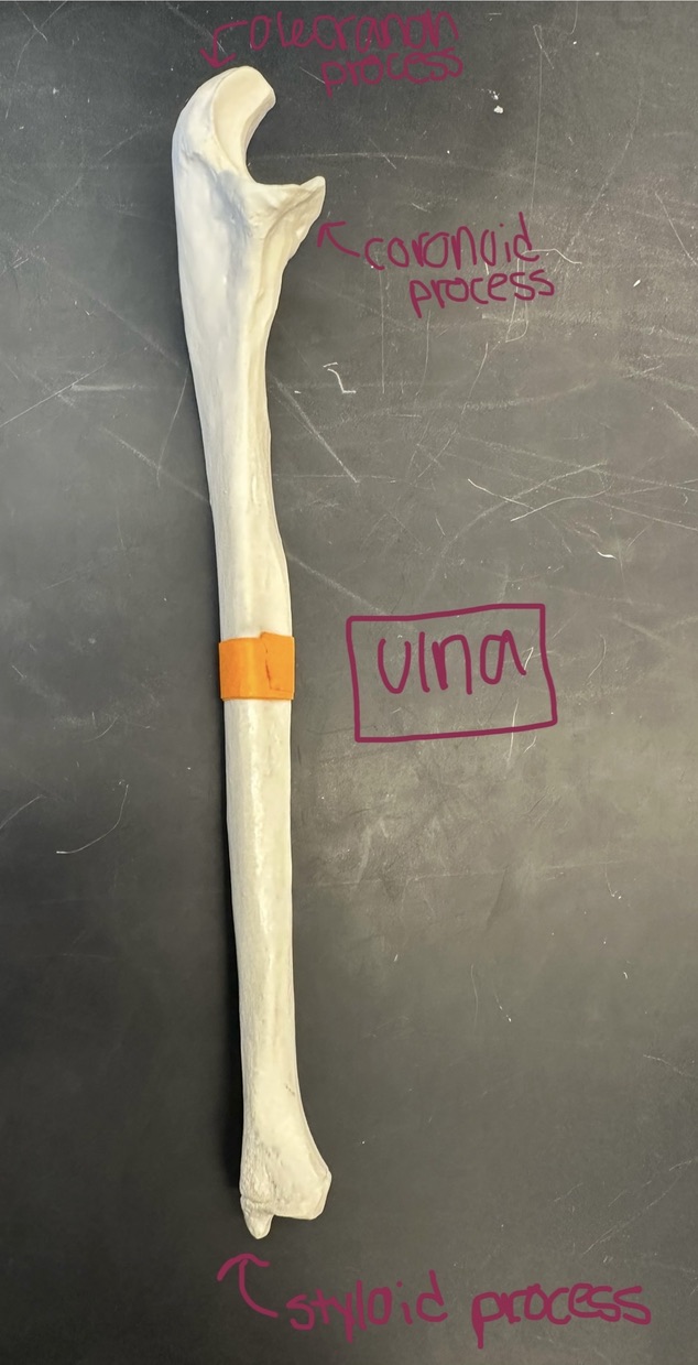
Ulna (“medial, pinky”, U-hook on the bone)
Olecranon process - bony projection on the top of the ulna and located on the posterior side
Coronoid process - a projection on the anterior surface of the ulna bone
Styloid process - bony projection at the distal end of the ulna
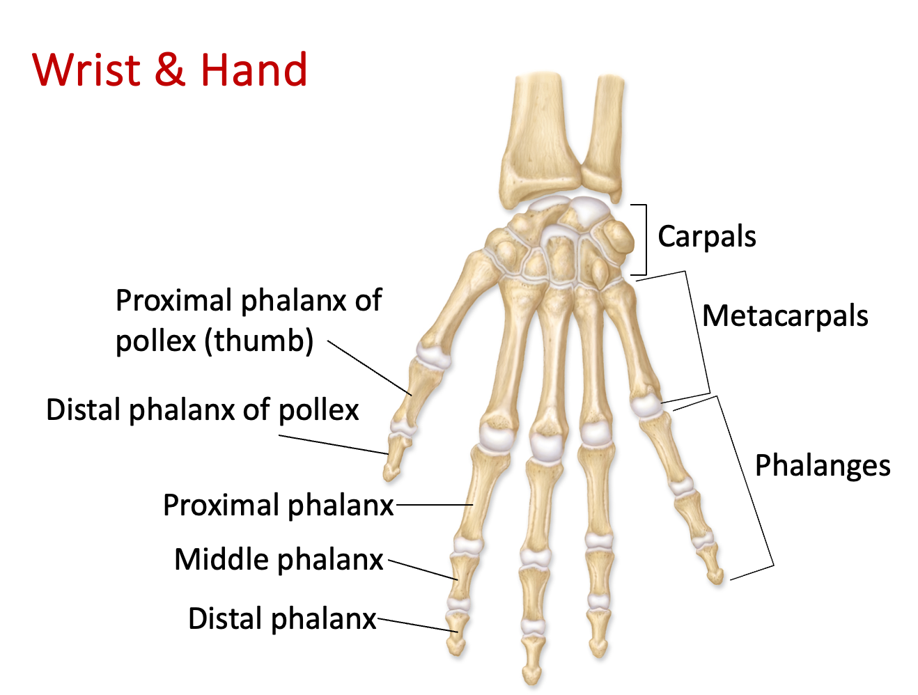
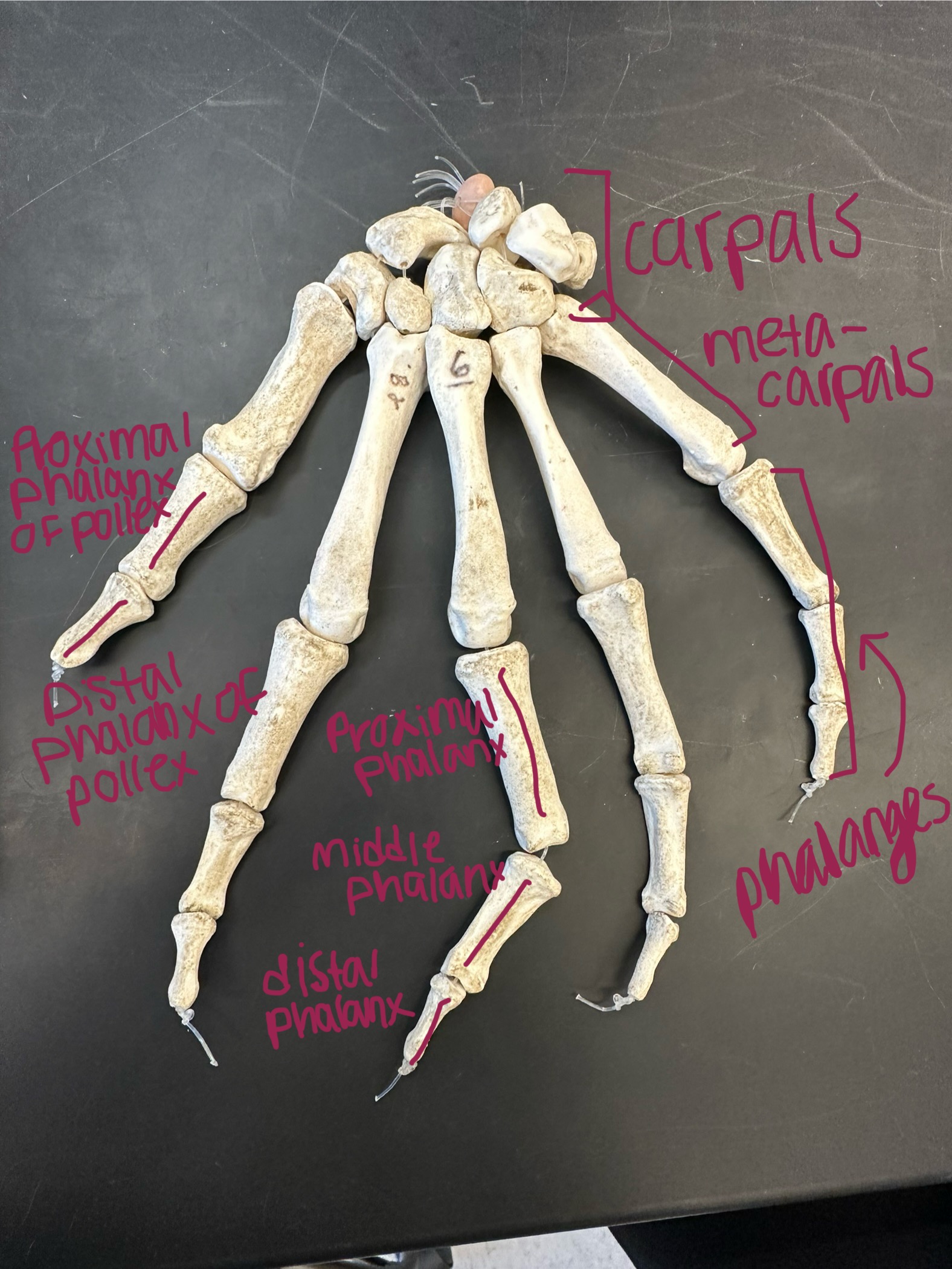
Carpals - 8 bones that make up the wrist in the upper extremity
Metacarpals - 5 long bones in the hand, located between the carpals and the phalanges
Phalanges - the bones in the fingers of the hand. Divided into three parts: proximal, middle, and distal phalanges.
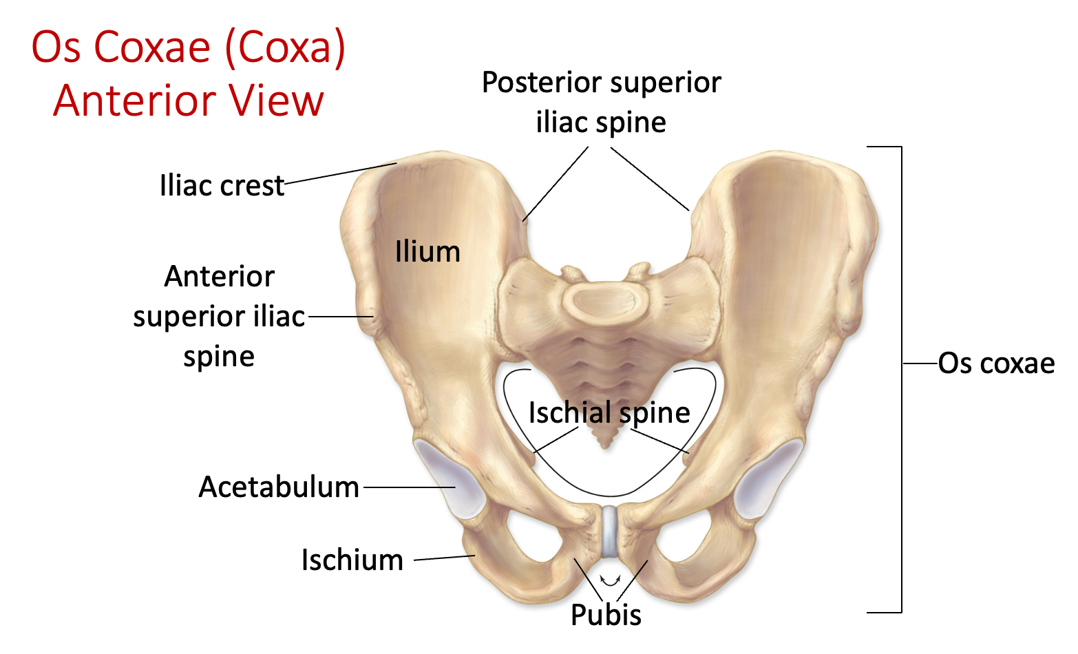
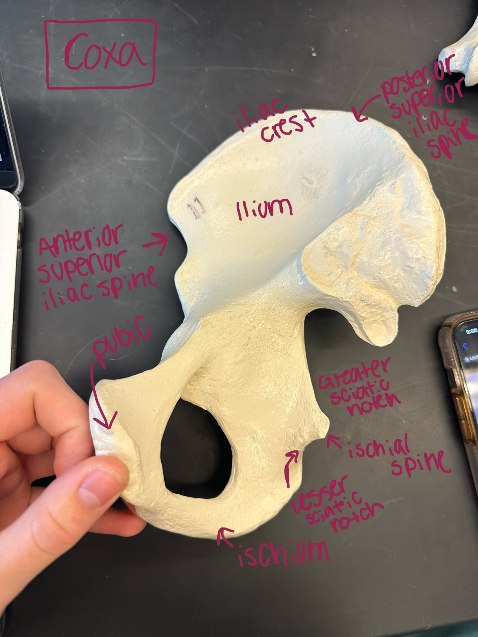
Pelvic girdle (male & female) - consists of acetabulum, ilium, ischium, iliac crest, and pubis bones
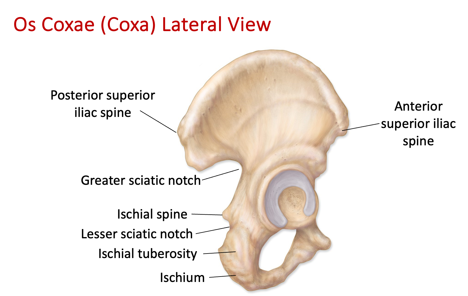
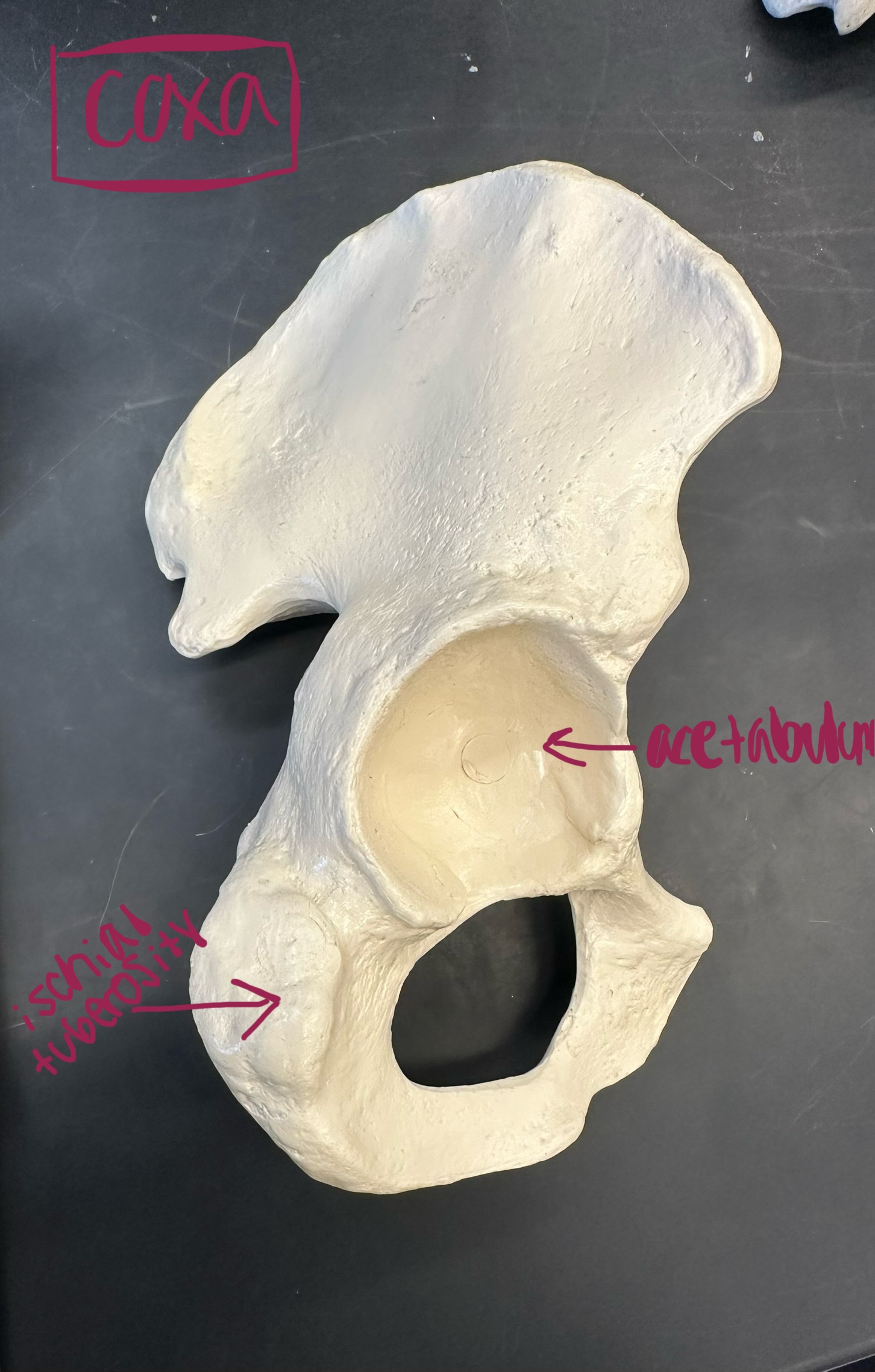
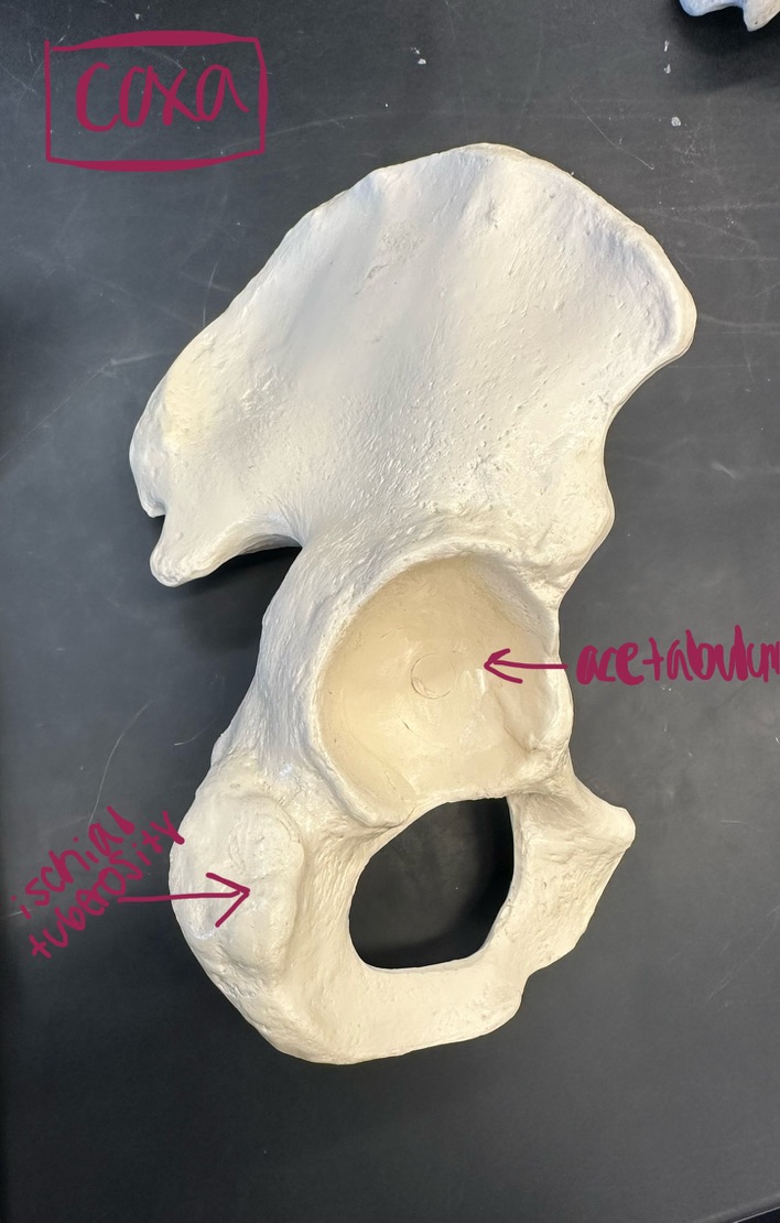
Coxa - the hip bone, consists of the acetabulum, ilium, ischium, iliac crest, and pubis bones, and forms the pelvic girdle and connects the lower limbs to the axial skeleton
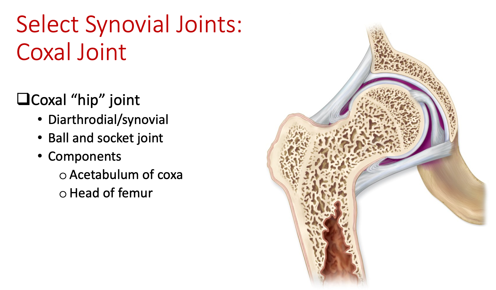
Acetabulum - creates the hip joint, depression on the lateral side of the coxa
Ilium - the uppermost and largest part of the pelvic girdle
Iliac crest - crest-like shape on the superior portion of the coxa, think of abdominal iliac region → hip
Anterior superior iliac spine - bony projection located at the anterior superior portion of the coxa, on the lateral side of coxa
Posterior superior iliac spine - bony landmark on the posterior aspect of the ilium
Greater sciatic notch - large opening in the pelvis, on the lateral side
Ischium - inferior and posterior part of the coxa
Ischial tuberosity - bony prominence on the inferior aspect of the coxa
Lesser sciatic notch - small notch on the posterior surface of the coxa, below the ischial spine
Ischial spine - bony projection on the ischium bone
Pubis - the front portion of the coxa, located below the ilium and ischium
*Anterior if pubis is facing front
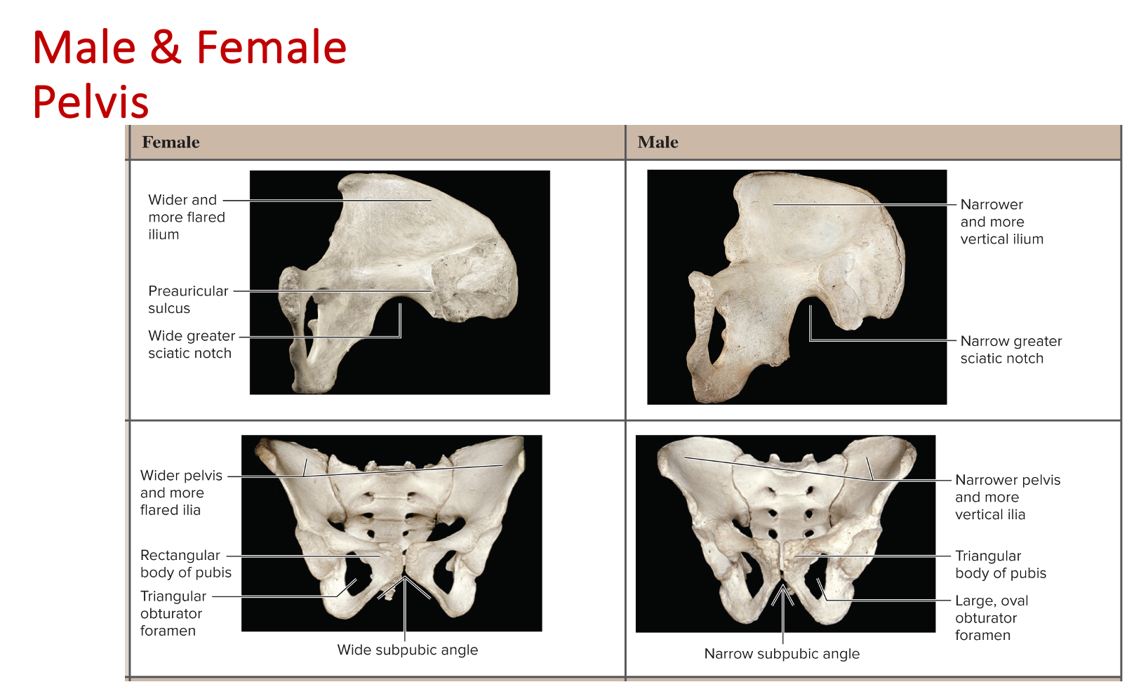 *Female = wider, shallower, and has a larger pelvic outlet
*Male = narrower, deeper, and has a smaller pelvic inlet
Lower extremity
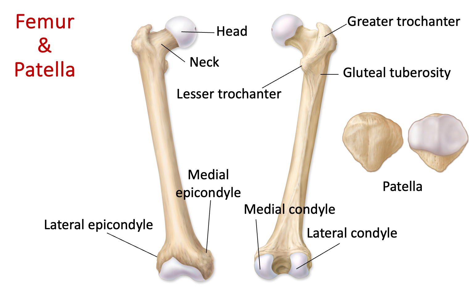
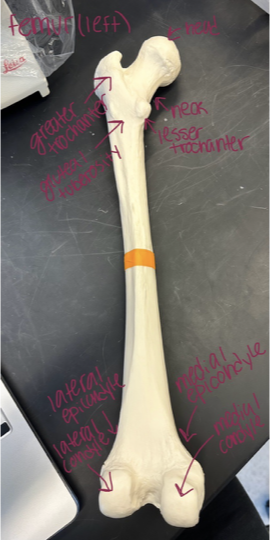
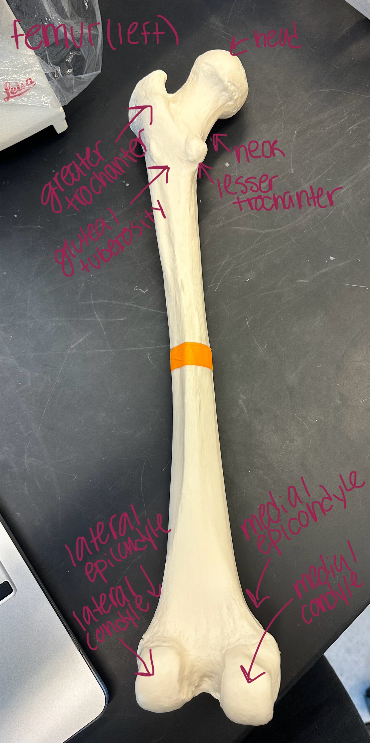
Femur (smooth is anterior, rough is posterior) - bone in the thigh, articulates with the hip bone at the hip joint and the tibia at the knee joint
Head - rounded upper end of the thigh bone that fits into the hip socket
Neck - connects the head of the femur to the shaft
Greater trochanter - bony prominence on the lateral side of the femur, sits superiorly to the lesser trochanter
Lesser trochanter - bony prominence on the medial side of the femur, sits inferiorly to the greater trochanter
Gluteal tuberosity - bony depression on the posterior side of the femur
Medial epicondyle - located at the distal, anterior, and medial end of the femur
Lateral epicondyle - located at the distal, anterior, and lateral end of the femur
Medial condyle - rounded projection at the medial, distal, and posterior end of the femur
Lateral condyle - rounded projection at the lateral, distal, and posterior end of the femur
Patella - sesamoid bone, located at the front of the knee joint
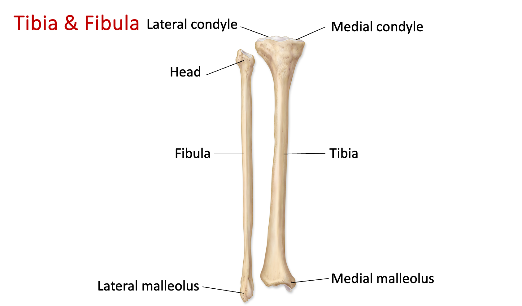
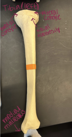
Tibia - the larger bone located in the lower leg, commonly known as the shinbone, connects the knee to the ankle
Medial tibial condyle - located on the medial side and superior region of the tibia
Lateral tibial condyle - located on the lateral side and superior region of the tibia
Tibial tuberosity - bony bump on the superior portion and anterior side of the tibia
Medial malleolus - bony process on the distal end and medial side of the femur
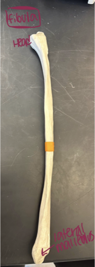
Fibula - bone in lower leg, lateral to tibia
Head - proximal end of the fibula bone that articulates with the tibia
Lateral malleolus - distal end of the fibula bone
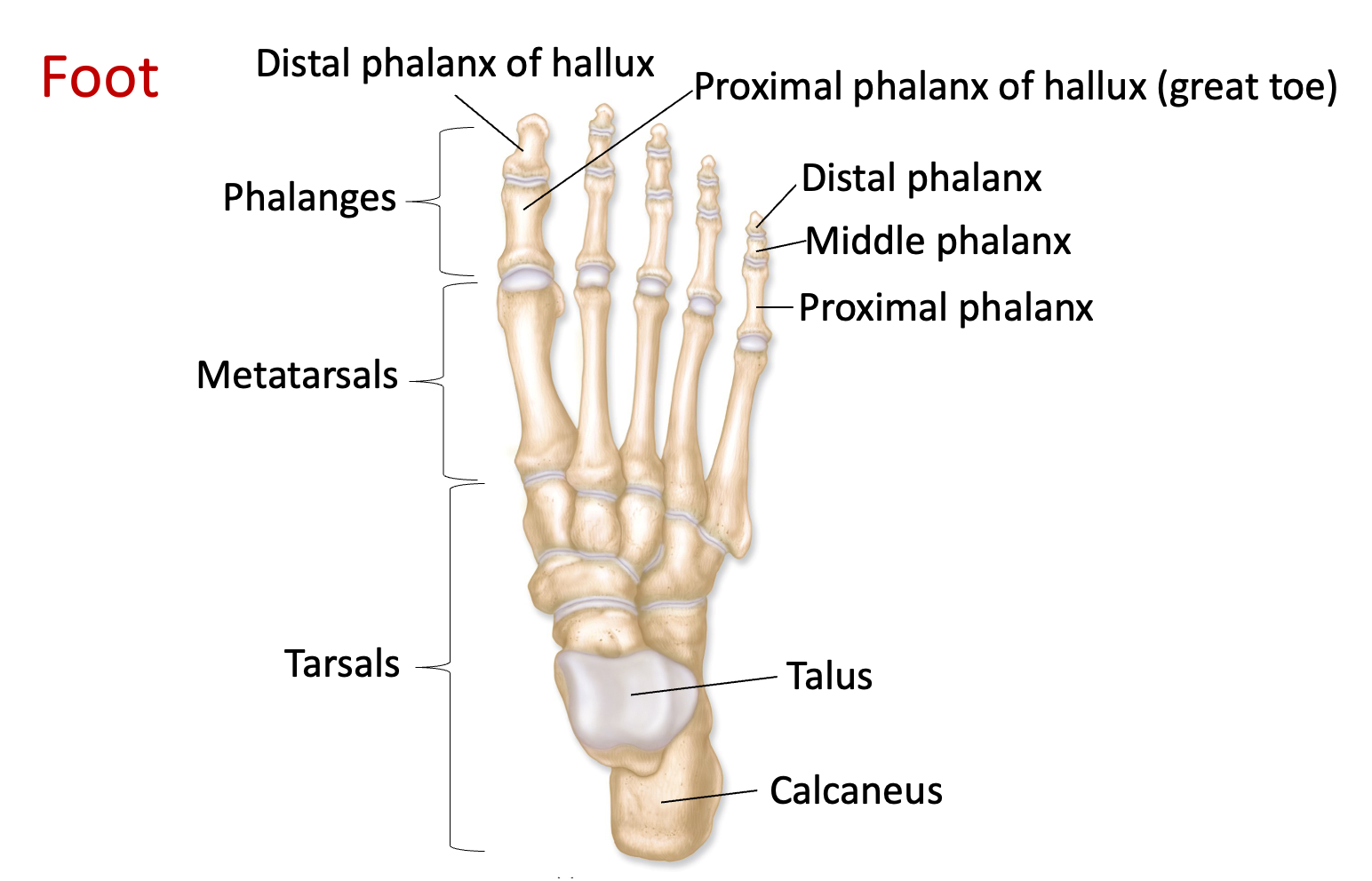
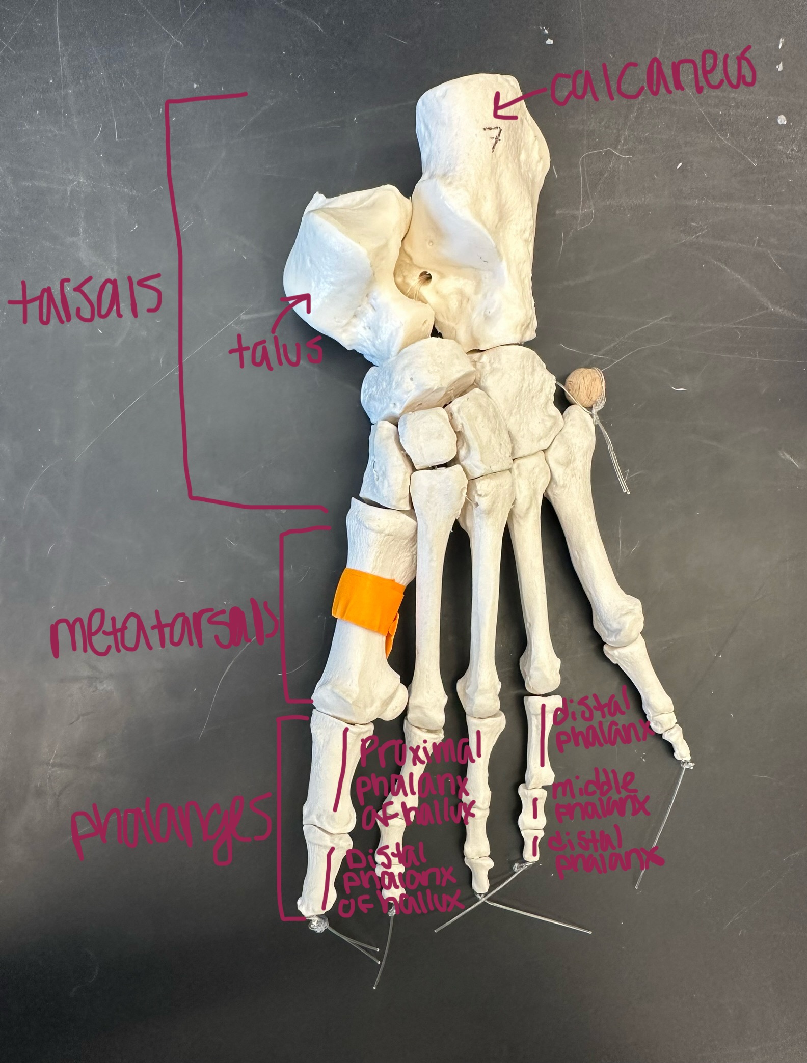
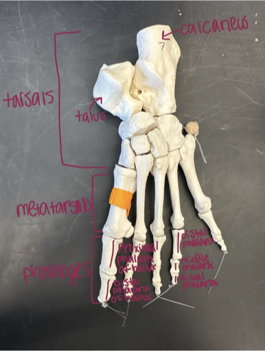
Tarsals - 7 bones in the ankle region of the foot, includes the calcaneus + talus
Calcaneus - largest tarsal bone located in the heel, forming the heel bone
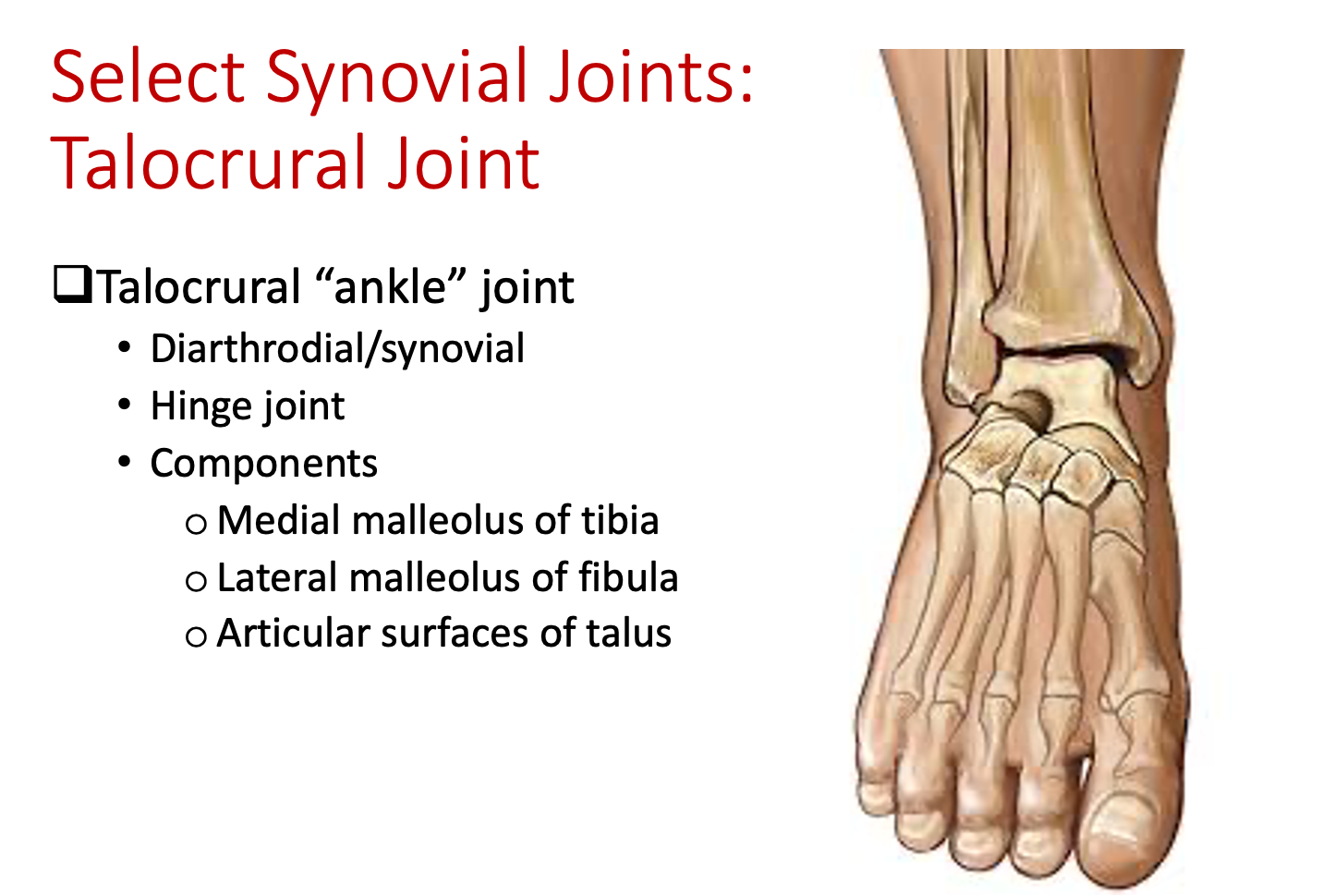
Talus - forms the ankle joint with the tibia and fibula
Metatarsals - 5 long bones in the foot
Phalanges - bones that make up the toes, divided into three parts: proximal, middle, and distal phalanges
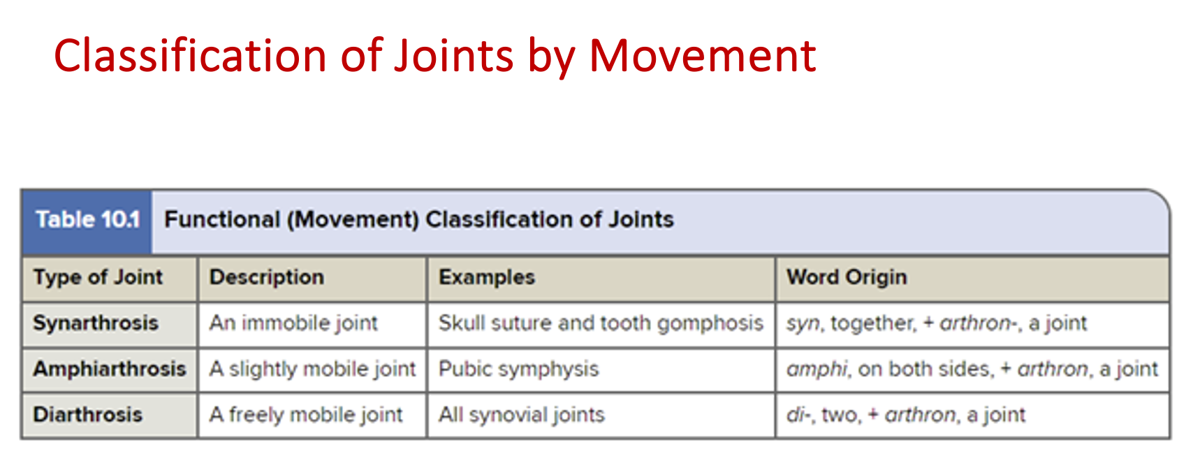
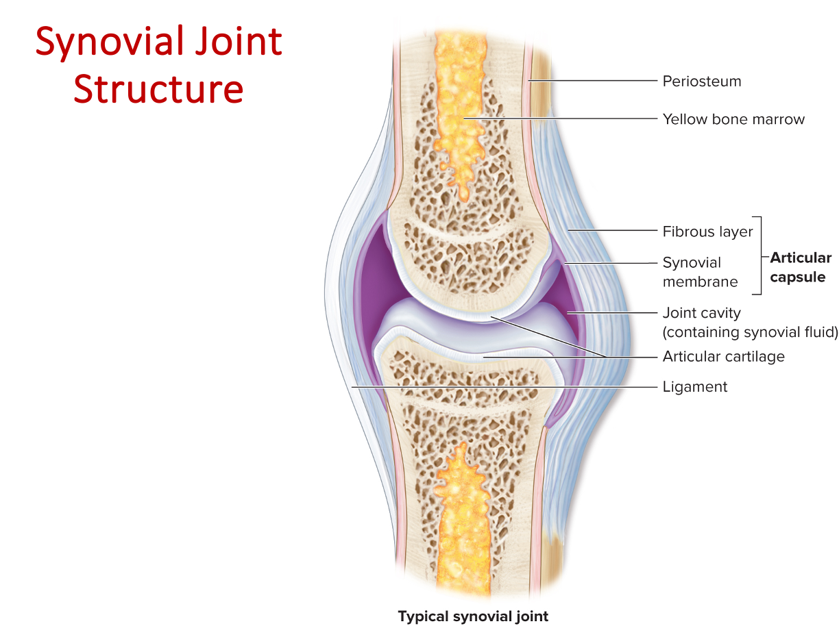
Joints classification
Functional classification
Synarthrosis
Amphiarthrosis
Diarthrosis
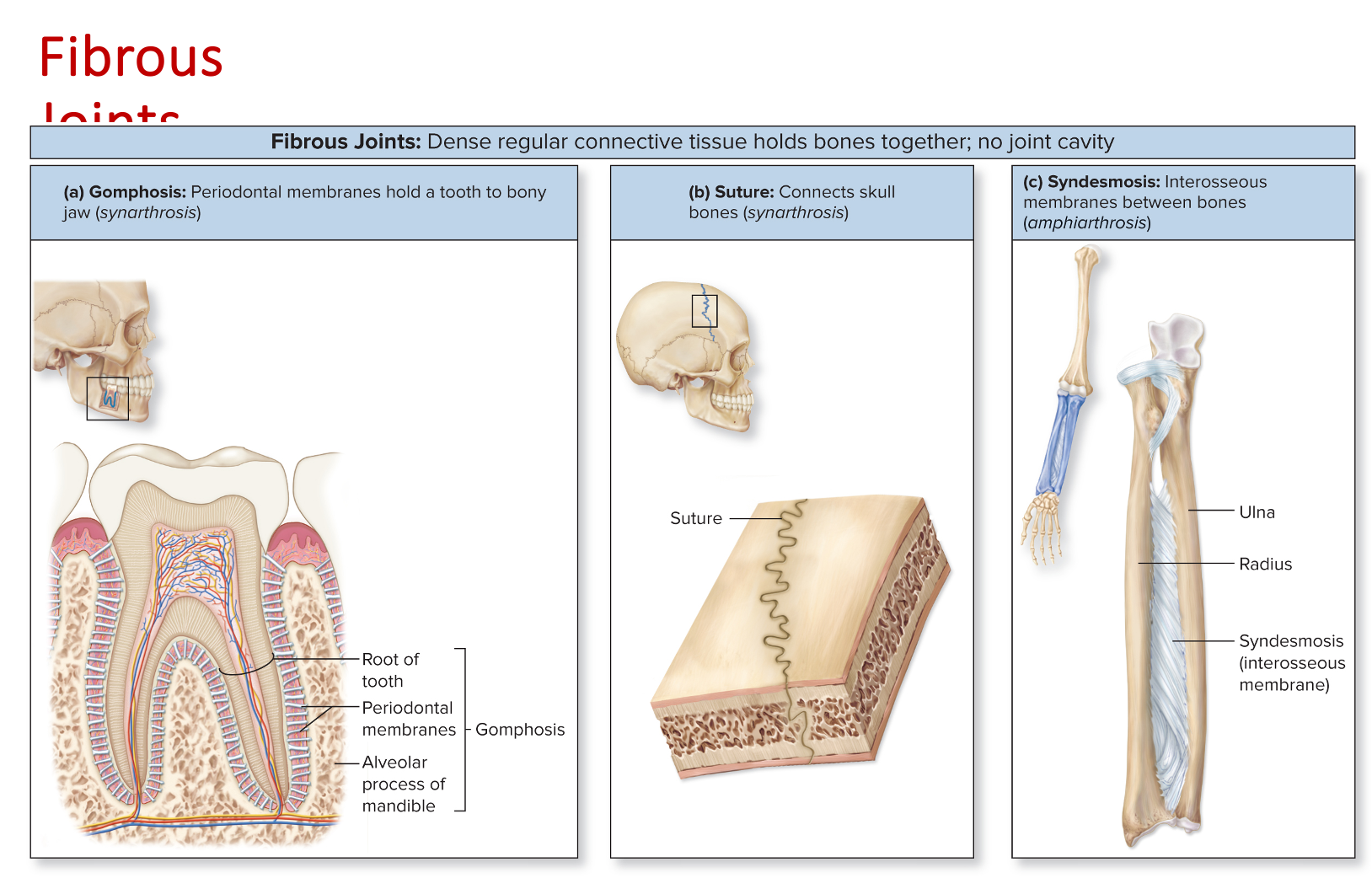
Structural classification
Fibrous
Gomphosis
Suture
Syndesmosis
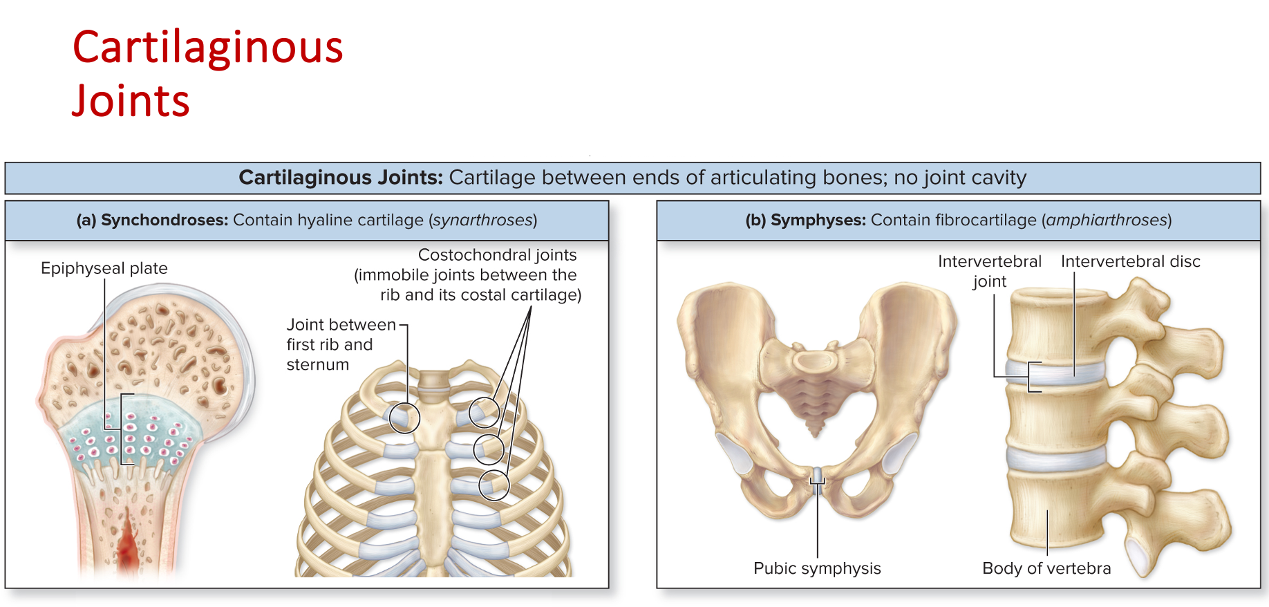
Cartilaginous
Synchondrosis
Symphysis
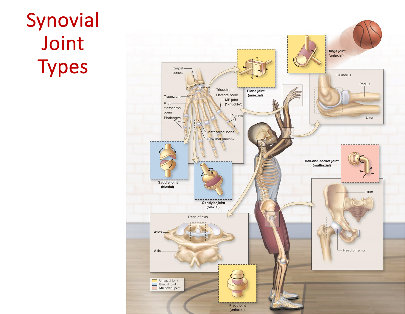
Synovial
Planar
Hinge
Pivot
Condylar
Saddle
Ball in socket
Specific joints
Glenohumeral joint
Humeroulnar joint
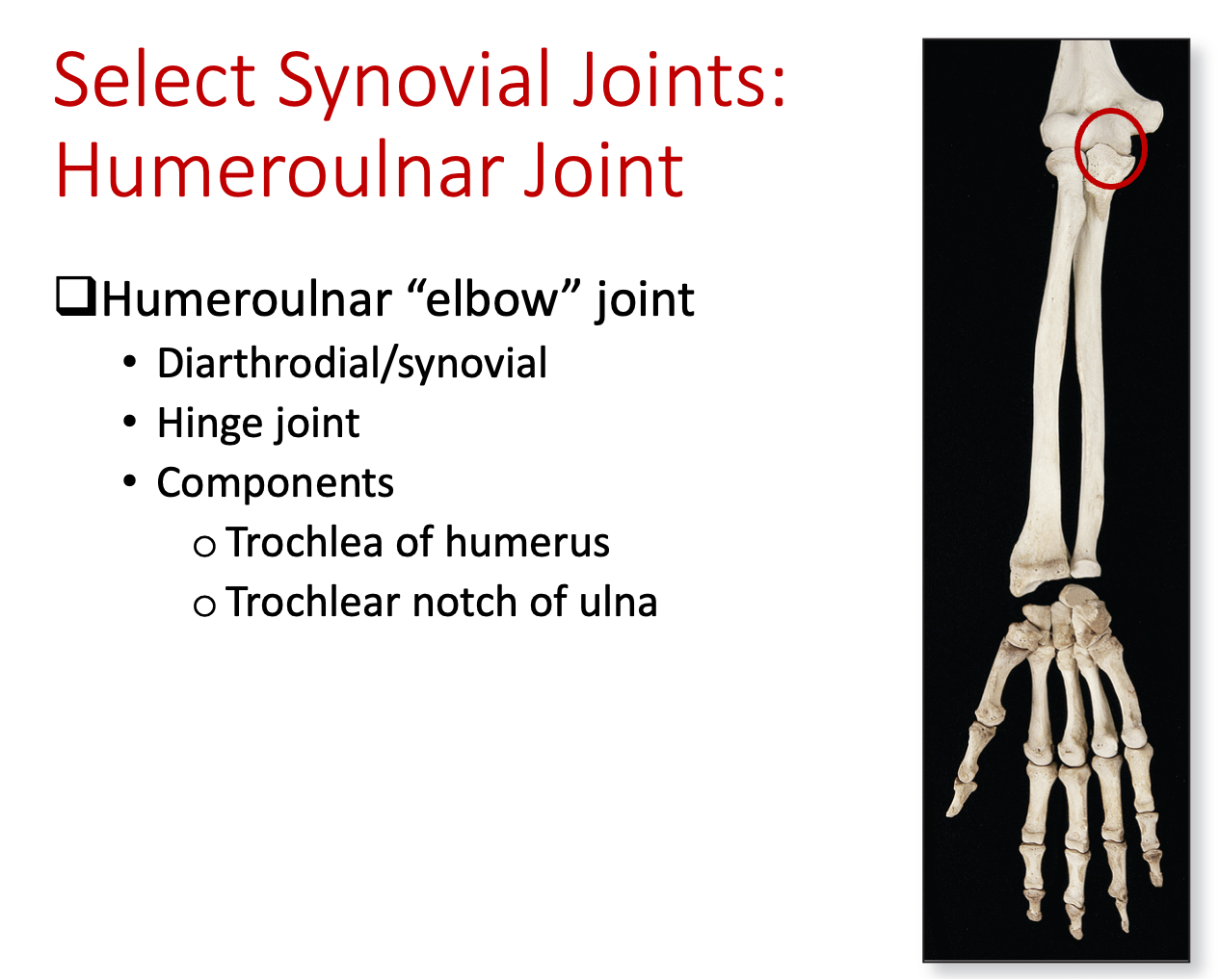
Humeroradial joint
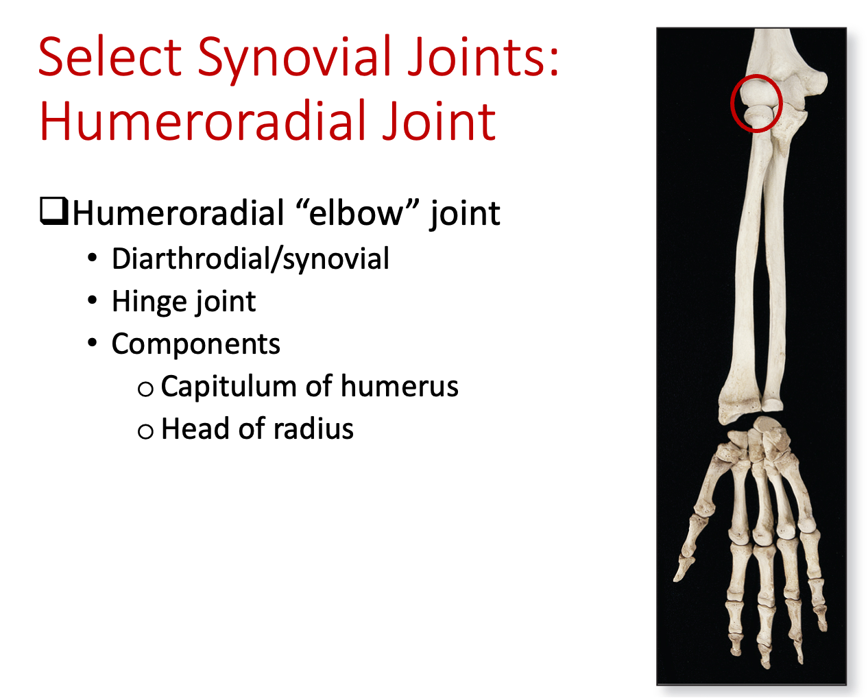
Proximal radioulnar joint
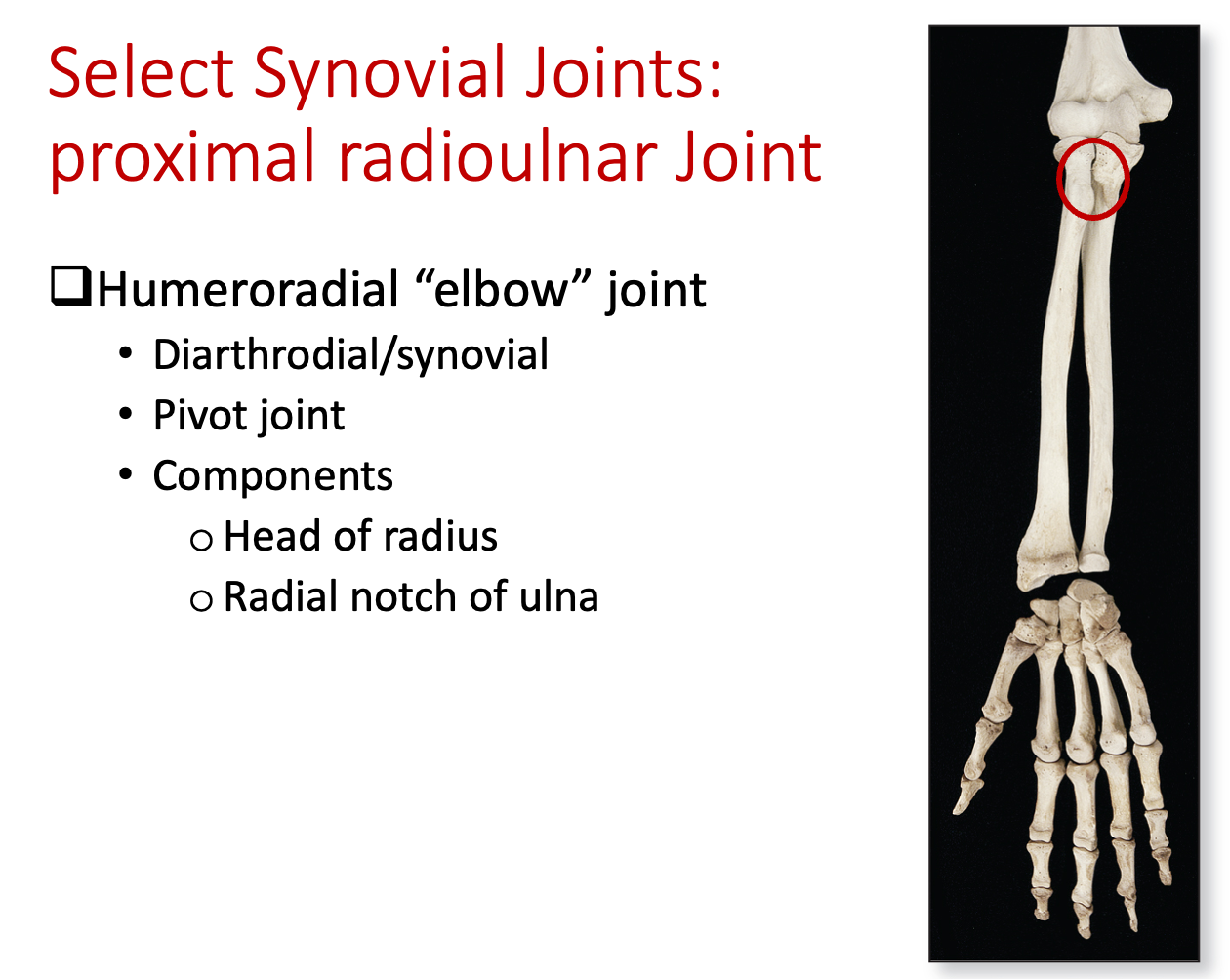
Intercarpal & intertarsal joints
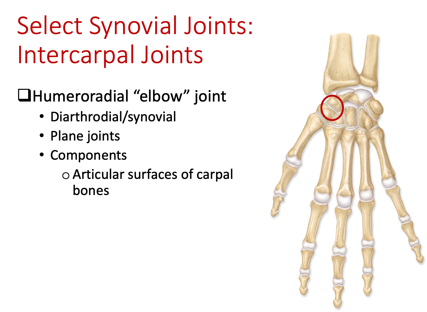
Coxal joint
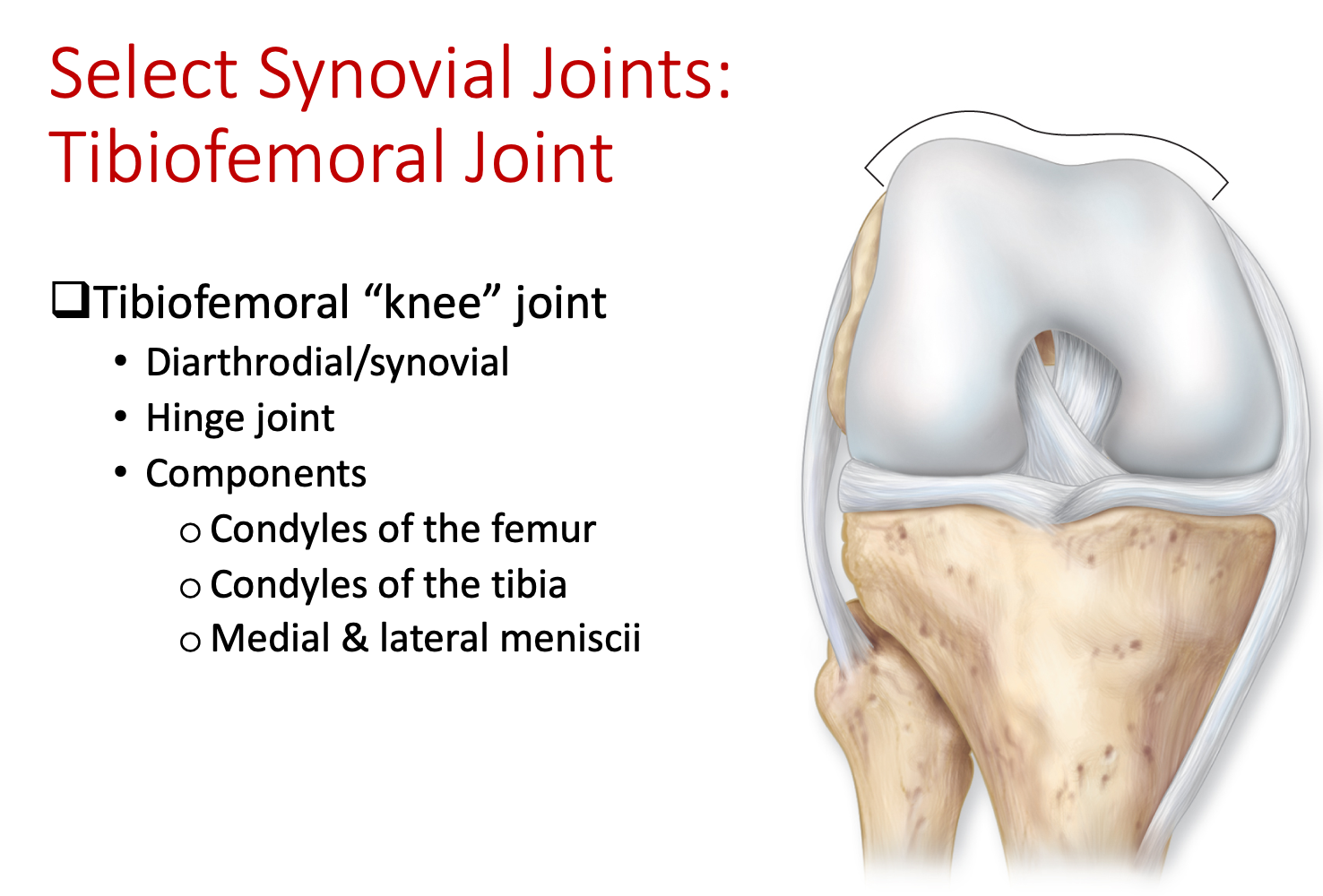
Tibiofemoral joint
Talocrural joint
Temporomandibular joint
*Questions about left or right bones will include these bones - humerus, scapula, femur, + coxa
Lab 6 (Muscle Tissue + Axial Muscles)
EXAM Objectives:
Structures and state functions for all histology slides ✅
Muscle tissue: skeletal, smooth, cardiac – identify and know characteristics ✅
Neuromuscular Junction: be able to identify muscle fibers, axons, axon terminals
Know basics of neuromuscular junction
Architecture of Skeletal Muscles
Skeletal muscles are different shapes due to fiber arrangement
Circular muscles - fibers form a circle
Convergent muscles - muscle is broad at origin, but converges to thin insertion
Parallel muscles - fibers are parallel, muscle is long and thin
Pennate muscles - have tendons that attach to fibers (unipennate, bipennate, multipennate)
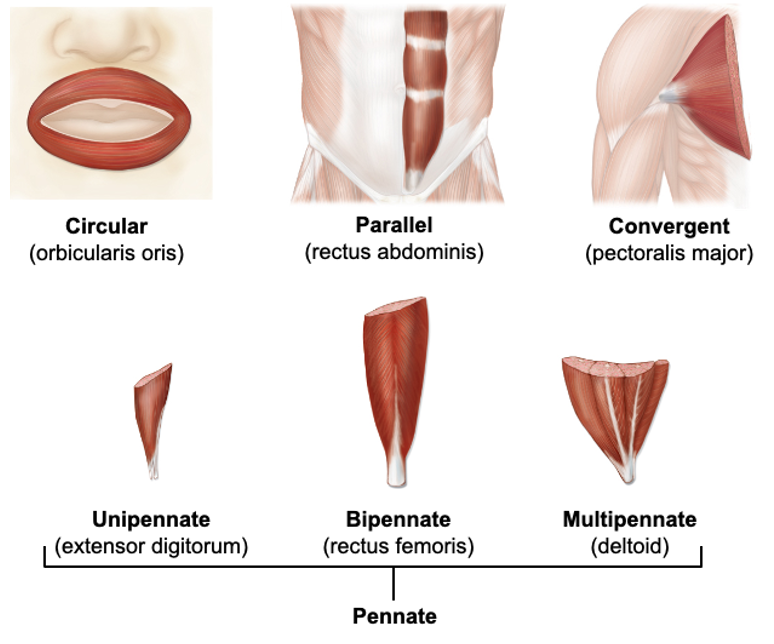 Naming of Skeletal Muscles
Skeletal muscles are commonly based on:
Shape/Size (rectus = straight)
Number of heads/bellies (biceps = two bellies)
Position/Depth (oris = mouth)
Action (levator = lifter)
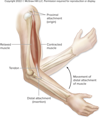
Insertion vs. Origin
Origin of a muscle is the less moveable end
Insertion of a muscle is the more moveable end
Example - the insertion of the biceps brachii is the radius, the origin is the scapula
Models: You should be able to identify the following structures on models on the lab benches and on the back bench. View lab manual for regions/tissues that each vessel supplies. Muscles marked with a “*” you need to know the origin, insertion and action for that muscle.
Muscles of the head, neck, and face
Sternocleidomastoid* - located in the neck
Origin - manubrium of sternum + clavicle
Insertion - mastoid process
Action - rotation, slight lateral flexion
Epicranius (frontal belly) - located on the forehead
Orbicularis oculi - surrounds the eye socket
Temporalis - located on temporal bone to mandible
Masseter - located in the jaw region
Orbicularis oris - located around the mouth
Buccinator - located in the cheeks
Zygomaticus (major and minor) - located in the face, zygomaticus major runs from the cheekbone to the corner of the mouth, zygomaticus minor extends from the cheekbone to the upper lip
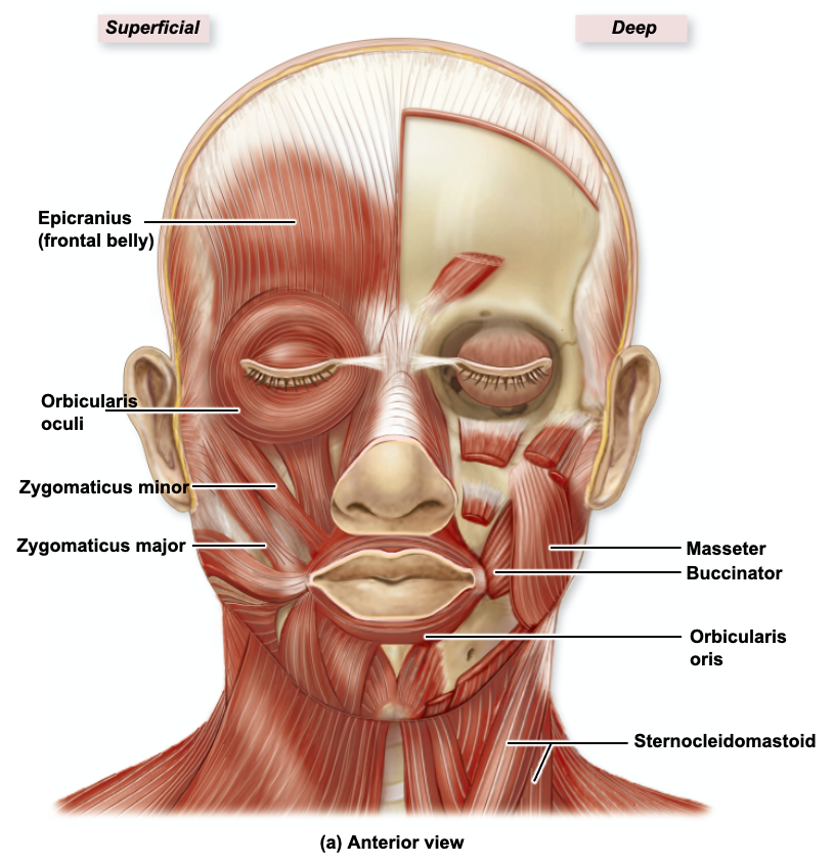
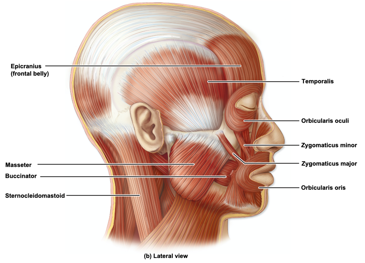 *Functions - head movement, mastication (chewing), facial expressions, and speech
Muscles of the trunk
Pectoralis major* - superficial, located on chest
Origin - costal cartilages of ribs 2-6; body of sternum
Insertion - humerus
Action - flexes arm, adducts + medially rotates arm
Pectoralis minor (deep to major)
Trapezius* - large, triangular muscle that extends from the occipital bone to the lower thoracic vertebrae
Origin - Clavicle; acromion process + spine of scapula
Insertion - occipital bone, ligamentum nuchae, spinous presses of C7-T12
Action - elevates scapula, rotates scapula superiorly, retracts scapula, depresses scapula
Latissimus dorsi* - large back muscle, extends from lower spine to upper arm
Origin - spinous processes of T7-T12; ribs 8-12; iliac crest; thoracolumbar fascia
Insertion - intertubercular sulcus of humerus
Action - extends arm, adducts arm, medially rotates arm
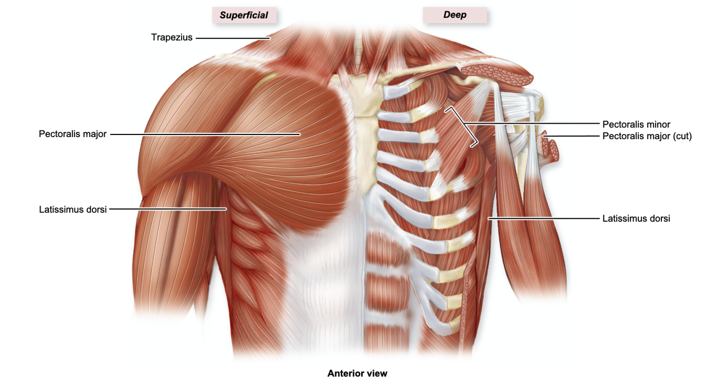 *Muscles of the chest and back - torso movement, but some help with arm movement
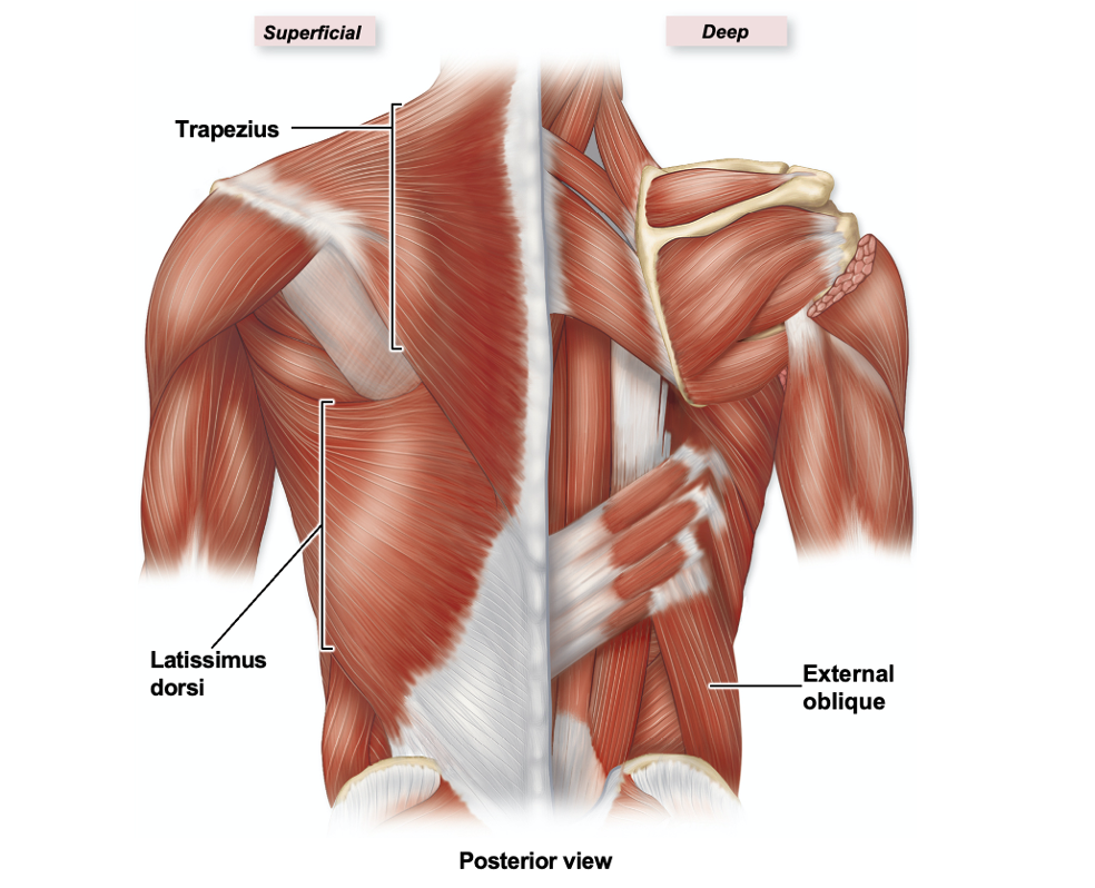
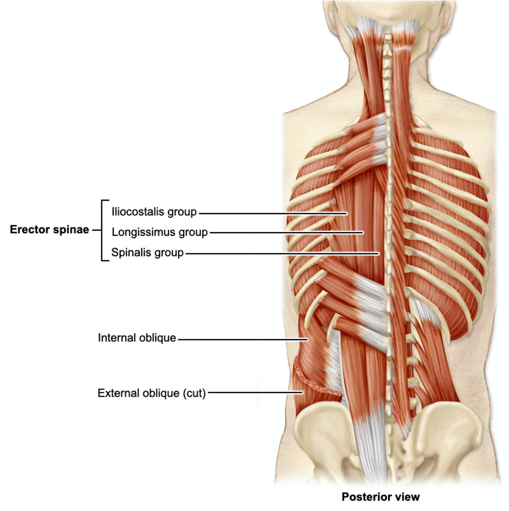 *Muscles of the back - torso movement, vertebral column movement
Rectus abdominis* (straight) - muscle located in the abdomen, running vertically from the pubic bone to the ribcage
Origin - pubic symphysis + crest
Insertion - xiphoid process + costal cartilages of ribs 5-7
Action - flexes trunk, compresses abdominal viscera, used in forced expiration
External and Internal obliques - muscles on the sides of the trunk, external is superficial to deep
Transversus abdominis (transverse cut) - deepest abdominal muscle, runs along transverse plane
Erector spinae - group of back muscles that run along the spine
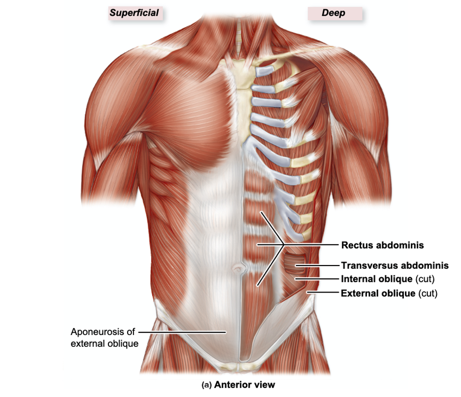
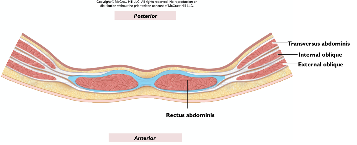 *Abdominal wall muscles - form several layers of muscle that contribute to abdominal movement + structure
*Superficial → Deep: External obliques, rectus abdominis (medial) and internal obliques (lateral), transversus abdominis
External and internal (between) intercostals - muscles between ribs, external intercostals are superficial to internal intercostals
Diaphragm - dome-shaped muscle located at the base of the ribcage, separating the thoracic and abdominal cavities
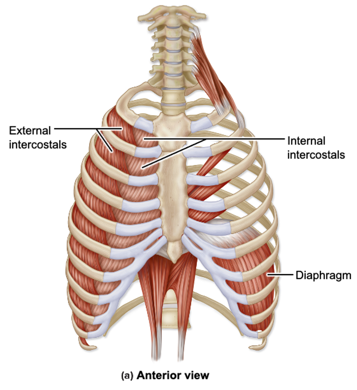 *Muscles of respiration - breathing in and out by changing pressure in the thorax
Histology: You should be able to identify the following structures on an image or microscope slide.
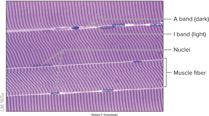
Skeletal Muscle(voluntary, striated, multinucleate)
Nuclei
Striations
Transverse vs. longitudinal section of muscle fiber
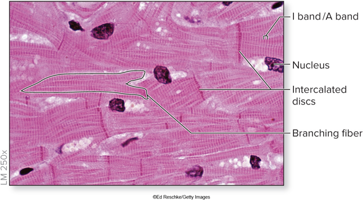
Cardiac Muscle(involuntary, striated, single nucleus)
Nucleus
Striations
Intercalated discs - allow muscle cells to contract so that heart can pump blood
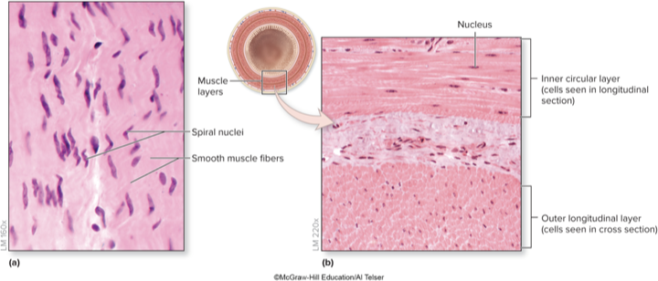
Smooth Muscle(involuntary, non-striated, single nucleus)
Nucleus
Longitudinal vs. cross-section
Lab 7 (Appendicular Muscles)
EXAM Objectives:
Identify muscles on Arm, leg & torso on the human body identifying these on pictures of models - Identify muscles, know action, origin & insertion: see muscle table in D2L ✅
Models: You should be able to identify the following structures on models on the lab benches and on the back bench. View lab manual for insertions and origins.
Note: for muscles marked with a “*” you need to know the origin, insertion and action for that muscle.
Muscles of the Shoulder, Arm, and Hand
Scapulohumeral Muscles
Originate on the scapula and insert at the humerus
They move + stabilize the shoulder joint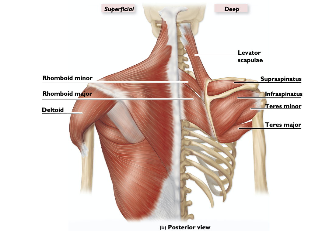
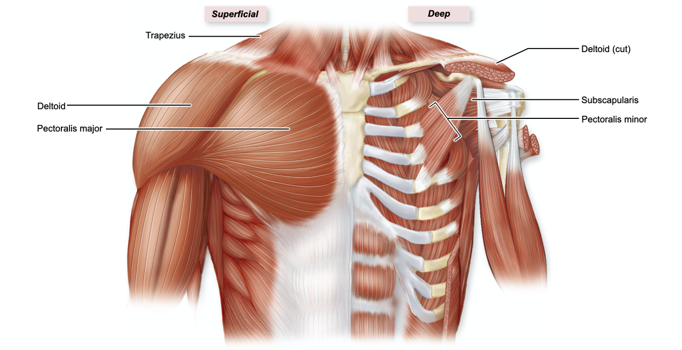
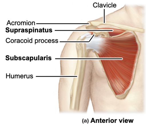
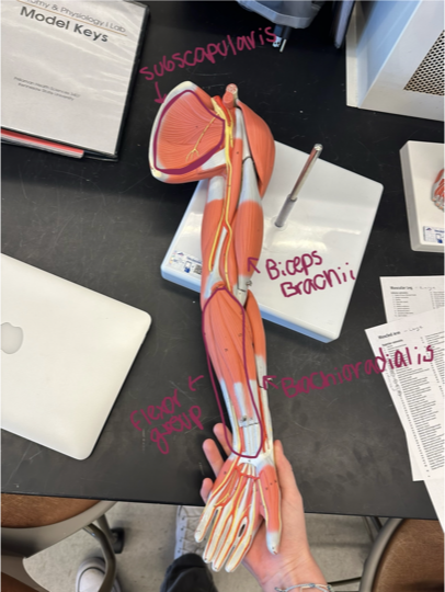
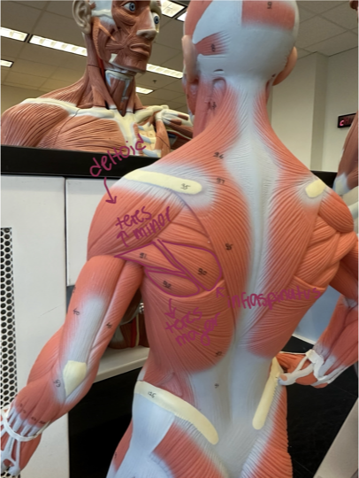
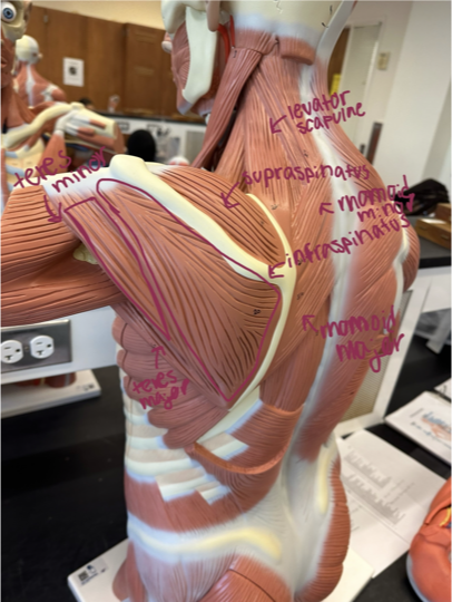
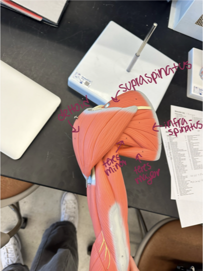
*SITS muscles make up the rotator cuff
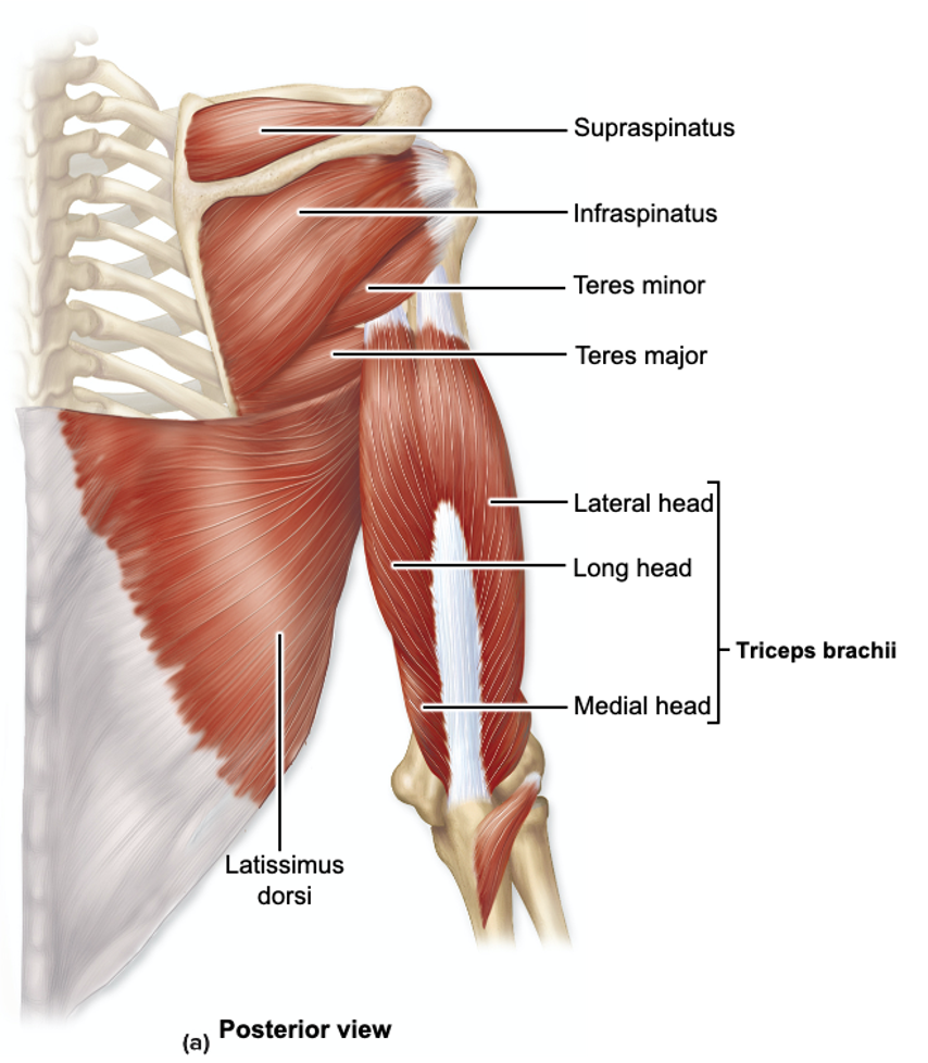
Supraspinatus (lies in supraspinatus fossa, above spine)
Infraspinatus (below supraspinatus)
Teres (major and minor) - located on the back, Teres Major attaches to the scapula, Teres Minor attaches to the humerus
Subscapularis (under scapula)
Rhomboids (major and minor) - large torso model ONLY, major = bigger, minor = smaller, originates on the spine, inserts on the medial border of the scapula
Levator scapulae - originates at the transverse processes of the first four cervical vertebrae, inserts into the superior part of the medial border of the scapula
Deltoid - looks like a triangle, covers shoulders
Anterior of Arm (Flexors)
Flexors - on the anterior side of the arm
Function to flex the arm at the elbow joint + wrist joint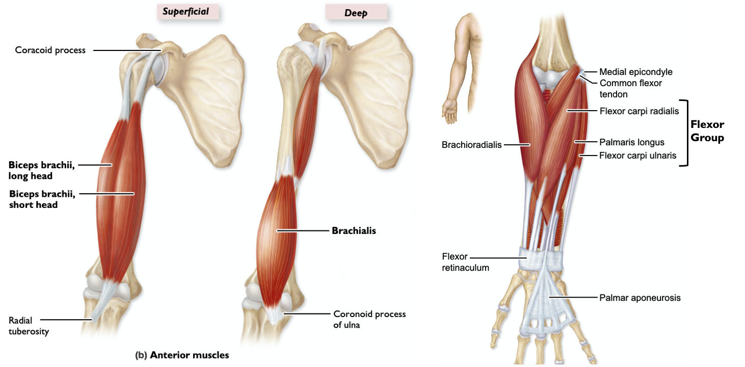
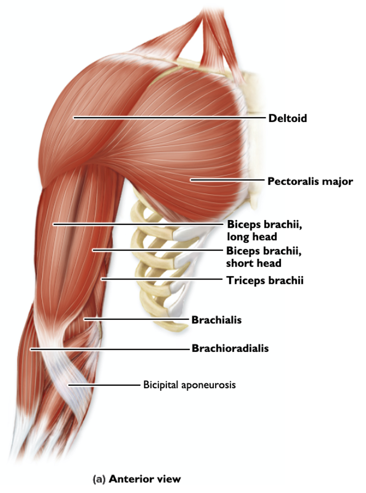
Biceps brachii* (thumb side up, anterior)
Origin - coracoid process of scapula
Insertion - radial tuberosity
Action - flexion of elbow
Brachioradialis - located in the forearm, runs from the lower arm to the wrist, positioned on the thumb + lateral side of the forearm
Brachialis* - located in the upper arm, lies underneath the biceps brachii, originates from the lower half of the humerus
Origin - humerus
Insertion - tuberosity and coronoid process of ulna
Action - flexes forearm (agonist)
Flexor group (anterior compartment of lower arm)
Posterior of Arm (Extensors)
Extensors - are on the posterior side of the arm
Function to extend the arm at the elbow and wrist joints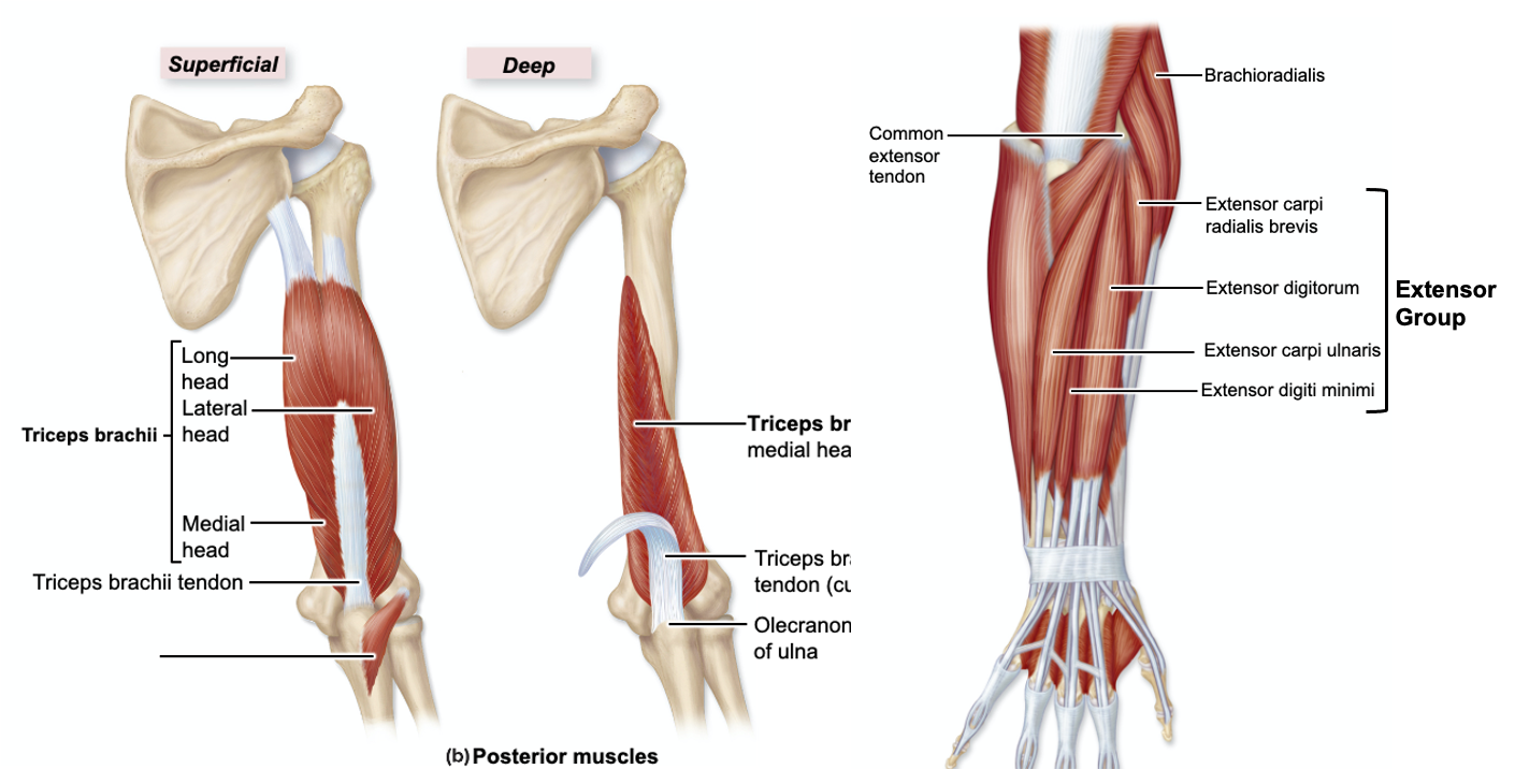
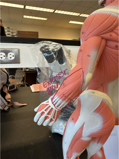
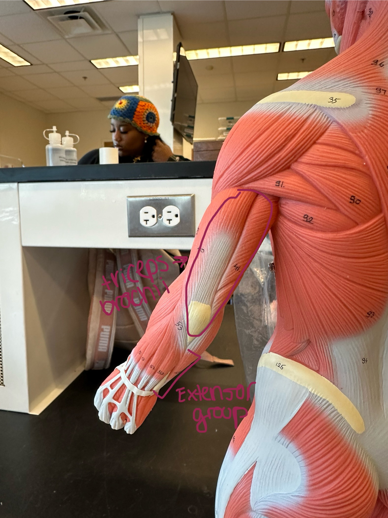
Extensor group (posterior compartment of lower arm)
Triceps brachii* (pinky side, posterior)
Origin - glenoid cavity and humerus
Insertion - olecranon of ulna
Action - extension of elbow
Muscles of the Pelvis, Leg, and Foot
Muscle of the Gluteal Region
Located on the posterior side of pelvis/upper leg
They primarily provide movement at the hip joint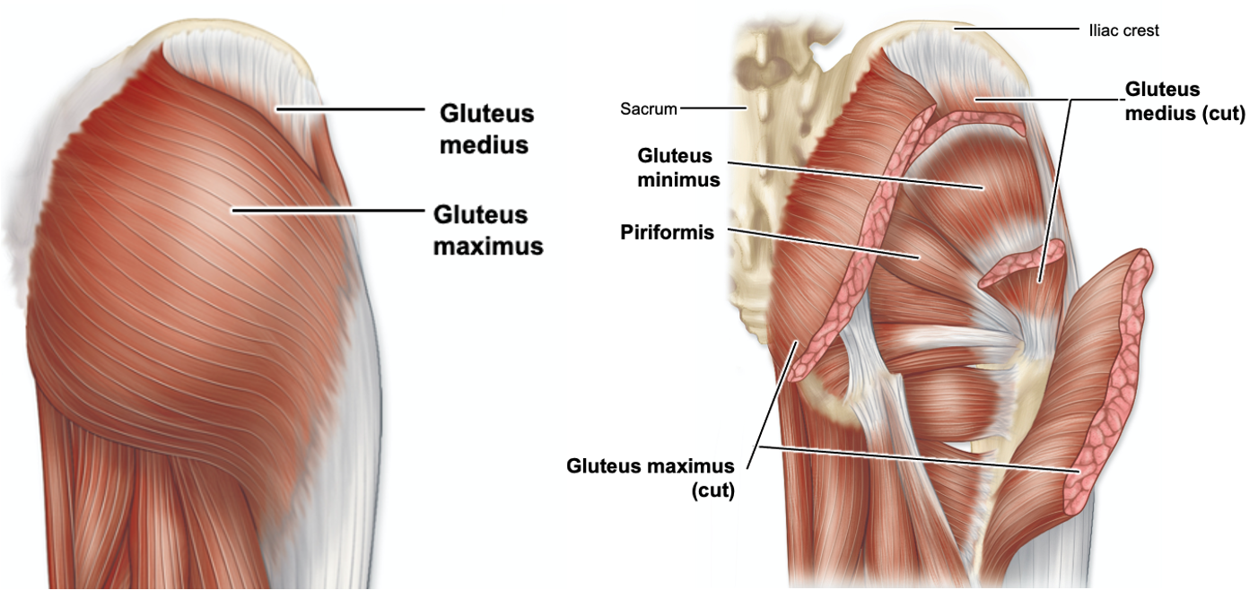
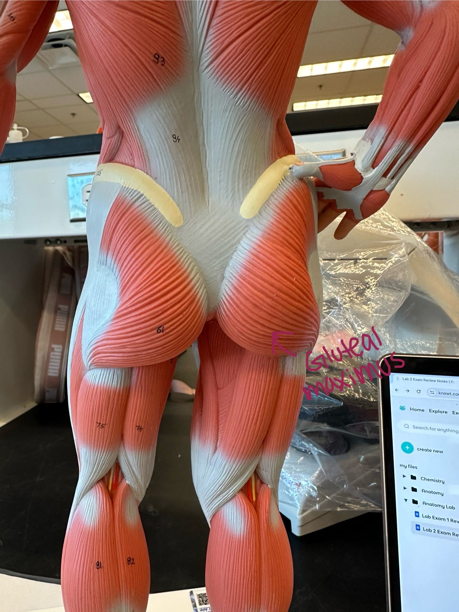
Gluteus maximus - muscle located in the buttocks, largest of the three gluteal muscles
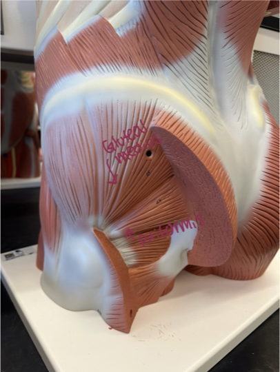
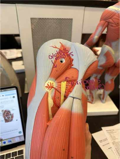
Gluteus medius - located on the outer surface of the pelvis, between the iliac crest and the greater trochanter of the femur
Piriformis (“Pinch - Piriformis”) - right above a nerve
Hamstring Muscle Group
Set of muscles on the posterior side of the leg that help flex the leg at the knee joint
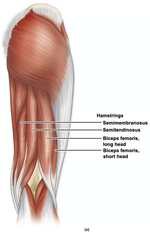
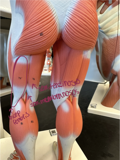
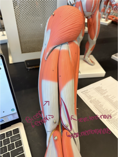
Biceps femoris* - most lateral
Origin - ischial tuberosity + femur
Insertion - head of fibula
Action - flexes the knee
Semitendinosus* - most medial
Origin - ischial tuberosity
Insertion - tibia
Action - extends thigh, flexes leg, medially rotates leg
Semimembranosus* - deep to semitendinosus
Origin - ischial tuberosity
Insertion - tibia
Action - extends thigh, flexes leg, medially rotates leg
Muscles of the Anterior Thigh
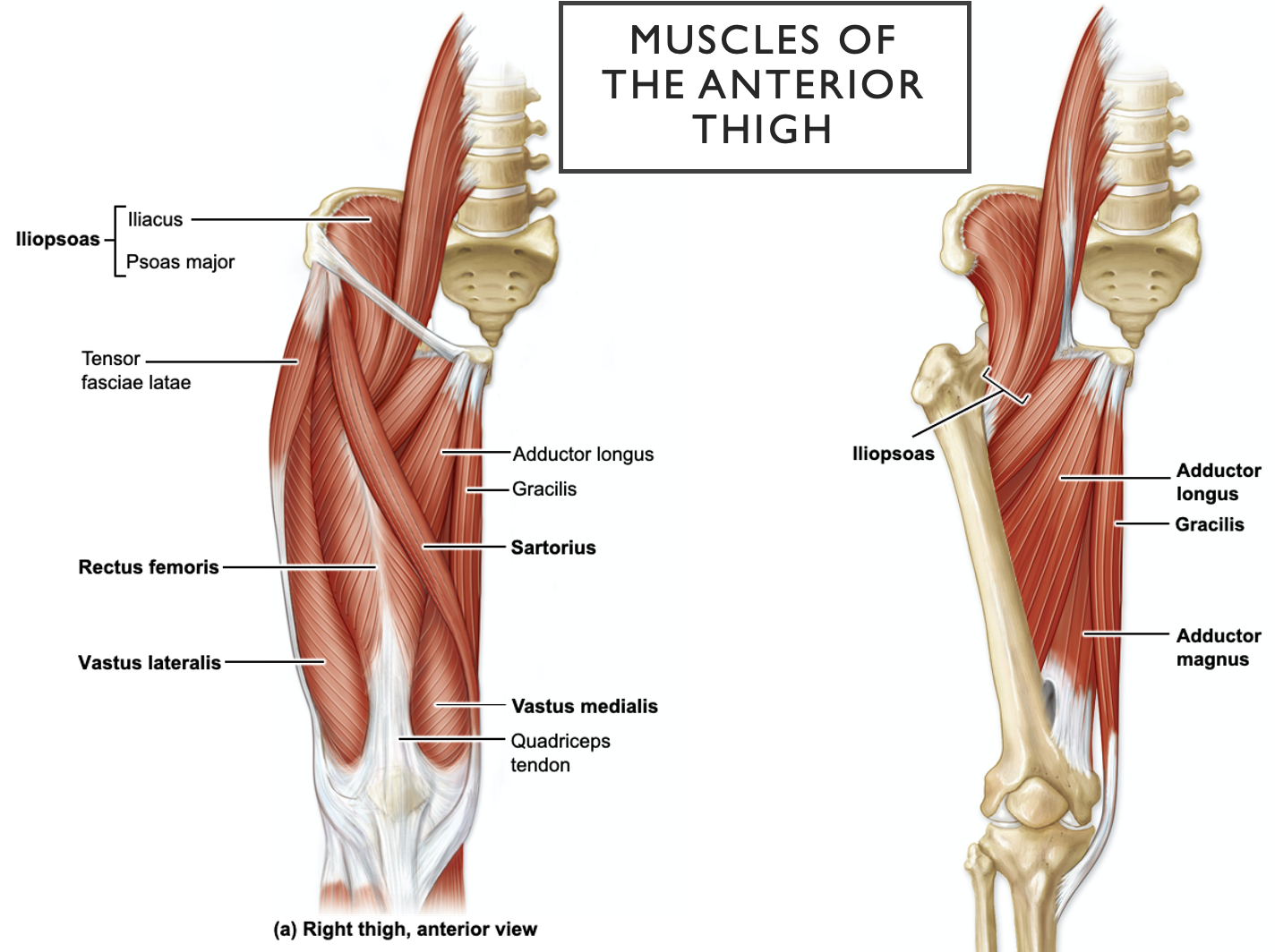
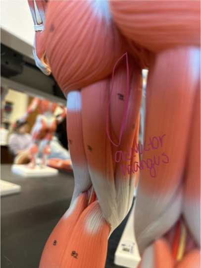
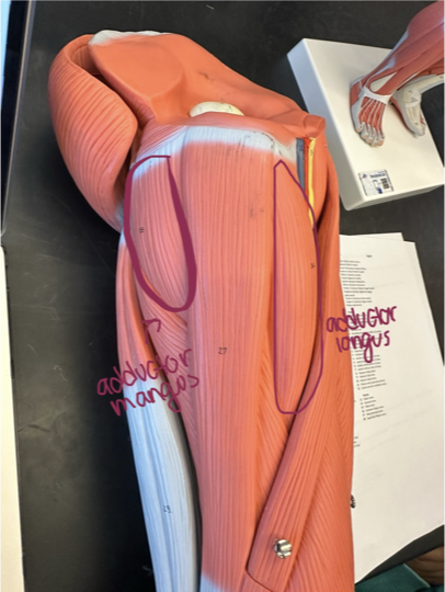
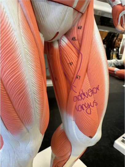
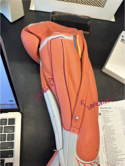
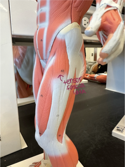
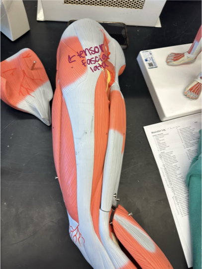
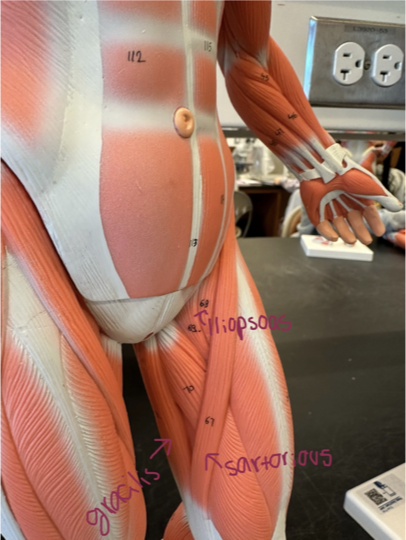
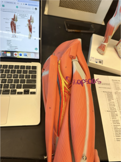
Iliopsoas - located in the anterior hip region
Sartorius - longest thin muscle, medial → lateral
Gracilis - located in the inner thighs
Adductors
Adductor longus - located above sartorius
Adductor magnus - located posteriorly to adductor longus
Tensor fascia lata - located in the outer hip region
Quadriceps Femoris Group
Quadriceps - set of 4 muscles on the anterior side of the upper leg that extends the leg at the knee joint
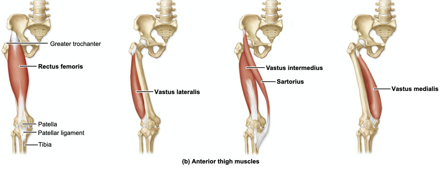
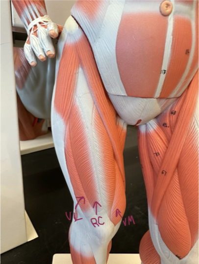
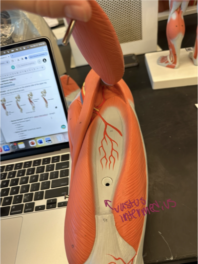
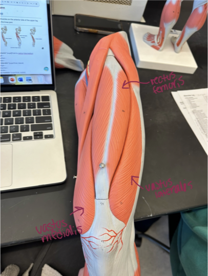
Rectus femoris* (superficial to vastus intermedius) - straight muscle on femoral region
Origin - ilium
Insertion - tibia
Action - flexes thigh + extends leg
Vastus lateralis* (superficial, lateral)
Origin - greater trochanter
Insertion - tibia
Action - extends leg
Vastus medialis* (superficial, medial)
Origin - femur
Insertion - tibia
Action - extends leg
Vastus Intermedius (deep to rectus femoris)
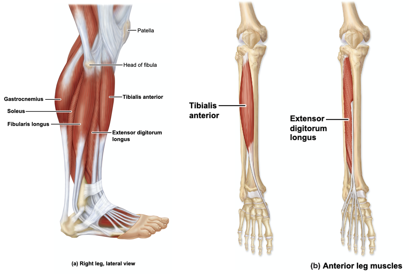
Muscles of the Lower Leg (Anterior)
Muscles of this region generally… dorsiflex foot + invert foot
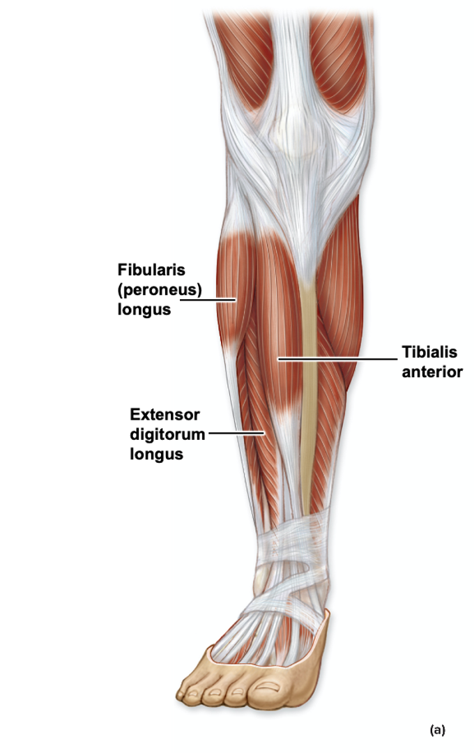
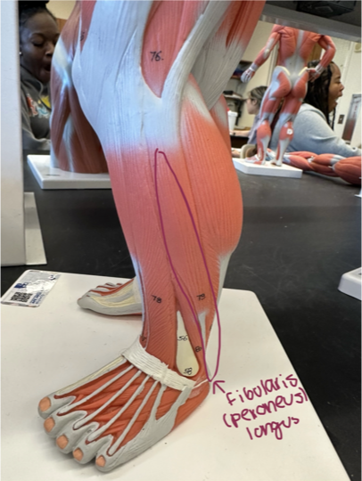
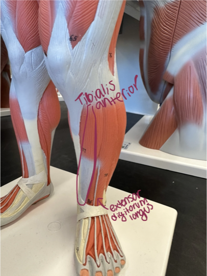
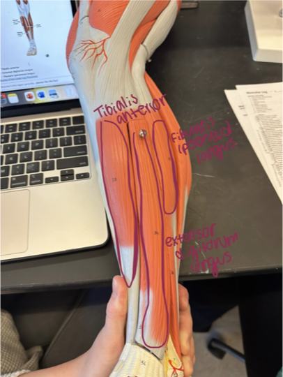
Tibialis anterior - located in the anterior compartment of the leg, just lateral to the tibia
Extensor digitorum longus - muscle located on the anterior surface of the lower leg
Fibularis (peroneus) longus - muscle located on the lateral side of the lower leg
Muscles of the Lower Leg (Posterior)
Muscles in this region generally… plantar flex the foot
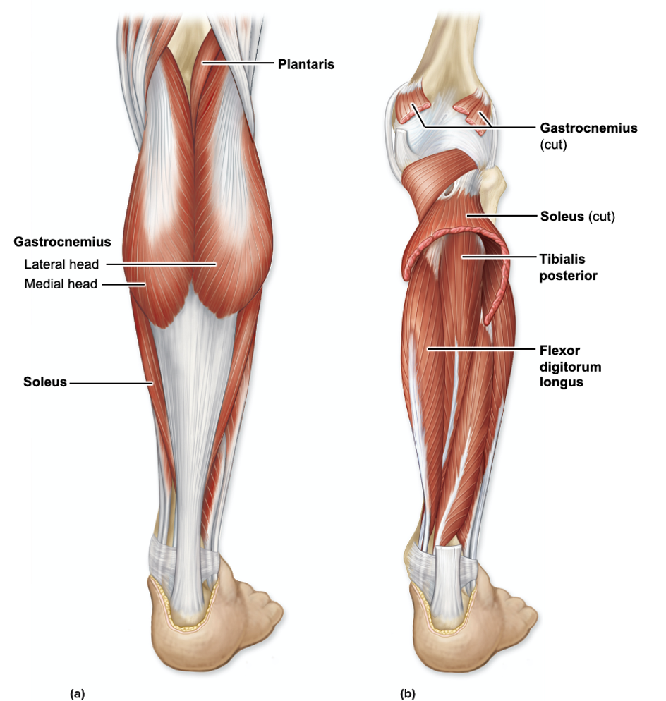
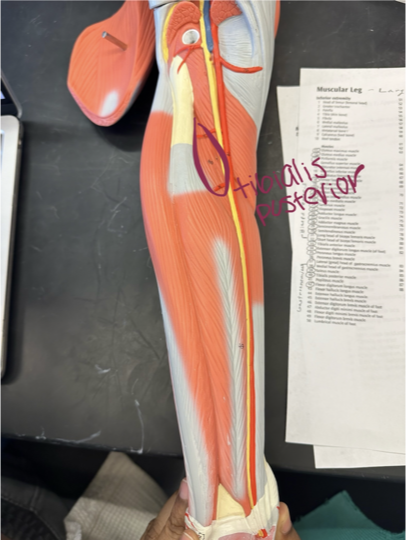
Tibialis posterior - muscle located in the back of the lower leg, deep to gastrocnemius
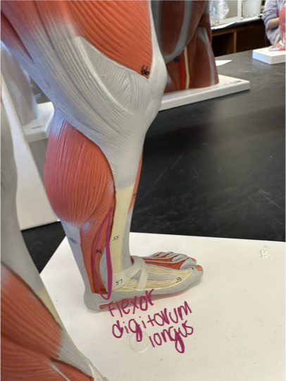
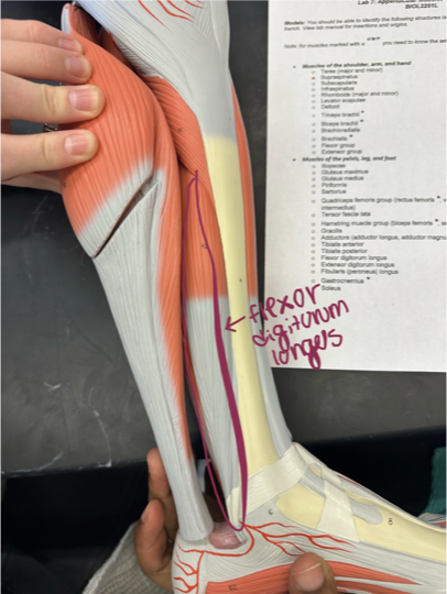
Flexor digitorum longus - muscle in the lower medial leg, located deep
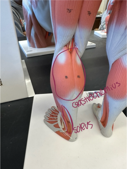
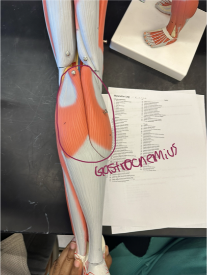
Gastrocnemius* - muscle located in the calf
Origin - femur
Insertion - calcaneus
Action - flexes leg, plantar flexes foot
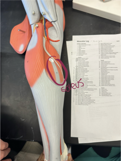
Soleus - calf muscle located beneath the gastrocnemius, on the lateral side
Muscles of the Lower Leg (Additional Views)