Connective tissue and Muscle staining
1/96
There's no tags or description
Looks like no tags are added yet.
Name | Mastery | Learn | Test | Matching | Spaced | Call with Kai |
|---|
No analytics yet
Send a link to your students to track their progress
97 Terms
Define connective tissue and its function
It is a fibrous material
Functions: support and protection, a media for exchange of nutrients, energy storage, insulation, and tissue repair
Three main components of CT
cells, fibers and extracellular matrix or ground substance
Most abundant fiber found in CT
Collagen
Define collagens make up and function
-Tough, thick, fibrous proteins that do not branch
-Provides great tensile strength giving the body form, firmness and resistance to outside forces
Staining characteristics
They are cationic (basic or positively charged)
Stain strongly with acid dyes (anionic or negatively charged)
Eosinophilic
Strongly birefringent under polarized light
Demonstrated most frequently with the Masson or Gomori Trichrome staining techniques
Diagnostic application of staining for CT
Plays a role in diagnosing collagenous CT diseases (cirrhosis and kidney-glom)
Plays a role in diagnosing soft tissue - tumors, cardiovascular
Most importantly- distinguishes muscle from CT
Mass Trichrome purpose
To identify an increase in collagenous connective tissue fibers such as in cirrhosis of the liver or to differentiate between collagen and muscle fibers
Mass Trichrome fixative, control tissue and microns
Fixative: Bouin, 10% NBF
Control tissue: Uterus, small intestine, appendix, or fallopian tube
Microns: 4-5
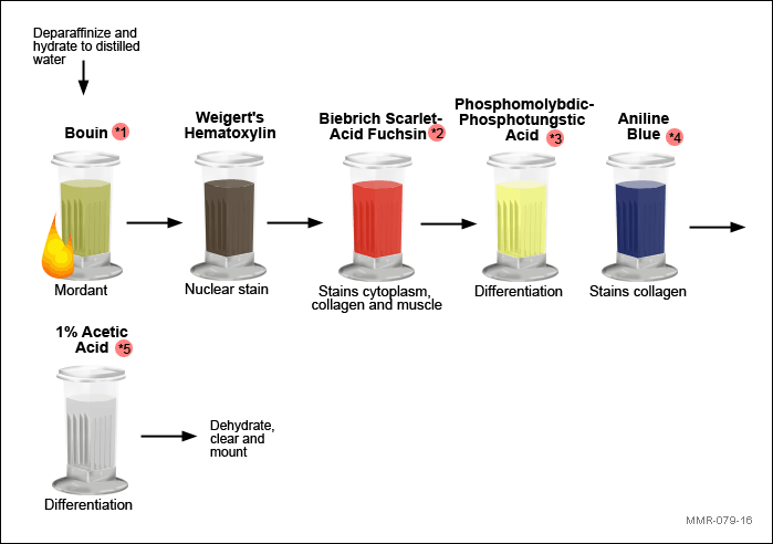
Masson Trichrome reagents and their purpose
Bouin | Mordant |
Weigert’s Hematoxylin | Nuclear Counterstain |
Biebrich Scarlet | Stains cytoplasm, collagen and muscle |
Phosphotungstic and/or phosphomolybdic acid | Differentiation |
Aniline Blue | Stains collagen |
1% Acetic Acid | Differentiation |
Masson Trichrome key quality Indicators
Key Quality Indicators:
Distinct blue/black nuclei
Strongly stained, brilliant blue or green collagen and red muscle
Excellent contrast between the collagen and muscle
Even staining throughout tissue section
Clean background, free of staining artifacts (e.g. precipitate, background staining)
Masson trichrome color results
Brilliant red… Cytoplasm, Keratin, Muscle fibers, Intercellular fibers, RBC’s
Blue…Collagen and mucin
Nuclei…… Black
Movat Pentchrome purpose
To demonstrate mucin, fibrin, elastic fibers, muscle, and collagen
Movat Pentachrome Fixative, thickness and control tissue
Fixative: 10% NBF
Thickness: 4-5 microns
Control Tissue: Lung, skin, aorta
Movat Pentachrome Reagents and their purpose
Alcian Blue 2.5 | Stains acid mucosubstances |
Alkaline Alcohol | Converts Alcian blue into insoluble monastral fast blue |
Verhoeff Working Solution | Stains elastic fibers |
2% Ferric Chloride | Differentiation |
5% Sodium Thiosulfate | Removes residual iodine |
Crocein scarlet-acid fuchsin working solution | Stains muscle and fibrin |
0.5% Acetic Acid | Lowers pH and safeguards phosphotungstic acid solution |
5% Phosphotungstic Acid | Differentiation |
0.5% Acetic Acid | Removes excess phosphotungstic acid |
Absolute Alcohol | Removes excess acid and dehydrates |
Alcoholic Spanish Saffron | Stains collagen and reticular fibers (counterstain) |
Movat Pentachrome Color results
Nuclei and Elastic fibers | Black |
Collagen and Reticular fibers | Yellow |
Ground substance, Mucin | Blue |
Fibrinoid, Fibrin | Intense red |
Muscle | Red |
C. Neoformans | Bright Blue |
Movat Pentachrome Key quality Indicators
Strongly stained, well demonstrated carbohydrates (mucin) and elastic fibers
Counterstain that enhances the desired structures
Excellent contrast between all structures to be demonstrated
Clean background, free of staining artifacts (e.g. precipitate, background staining)
Reticulin Purpose
Reticulin Fixative, Thickness, and control tissue
Fixative: 10% NBF
Thickness: 4-5 microns
Control: Liver
Reticulin Reagents and their purpose
Acetified Potassium Permanganate | Oxidation to aldehydes |
1% Oxalic Acid | Bleaches potassium permanganate |
2% Ferric Ammonium Sulfate | Sensitizer |
Diamine Silver | Impregnation |
10% Formalin | Reduces silver to a visible metallic form (develop) |
0.2% Gold Chloride | Tones silver |
5% Sodium Thiosulfate | Removes unreduced silver |
Nuclear Red Fast | Nuclear counterstain |
Reticulin color Results
Reticular fibers | Black |
Background | Pink |
Reticulin Key Quality Indicators
Well defined, linear (rather than a granular) pattern of reticular fibers
Strongly stained black reticular fibers
Counterstain enhances reticular fibers
Nuclear staining the color of the counterstain (not the silver)
Clean background, free of staining artifacts (e.g. precipitate, nonspecific or background staining)
8 steps of silver
Oxidation
Removal of excess potassium permanganate
Sensitization
Impregnation
Reduction
Tone
Remove unreacted silver
Counterstain (optional)
Three theories of staining method for Trichrome
Size of dye molecule (diffusion)
Varying affinity of anionic dyes to cationic tissue components
Mordants
Size of dye molecule theory- trichrome
Biebrich scarlet is relatively small and can therefore penetrate into the compact spaces within muscle fibers
Aniline blue is relatively large and can only penetrate into the more open spaces of connective tissue
Mordant theory- trichrome
Phosphotungstic and Phosphomolybdic act as mordants in binding aniline blue to the collage
Affinity theory- Trichrome
Biebrich scarlet and aniline blue are believed to have varying affinity for the various tissue components
Why is Bouin used in Masson Trichrome
Mordant - enhance the staining intensity and brilliancy of the subsequent colors
What type of dye is Biebrich and what tissue is it affinity for
Acid dye- will dye acidophilic tissues (cytoplasm, muscle, and collagen)
Which tissue region does phosphotungstic cause Biebrich to diffuse out of and why?
Biebrich will diffuse out of the collagen but no the cytoplasm or muscle because collagen has higher permeability (looser configuration)
Phosphotungstic and phosphomolybdic acids are mordants
True - acidic properties that act as a link (mordant) between the decolorized collagen and aniline blue
How does the pH of phosphotungstic and phosphomolybdic acids affect staining
Increases selective collagen staining and aids in the diffusion or removal of Biebrich scarlet allowing for more binding sites for the aniline blue
Analine blue is _______ charged and is ______ in size, allowing for it to permeate collagen
Negatively, larger
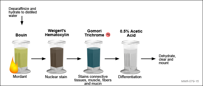
Gomori One-Step Trichrome purpose
Identify an increase in collagenous connective tissue fibers in diseases such as cirrhosis of the liver or to differentiate between collagen and muscle fibers.
Same as Masson
Gomori One-Step Trichrome reagents and their purpose
Define elastic fibers
Thin, small, branching fibers that allow for stretching and flexibility of tissue
Found in CT - lungs, skin and the cardiovascular system
Which tissue component has less tensile strength than collagen and are composed of microfibrils and the protein elastin
Elastic fibers
Diagnostic purposes of staining for elastic fibers (4)
Atrophy of elastic fibers can be an indicator of emphysema
Thinning or loss of fibers can be an indicator of atherosclerosis
Reduplication, breaks, or splitting can be an indicator of vascular disease
Tumors can also be detected in the blood vessels by staining for fibers
VVG demonstrates
Detect changes in elastic fibers
VVG fixative, thickness and control tissue
Fixative: 10% NBF, zenker
Thickness: 4-5 microns
Control: Aorta on edge, cross section of artery
VVG reagents and their purpose
Verhoeff Working Solution | Stains elastic fibers and nuclei |
2% Ferric Chloride | Differentiation |
95% Alcohol | Prepares slide for Van Gieson |
Van Gieson | Stains collagen, muscle and cytoplasm |
Methanol | Dehydration |
Acetone | Dehydration |
Acetone | Dehydration |
VVG color results
Elastic fibers.....blue-black to black
Nuclei.....blue to black
Collagen.....red
Other tissue elements.....yellow
Verhoeff Elastic solution is a _____ dye lake that consists of what reagents
soluble
Hematoxylin, ferric chloride and iodine
What are the two mordants in Verhoeff Elastic solution?
What other purpose do they serve?
Iodine and ferric chloride
They are also oxidizers that convert hematoxylin to hematein
How does Verhoeff Elastic solution dye bind to the tissue
formation of hydrogen bonds between the tissue and dye
Is VVG regressive or progressive? why?
Regressive- overstained with Verhoeff Elastic solution then differentiated with an excess mordnat, ferric chloride
Van Gieson in VVG is made up of what reagents
picric acid and acid fuchsin
How does binding work with picric acid and acid fuchsin in a VVG
small molecules of picric acid penetrate all of the tissues rapidly, but are only firmly retained in the close textured red blood cells and muscle
The larger molecules of acid fuchsin (an acidic aniline dye) displace picric acid molecules from collagen fibers, which have larger pores, and allow the larger molecules to enter.
What’s important that is provided by picric acid in Van Gieson
Low pH- allows for selective staining of collagen
What happens if you prolong staining in Van Gieson
picric acid can differentiate the elastic fibers stain
Why is the dehydration run down different in VVG than standard staining methods
Dehydrated- methanol
Cleared- acetone
Regular run down can wash the dye out (decolorize) of the tissue
Define reticular fibers
a type of thin, branching connective tissue fiber composed of type III collagen- creates a mesh network- found in the liver, lymph nodes, spleen, and bone marrow
Diagnostic applications of reticular fibers - how are they demonstrated in carcinomas, sarcomas and lymphosarcomas
Tumor identification, differential diagnosis of certain tumor types and any changes in reticular fiber patterns
Carcinomas- reticulum surrounds nests of tumor cells and supports the outer surface
Sarcomas- mesh-like pattern is demonstrated with each cell surrounded by some reticulum
Lymphosacrcomas- fibers can be found between individual cells within lymph node
Reticulin demonstrates
Demonstration of reticular fibers
Reticulin reagents and their purpose
Acetified Potassium Permanganate | Oxidation to aldehydes |
1% Oxalic Acid | Bleaches potassium permanganate |
2% Ferric Ammonium Sulfate | Sensitizer |
Diamine Silver | Impregnation |
10% Formalin | Reduces silver to a visible metallic form (develop) |
0.2% Gold Chloride | Tones silver |
5% Sodium Thiosulfate | Removes unreduced silver |
Nuclear Red Fast | Nuclear counterstain |
Reticulin color results
Reticular fibers | Black |
Background | Pink |
Reticulin Fixative, thickness and control tissues
Fixative: 10% NBF
Thickness: 4-5
Controls: Liver
8 steps of staining with sliver stains
Oxidation
Removal of excess potassium permanganate
Sensitization
Impregnation
Reduction
Tone
Remove unreacted silver
Counterstain (optional)
Walk through the oxidation step of reticulin
Glycol groups of the hexose sugars in the reticular fibers are first oxidized to aldehydes by acetified potassium permanganate
Why is acetified potassium permanganate used as the oxidizer for reticulin
It prevents silver staining of the nuclei
What is used to remove or “bleach” excess potassium permanganate in Retic
Oxalic acid
Walk through the sensitizer step of Retic
Ferric ammonium sulfate, due its low affinity to silver, it enhances reticular staining- forms a metal organic compound with the tissue
Walk through the impregnation step in Retic
Diamine silver replaces the metal organic compound made by ferric ammonium sulfate - the deposition of silver or gold ON or around the tissue, not in the tissue
What can cause the tissue to detach from the slide in a retic stain, how can it be avoided
Diamine silver is alkaline and has a pH of 11-12- the basicity of the dye can cause the tissue to detach
Silanized, charged, or coated slides may need to be used to avoid it
Walk through the reduction step of retic - differentiate argyrophill and argentaffin
Formalin is used as a reducing agent to reduce or developed the silver that has been deposited
Argyrophill - an external reagent is used as a reducer (formalin)
Argentaffin - Stains that does not require an external reducer
Walk through the toning step of Retic- why is this step important
Gold chloride is used - toning is the process when the metallic silver is replaced with metallic gold
allows for clarity and better contrast of reticular fibers - gold is also more stable
How do we remove unreacted silver in Retic
Sodium thiosulfate removes any unreacted silver that remains in the tissue to prevent any nonspecific reduction of silver by natural light
Define fibrin
a fibrous protein involved in blood clotting that is polymerized to create a “mesh” that forms a plug or clot over a wound site- seen in tissue with tissue damage or acute inflammation
Diagnostic applications of staining for fibrin
Excess fibrin can lead to thrombosis= blood clot
Disease of the liver can decrease fibrin production
The consequences of reduced, absent, or dysfunctional fibrin is likely to render patients as hemophiliacs
Movat Pentachrome demonstrates
To demonstrate mucin, fibrin, elastic fibers, muscle, and collagen
Movat Pentachrome thickness, fixative and control tissue
Fixative: 10% NBF
Thickness: 4-5 microns
Control tissue: Lung, skin, aorta
Movat Pentachrome reagents and their importance
Alcian Blue 2.5 | Stains acid mucosubstances |
Alkaline Alcohol | Converts Alcian blue into insoluble monastral fast blue |
Verhoeff Working Solution | Stains elastic fibers |
2% Ferric Chloride | Differentiation |
5% Sodium Thiosulfate | Removes residual iodine |
Crocein scarlet-acid fuchsin working solution | Stains muscle and fibrin |
0.5% Acetic Acid | Lowers pH and safeguards phosphotungstic acid solution |
5% Phosphotungstic Acid | Differentiation of crocein scarlet acid fuchsin WS |
0.5% Acetic Acid | Removes excess phosphotungstic acid |
Absolute Alcohol | Removes excess acid and dehydrates |
Alcoholic Spanish Saffron | Stains collagen and reticular fibers (counterstain) |
Movat Pentachrome color results
Nuclei and Elastic fibers | Black |
Collagen and Reticular fibers | Yellow |
Ground substance, Mucin | Blue |
Fibrinoid, Fibrin | Intense red |
Muscle | Red |
C. Neoformans | Bright Blue |
Define lipid
Insoluble in water
Function as structural components of cell membranes, serve as a metabolic energy reserve and protect numerous organs
Simple lipids which include fatty acids, waxes, triglycerides and sterols
Diagnostic application of staining for lipids
Fixative used for lipids
Osmium tetroxide, uncommon so we opt for frozen sections
Oil red O demonstrates
To demonstrate neutral (non-charged) lipids
Oil red O reagents and their purpose
10% NBF | Fixation |
50% Isopropyl alcohol | Dehydration |
Oil Red O | Stains fat |
50% Isopropyl alcohol | Prevents nonspecific staining |
Hematoxylin | Nuclear stain |
Saturated Lithium Carbonate | Bluing |
Oil Red O color results
Fat | Red |
Nuclei | Blue |
Oil Red O Fixative, Thickness and control
Fixative: None (frozen)
Thickness: 10 microns
Control: skin
Is Oil Red O a physical method of staining or is it a chemical method- what does this mean?
PHYSICAL - physically moves out of the staining solution and into the fat
What four features should a stain have to stain with fat
Be more soluble in the tissue lipid than in the solvent in which it is dissolved
Must not be water soluble
Must be strongly colored
Must act with tissue constituents only by solution. Note: the solvent used is critical, with isopropanol removing a minimal amount of lipid and propylene glycol not extracting any lipid.
What are two dyes that work similarly to Oil Red O
Sudan IV (solvent 70%) and Sudan Black B (solvent propylene glycol)
What mounting media is used with Oil Red O
Aqueous mounting media
What cells are found in connective tissue (8)
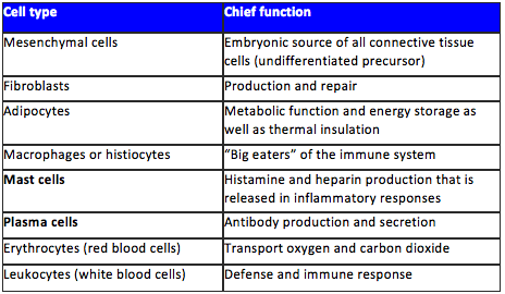
Diagnostic application of staining for connective tissue
Mast and plasma cells, components of the immune system, are indicative of disease
Mast cells are metachromatic
true - they will stain a different color from the dye solution and the rest of the tissue
TBO demonstrates
Mast cells
TBO reagents and their purpose
Toluidine Blue Solution | Stains mast cells |
0.2% acetic acid | Differentiation |
TBO color results
Mast cells | Deep Violet |
Background | Blue |
Nuclei, Nissl granules | Dark blue |
Cytoplasm, muscle | Light blue |
Mucin | Red/purple |
Red blood cells | Green |
TBO fixative, thickness and control tissue
Fixative: 10% NBF
Thickness: 4-5 microns
Controls: Mast cell sections (Spleen, liver)
Define Basement membranes
provide structural support to the epithelium and act as a barrier from the underlying connective tissue
Diagnostic applications of staining basement membranes
Important for kidney disease- basement membrane of gloms
Periodic Acid Methenamine demonstrates
To demonstrate glomerular basement membranes in kidney
Periodic Acid Methenamine reagents and their purpose
1% Periodic acid | Oxidation to aldehydes |
Methenamine Silver | Impregnation |
0.2% Gold chloride | Tones silver |
2% Sodium Thiosulfate (Hypo) | Removes unreduced silver |
Light Green | Counterstain |
Periodic Acid Methenamine color results
Basement membrane | Black |
Background | Green |
Periodic Acid Methenamine Fixative, Thickness and controls
Fixative: 10% NBF (avoid mercury- Zenker and B-5)
Thickness: 2 microns
Controls: Kidney