B2L1+L2: Anatomical/Directional terms + intro to locomotor apparatus
1/75
There's no tags or description
Looks like no tags are added yet.
Name | Mastery | Learn | Test | Matching | Spaced | Call with Kai |
|---|
No analytics yet
Send a link to your students to track their progress
76 Terms
Median (midsagittal) Plane
divides body equally into equal left and right halves
Sagittal Plane
divide body into UN-equal halves
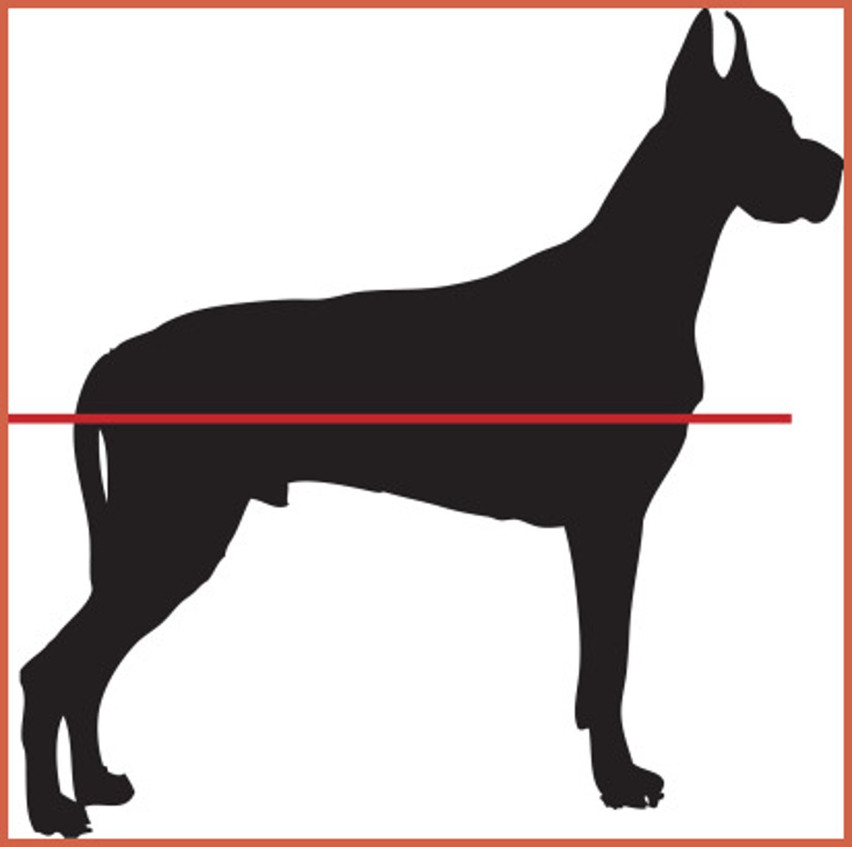
What is the transverse plane?
A plane that is perpendicular to the axis of the trunk, head, and limbs.
What body segments does the transverse plane divide?
Cranial/caudal or rostral/caudal.
How does the transverse plane divide limbs?
Into proximal and distal.
Dorsal Plane
divides body into dorsal and ventral parts
Dorsal
in the direction toward the back
Ventral
in the direction toward the belly
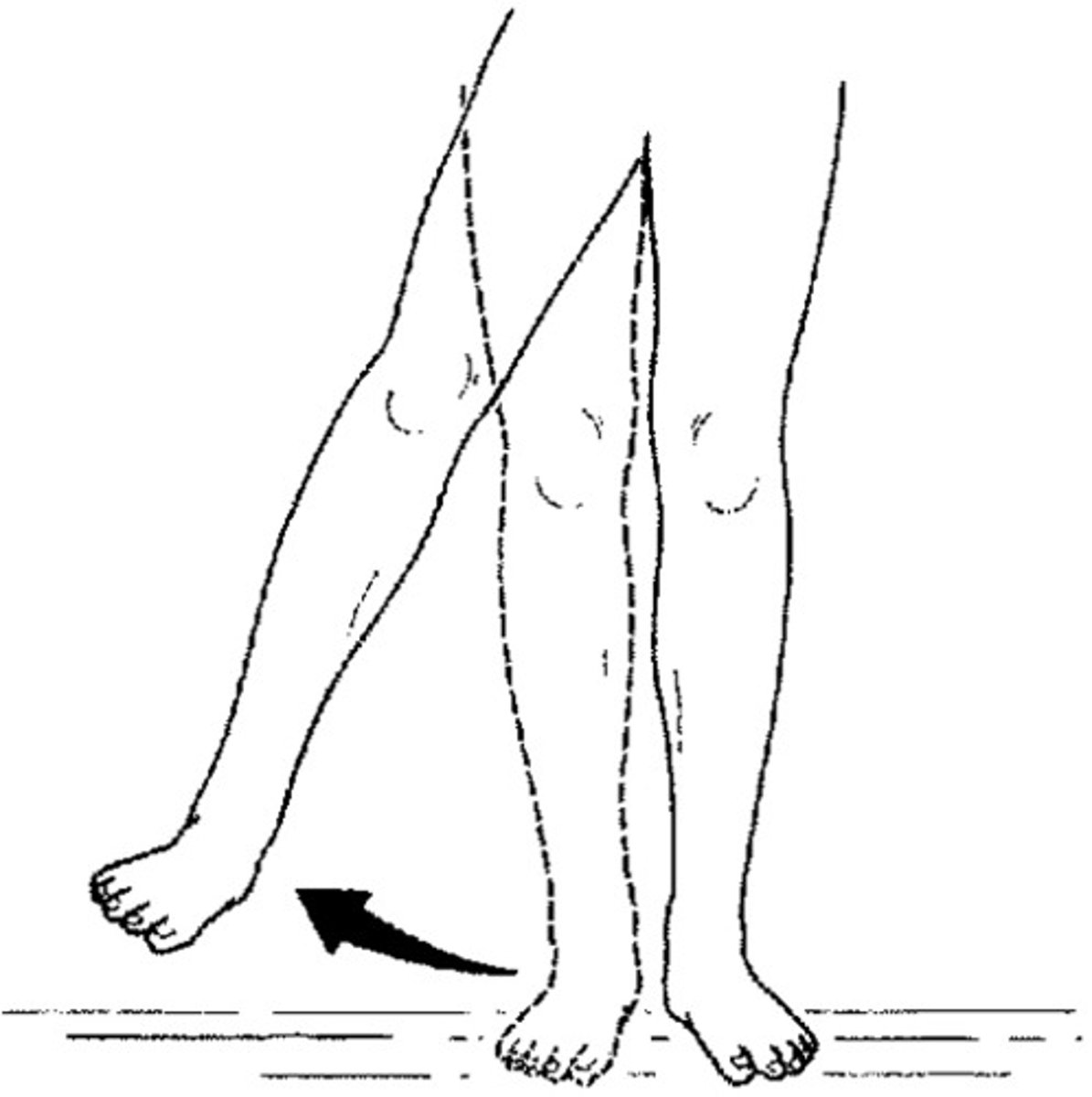
Medial
Toward the midline of the body
Lateral
Away from the midline of the body
Cranial
toward the head
Caudal
toward the tail
Rostral
toward the nose
Internal
Located within the body
External
Located outside the body
Superficial
near the surface of the body
Deep
Away from the body surface; more internal
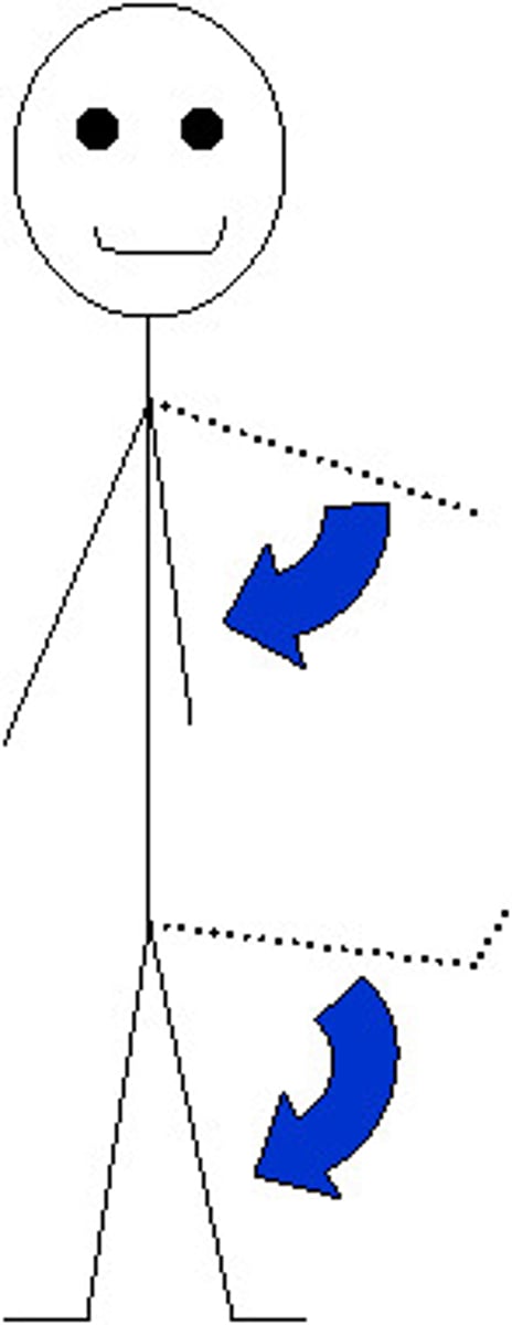
Proximal
Nearer to the trunk of the body
Distal
Farther from the trunk of the body
Axial
directed toward the longitudinal central axis of the limb
Abaxial
directed away from the longitudinal central axis of the limb
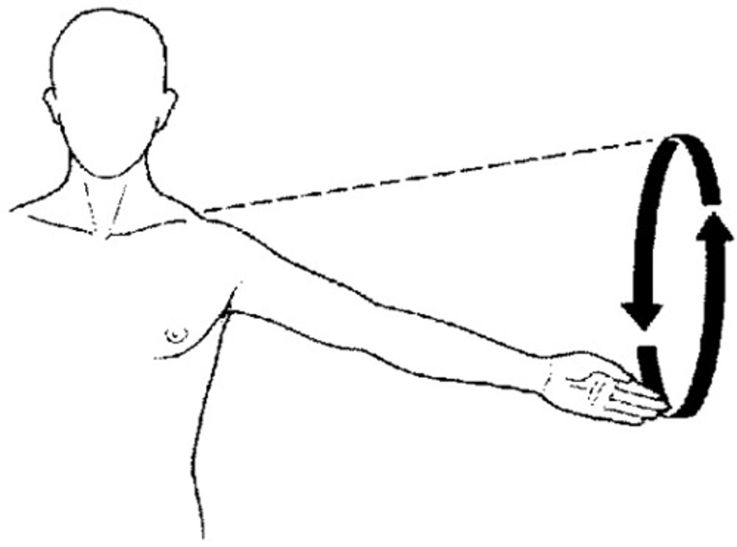
Flexion
decreasing angle between limb segments
Extension
increasing angle between limb segments
Abduction
moving away from median plane
Adduction
moving towards median plane
Circumduction
movement circumscribing a cone shape
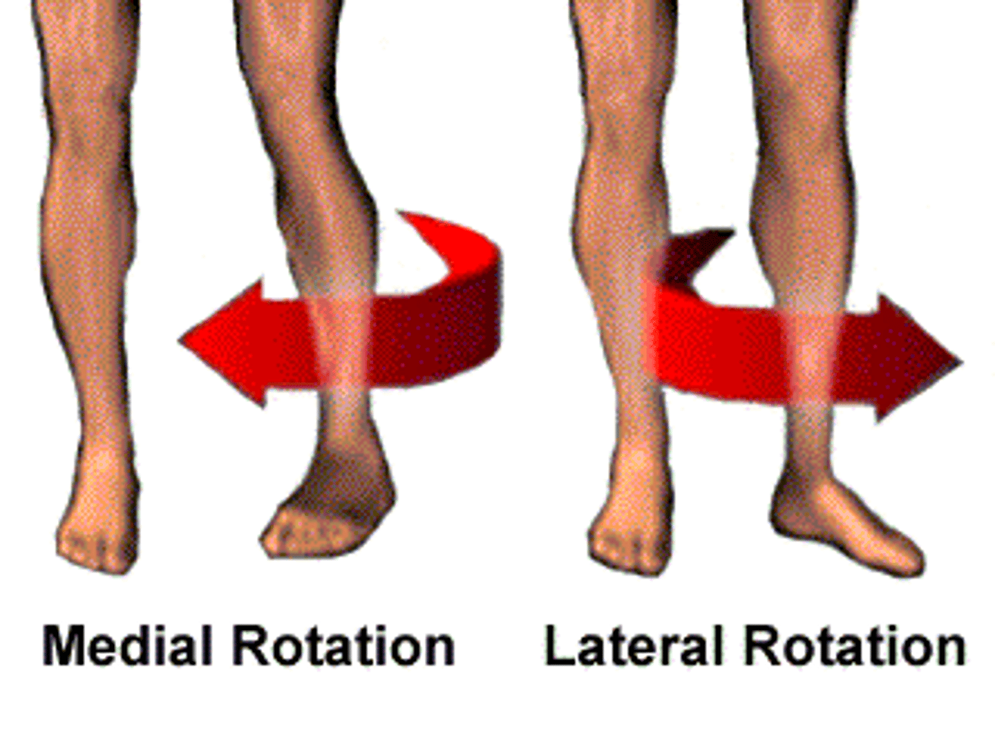
Rotation
rolling pin movement on the axis of limb
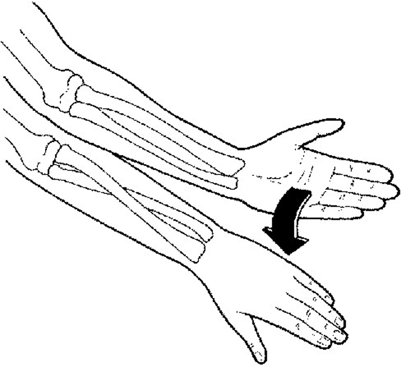
Supination
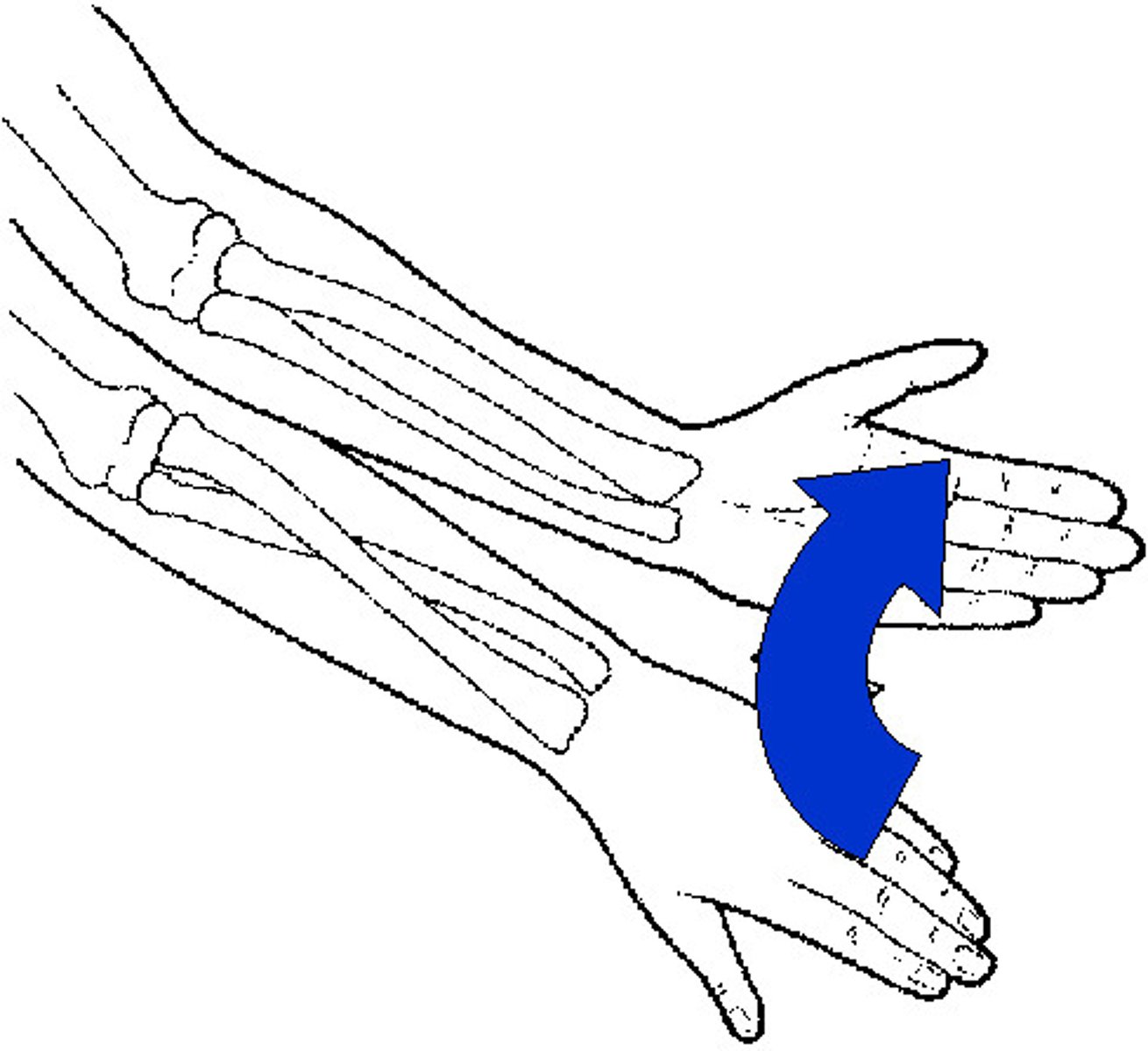
Pronation
Brachium
supported by humerus
Antebrachium
supported by radius and ulna
Crus
Manus
Front paw = Carpus + Metacarpus + Digits
Flat bone
scapula
Long bone
humerus, radius, ulna
Short bones
carpal bones
Sesamoids
special type of short bones within tendons
Diaphysis
bone shaft
Medullary cavity
contains yellow marrow
Epiphysis
end regions of bone
Physeal growth plate
located between epiphyses and diaphysis in young animals, comprised of cartilage cells
Wolf's law
form follows function
- bones get remodeled in response to stress placed upon them
Non-articular prominences
attachment sites for muscles, passages of vessels and nerves
Ex: spine of scapula
Articular promiences
make up joints
Ex: glenoid cavity
What type of connective tissue unites the articular surfaces of bones in fibrous joints?
Strong connective tissue
What is the term for the fusion of bones in fibrous joints?
Synostosis
What is the movement capability of bones in fibrous joints?
Almost no movement, or none at all
Cartilaginous joints
cartilage unites articular surfaces of bones >> limited movement
Synovial joints
joint cavity between articular surfaces of bones filled with synovial fluid >> most movable
Articular surfaces
- protected by articular cartilage
- enclosed within a fluid-filled joint cavity
Joint capsule is composed of three parts
- synovial layer
- fibrous layer
- joint cavity
Synovial layer
- produces synovial fluid
- highly vascularized and innervated
Fibrous layer
provides strenght and resistance
Joint cavity
for lubrication and nutrition of the articular cartilages
Accessory structures of synovial joints
- ligaments
- meniscus
Ligaments
bands of tough fibrous connective tissue
Meniscus
fibrocartilages located within a synovial cavity, allow articular surfaces to fit together
2 articulating bones
- simple
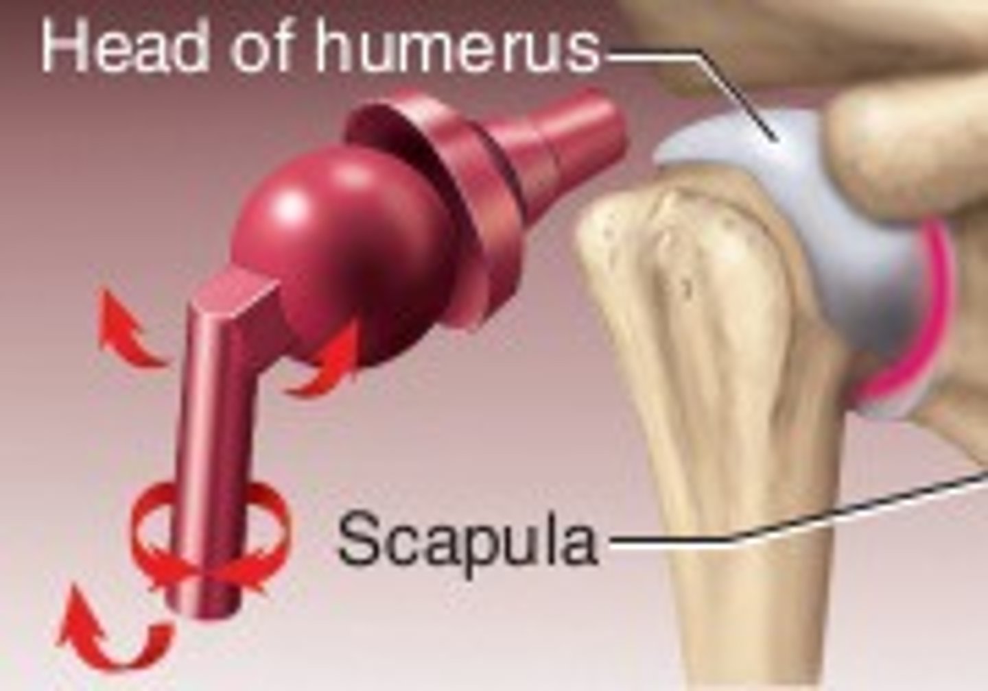
Ex: glenohumeral (shoulder) joint
More than 2 articulating bones
- compound
Ex: humeroradioulnar (elbow) joint, carpal joint
Congruent joint
articular surfaces fit well together
Ex: elbow joint
incongruent joint
articular surfaces do not fit well together
Ex: knee joint
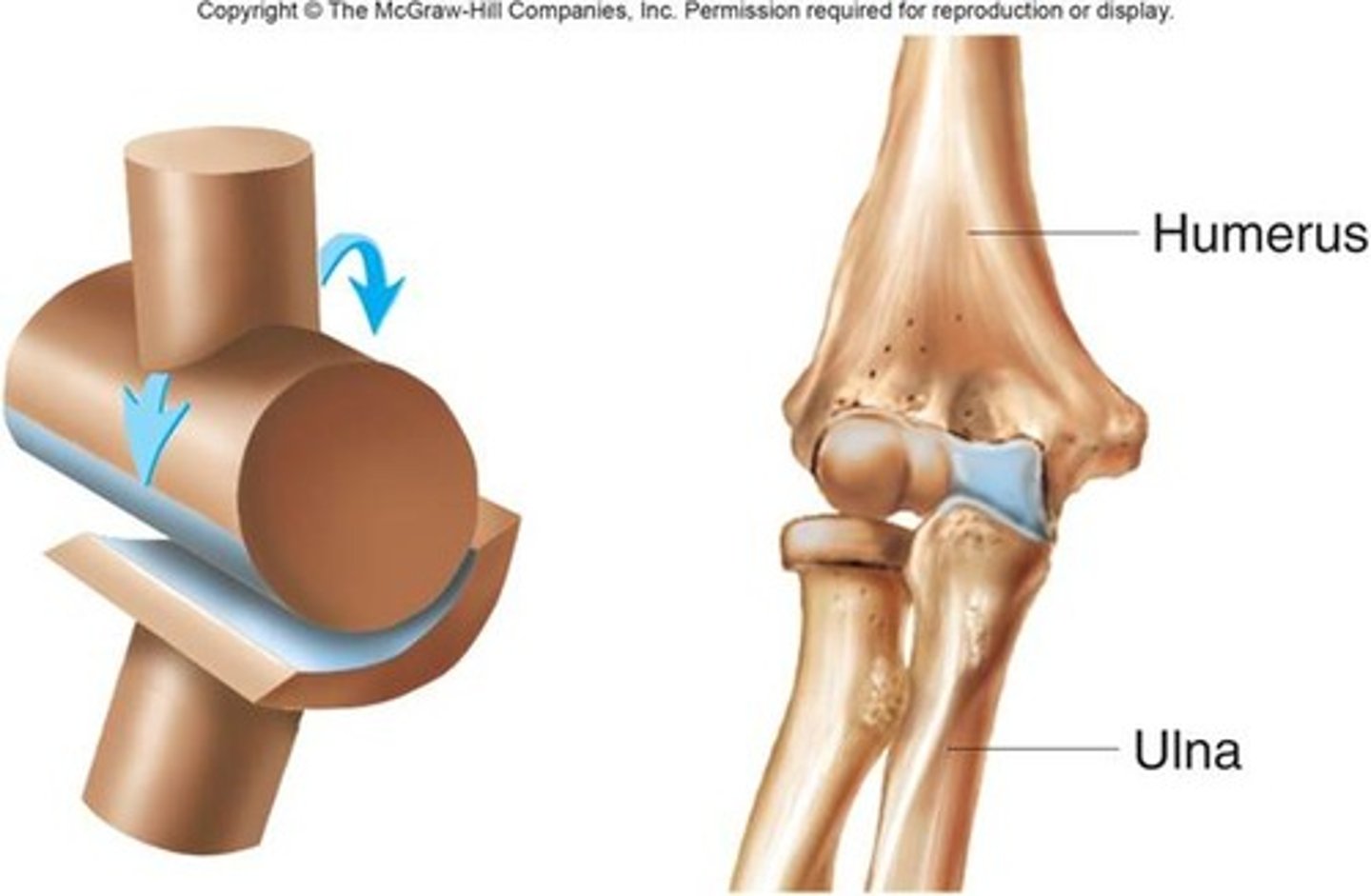
Hinge joint
- least versatile
- flexion and extension only
Ex: elbow
Spheroidal (ball &socket) joint
- most versatile
- all rand of movements
Ex: shoulder joint, hip joint
Tendon of origin
- proximal attachment
- relatively fixed point, less movement here than at the insertion end
Muscle head or belly
the part that contracts
Tendon of insertion
- distal attachment
- relatively more motile, more movement at the insertion end that at the origin end of the muscle
Aponeurosis
a flat, sheet-like tendon, allows muscle to have a broader attachment
Synovial burse
synovial fluid-filled "balloon" protecting a tendon from a bony surface
Synovial tendon sheath
synovial fluid-filled "sleeve" completely surrounding a tendon, easing its gliding between a retinaculum and bone
Retinaculum
fibrous band holding down tendon(s) to a bone
Ligaments
attach bone to bone
Tendons
attach muscle to bone
Fasciae
fibrous layers enveloping and isolating muscle groups and individual muscles
Superficial fascia
loose connective tissue attaching skin to underlying mm
Deep fascia
leaf of dense connective tissue from which some muscles may originate or insert, separates muscle groups/layers into fascial planes
Still learning (7)
You've started learning these terms. Keep it up!