Mycology - Fungal Infections
 Call Kai
 Learn
Learn Practice Test
 Spaced Repetition
 Match
Match Flashcards
 Knowt Play
1/33

There's no tags or description
Looks like no tags are added yet.
Name | Mastery | Learn | Test | Matching | Spaced | Call with Kai |
|---|
No analytics yet
Send a link to your students to track their progress
34 Terms
Describe the key features of fungi, what are they, what are they made of, how do they reproduce?
• Uni or multicellular organisms, aerobic eukaryotes with defined nuclei and cell walls composed of carbohydrate and chitin, ergosterol in plasma membrane
• Saprophytic (organisms and nutrition where a living thing obtains food by absorbing dissolved organic material from dead or decaying matter, rather than producing its own food) or parasitic
• Sexual and asexual reproduction
Asexual spores by mitosis - infectious forms of hyphal fungi
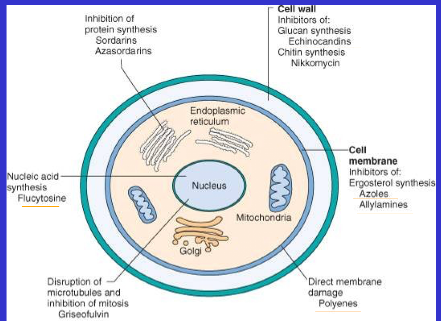
What is antifungal chemotherapy?
Relies on selective toxicity, parts of fungus which are different than eukaryotic cells (To protect host)
Cell wall, nuclear division, protein synthesis
What is the significance of fungi in veterinary medicine?
• Animals which are allergic (to fungal spores)
• Cutaneous and superficial mucous membranes (Dermatophytes, Malessezia, Candida infection)
• Subcutaneous (Sporotrichosis)
Subcutaneous fungi infect the skin when a fungal spore is introduced through a wound, such as a cut or scratch from contaminated soil, plants, or thorns.
• Systemic - worst ones
primary pathogens (Histoplasma)
opportunistic pathogens (Aspergillus)
• Toxins (mycotoxins)
What are some of the beneficial and harmful fungi products?
Fungal spoilage affects….
• Fungal products: Beneficial (antibiotics) Harmful (produce mycotoxins - biological effects)
Animal foods, forages, animal products such as meat and hides
In what ways can laboratory techniques be applied to diagnose fungi?
• Clinical specimens - skin, hair, nails, swabs from mucous membranes, blood etc
Direct microscopy - skin, hair, nails - KOH preps, calcofluor white (Binds to chitin in the fungal cells, which stains white), Gram stain on colonies, Giemsa
Culture - Sabarauds dextrose agar, chromagar, specialised agars - days or weeks
Identification by morphology of hyphae, production of spores
PCR
Histology of tissue in invasive disease - silver stains
To diagnose, a culture from site of infection and histological diagnosis is important (Because fungi are ubiquitous in the environment, may contaminate samples)
What are yeasts?
Unicellular organisms that reproduce by budding - grow on skin, usually cause infection in mucous surfaces and in the body
What are moulds?
Multicellular organisms that produce hyphae and mycelium and spores (eg - Dermatophytes - hyphae grow in skin, hair and nails, thus producing infections here)
What are dimorphic fungi?
Organisms that can produce both hyphae and yeast- like forms. Hyphae in environment (+ spores) and form yeasts in the infected host - geographically restricted
Pathogenic form is the yeast
Only present in countries of higher ambient temperature
How do yeasts reproduce?
What are a few different examples?
• Unicellular fungi - single cells
• Grow by division
Budding
Binary fission
• Some times daughter cells may not detach themselves - the result is a pseudohyphae (pseudo and true hyphae - Candida albicans)
• 3 Important species: Candida, Malassezia, Cryptococcus
What are the key features of Candida Albicans?
Ensure to note what three forms this yeast can exist in.
• 3-6 mu, oval cells
• Gram positive
• Yeasts, pseudohyphae, hyphae in tissue (Candida albicans)
Exist in the yeast form, hyphae form and pseudohyphae form, not true hyphae due to pinch points
• Germ tubes
Grow organism, place in serum, and you will see germ tubes
• Chlamydospores (thick-walled asexual spores)
• Grows at 37°C on Sabouraud's dextrose agar/Chromogenic agar
• Creamy white 2mm colonies
• API or Auxacolour system for identification of species
Relevant for treatment, especially if presence of other fungi
What conditions are caused by candida albicans?
• Cattle - Mycotic abortion
Rumenal infections
Mastitis
• Pigs - dermatitis
• Dogs - chronic enteritis and dermatitis, vaginitis/vulvitis in diabetes
• Birds - crop infections, enteritis
• Man - mucosal and systemic infection
What are the key features Malassezia?
• Bottle shaped yeasts
• Malassezia pachydermatis, causative agent of external ear infection in dogs
• Grows on Sabouraud's dextrose agar at 37°C
• Lipase, urease
• Otitis externa
• Dermatitis
What are the key features of cryptococcus?
• C. neoformans (Commonest - affects immunocompromised individuals), C. grubii, C gattii
Inhalation of yeast cells in dust - pigeon faeces, can cause lung infections
Opportunistic infections - defective cell mediated immunity - fungal meningitis
Nasal cavity of cats with chronic rhinitis
• Nasal, cutaneous, neural and ocular disease in cats
• Mucilaginous capsule - india ink stain - virulence factor, 20 size mu total
Prevents phagocytosis by WBCs
What are the key features of filamentous hyphal fungi (moulds)?
• Multicellular, multinucleate
• Basic unit is the hyphae, a long thin filament
• Hyphae branch and cross link to forma mat (mycelium)
• Growth occurs at hyphal tip
• Hyphae may be divided by cross-walls (septa) into uninuclear units
• Non septate hyphae: coenocytic
• Septa contain pore. All cytoplasm is connected.
What are the dermatophytes - microsporum, trichophyton sp.
• Septate branching hyphae
• Anthropophilic (Human to human), geophilic (Environment to human/animal transmission), zoophilic (Animal to human)
• Digest keratin - infect skin, hair and nails
• Arthrospores in clinical specimens (need a live species)
• Grow on Sabouraud's within 7-14 days at 28°C
microconidia and macroconidia in lab culture
• Identify by surface appearance and colour of underside
• Confirm by shape of macroconidia
• Microsporum, Trichophyton
(Epidermophyton - man only)
What is microsporum?
Type of Dermatophyte
• Ringworm in man and animals
• Anthropophilic, zoophilic and geophilic
• Microconidia en thyrse (along sides of hyphae)
Produce Macroconidium (Large spores)
Microconidium (Small Spores)
• May fluoresce under Wood's light
What is M. canis?
• Ringworm in cats and dogs transmissible to man
• Grows on hair with arthrospores - ectothrix infection - fungi on the outisde
• Microconidia relatively common
• Macroconidia elliptical with up to 14 divisions, rare on isolation
• Fluoresces
• Colonies smooth white surface, yellow underside
What are some other notable microsporum species afflicting other animals?
• M. gypseum - many macroconidia, dog, geophilic
• M. nanum - pigs (not uk)
What are the key features of Trichophyton?
• Zoophilic (Animals to people)
• Ringworm in man and animals
• Club-shaped macroconidia
• Microconidia en thyrse and en grappe
• Spiral hyphae
• No fluorescence
What are the main features of the species of trichophyton?
What are the species affecting, dog, horse, and fowl?
• T. verrucosum - Ringworm in cattle transmissible to man
• Abundant chlamydospores
• Large spore ectothrix on hair
• Colonies slow growing, grow at 37C, all other are grown at 27ish
• Deep in agar
• T. mentagrophytes - dog, horse
White top, powdery green, burnt orange underside
Characteristic feature are spiral hyphae
• T. equinum - horse
• T. gallinae - fowl
What are the key features of aspergillus?
Ubiquitous in the environent
• Septate branching hyphae
• Sporing heads or conidia in oxygen
• Conidiophore and sterigmae
• Use colonial appearance, size and details of conidiophore to identify
What are the key features of aspergillus fumigatus?
• On food, fodder
• Spores infect young non-immune or immunosuppressed animals
• Grows best on Sabouraud's at 24 - 28°C
• Star shaped colonies, Green-blue with sporing heads on culture
• Histopathological evidence of tissue invasion
What is aspergillus disease?
• Mainly respiratory infection - spore inhalation
• Pneumonia in newly hatched chicks
• Air sacculitis in birds
• Guttoral pouch mycosis in horses
• Nasal aspergillosis in dogs
• Mycotic abortion in cows - poor quality feed
• Man - allergic disease and severe infection in the immunocompromised to spores
What are the host defences against aspergillus?
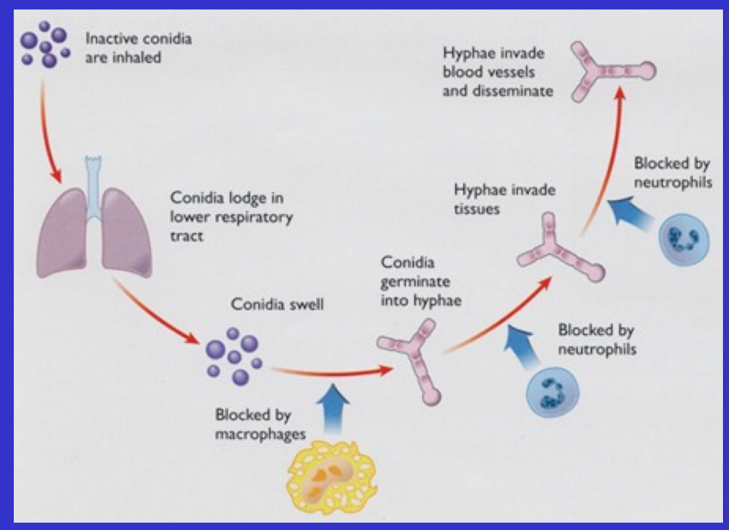
Consider that we do not get Aspergillus lung infection despite breathing in spores
Hyphae WANT to invade blood vessels and disseminate
Macrophages block transition of spore into hyphae
AND they can block hyphae invading tissue
AND they can block hyphae invading the blood vessels
WHICH IS WHY THIS IS DISEASE OF IMMUNOCOMPROMISED PATIENTS
What are the features of dimorphic fungi?
• Geographically restricted
• 2 distinct forms - mould and yeast
• Moulds in the environment, yeast in animal tissue
yeast is the pathogenic form
• Spores of these fungi enter host by respiratory route (or skin)
• Species most associated with disease - Histoplasma capsulatum, Blastomyces dermatitidis, Coccidioides immitis and Sporothrix schenckii
What are key features of histoplasma capsulatum?
• Endemic in Mississippi and Ohio river valleys
Usually found in the soil, enriched or growth encouraged by bat and bird feces
• Affects Man, Dogs and cats
• Impaired CMI (Cell-Mediated Immunity), but can affect normally functioning animals
• Granulomas in lungs
• Infection of macrophages (Intracellular infection)
What are the key features of histoplasma capsulatum farcinimosum?
• Epizootic lymphangitis
• Spores enter through skin
• Ulcers over lymphatics and lymph node
• Horses
• Notifiable, Category 3
• India, Africa, Middle east
• Grows at 30°C in serum-rich media
What is sporotrichosis (Sporothrix schenckii)?
Subcutaneous disease
Natural infections of animals (cats) and humans - cutaneous and systemic
Infection caused by penetration of the skin
What are the key features Zygomycetes?
• Broad hyphae non-septate
• Asexual spores in a sporangium
• Sexual spores oospores or zygospores
• Mucor, Rhizopus, Absidia, Saprolegnia (Goldfish)
• Infection uncommon in healthy immunocompetent animals
• Immunodeficiency, steroids, antibiotics
• Abortion in cattle
• Rumenitis
What are mycotoxins and how are they involved in producing illness?
Secondary metabolites of fungal species in crops, pasture or stored feed - low MWT, heat stable, non antigenic, immunosuppression, mutagenic, teratogenic, carcinogenic
Acute or chronic intoxication - Penicilliumn Aspergillus, Fusarium
Clinical disease - Period of exposure, amount ingested
What are three examples of clinical disease caused by mycotoxins?
Aflatoxin - difuranocoumarins - Aspergillus sp. Aflatoxicosis - absorption from GI tract, metabolised by liver to toxic product - carcinogenic
Associated with moldy peanuts
Ergotism - ergopeptide alkaliods - Claviceps purpurea - domestic animals and humans - seed heads of grasses and cereals - ergotamine and ergometrine - affect nerves supplying arteriolar smooth muscles
Mycotic ostrogenism - Zearalenone - potent non steroidal ostrogen produced by Fusarium sp. - maize other cereals - reproductive problems in cattle and sheep
What is the difference between septate and non-septate hyphae?
Septate hyphae are divided into individual cells by internal cross-walls called septa, while nonseptate (or coenocytic) hyphae lack these walls and are a continuous, multinucleated tube.
What is the difference between extothrix and endothrix infections?
Ectothrix refers to a dermatophyte (fungal) infection where fungal spores and hyphae form on the outside of the hair shaft, often destroying the hair's cuticle, as seen with Microsporum species. In contrast, Endothrix infection involves the fungus growing inside the hair shaft, retaining the hair cuticle, and is characteristic of Trichophyton tonsurans and other Trichophyton species. This difference in location is a key diagnostic feature for tinea capitis, and ectothrix infections may fluoresce under a Wood's lamp, while endothrix infections do not.
Why do ectothrix infections “glow” under a wood’s lamp?
Ectothrix infections glow under a Wood's lamp because the specific fungal species causing the infection, most commonly Microsporum species, produce compounds called fluorochromes. These compounds absorb the ultraviolet (UV) light from the lamp and re-emit it at a visible wavelength, causing the infected hairs to fluoresce a bright greenish-yellow color.
Infection Type (Ectothrix vs. Endothrix): Microsporum species predominantly cause ectothrix infections, meaning the fungal spores (arthroconidia) heavily encase the outside of the hair shaft. This creates the perfect environment for the fluorescent metabolic byproduct to coat the hair and glow.
Why Trichophyton lacks the glow: Most Trichophyton species primarily cause endothrix infections, where the spores develop on the inside of the hair shaft. Because the fungus does not leave a thick spore coating mixed with metabolites on the outside of the hair, it typically fails to fluoresce. [1, 2, 3, 4, 5]