Caren test 3
1/80
There's no tags or description
Looks like no tags are added yet.
Name | Mastery | Learn | Test | Matching | Spaced | Call with Kai |
|---|
No analytics yet
Send a link to your students to track their progress
81 Terms
What does the P wave represent?
Atrial depolarization.
What does the QRS complex represent?
Ventricular depolarization (ventricular contraction).
What does the T wave represent?
Ventricular repolarization — the recovery phase of the ventricles.
What is the correct conduction pathway?
SA node → AV node → Bundle of His → Right & Left Bundle Branches → Purkinje fibers.
What is another name for the SA node?
The pacemaker of the heart.
What is the normal rate of the SA node?
60–100 bpm.
What is the normal rate of the AV node?
40–60 bpm.
What is the normal rate of the Purkinje fibers?
20–40 bpm.
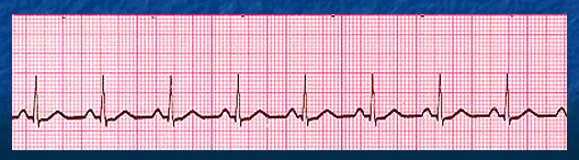
What is this?
Sinus Rhythm meaning functioning normally.
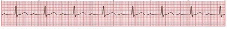
What is this?
1st‑degree AV block
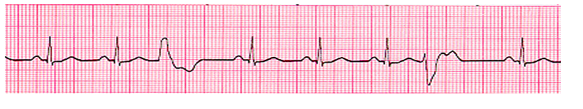
How do you identify First‑Degree AV Block?
Regular rhythm
Normal P waves
PR interval greater than .20 seconds
QRS normal (.06–.10 sec)
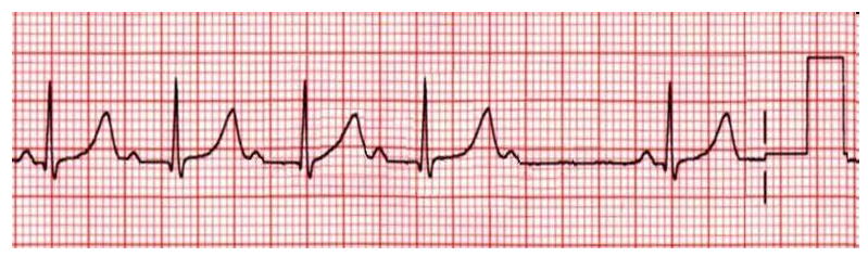
What is this?
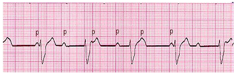
2nd Degree AV Block Mobitz I (Wenckebach)
How do you identify Mobitz I (Wenckebach)?
the PR interval is greater than 0.20 seconds and progressively lengthens until a QRS is dropped, producing an irregular R‑R interval because some SA node impulses fail to conduct through the AV node, resulting in a normal atrial rate, a slower ventricular rate, and a repeating cycle that resets after each dropped beat. is dropped, then the cycle is restarted
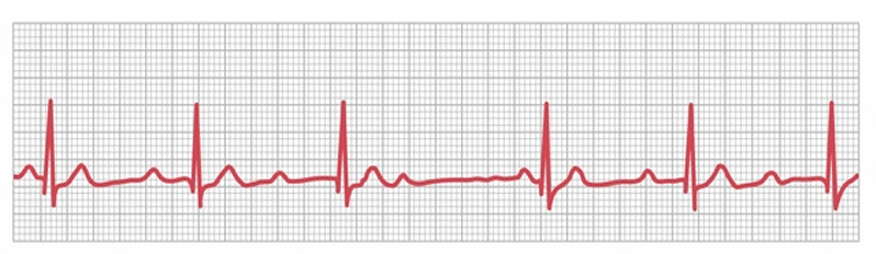
What is this?
Second Degree AV Block, Mobitz II (Missing QRS)
How do you identify Mobitz II?
is the classical heart block in which the AV node unpredictably blocks impulses—dropping QRS complexes without a pattern—often progressing to third‑degree heart block, with a normal atrial rate and a ventricular rate that is slower than the atrial rate.
How Patient is Affected by 2nd Degree (Wenckebach)?
Condition can progress within seconds to third degree AV block or complete heart block
What is this?
3rd Degree AV Block (Complete Heart Block)
How do you identify Third‑Degree Heart Block?
AKA complete heart block (CHB)
Ventricular is 20-40 beats per minute .
A long PR interval followed by a short PR interval indicates complete heart block .
How do you identify a BBB?
longer contraction time reflected in wider QRS complex
QRS duration - .12 and greater
What is this?
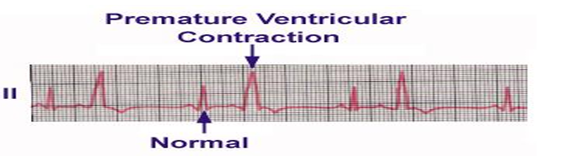
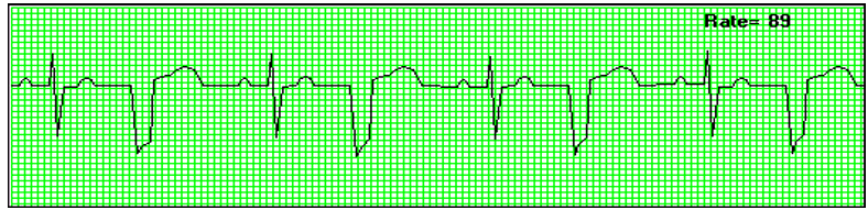
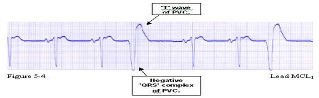
Premature Ventricular Contraction (PVC)
How do you identify a PVC?
has an early beat that makes the ventricular rhythm faster than normal and a wide early QRS greater than 0.12 seconds.
What is this?
unifocal
What is a unifocal PVC?
early beat (has similar shape only one irritable focus present)
What is this?
Multifocal PVC (more than one type of PVC.)
What is a multifocal PVC?
varied shapes and forms of the PVC’s
What is Interpolated?
PVC occurs during the normal R-R interval without interrupting the normal cycle
What is Occasional?
more than one to four PVCs
What is frequent?
more than five to seven PVCs per minute
What is this?
bigeminy PVC’s (Every other beat is a PVC)
What is this?
trigeminy PVC’s (Every third beat is a PVC.)
What is this?
quadrigeminy (Every fourth beat is a PVC.)
R on T PVC’s
PVC occurs on the T wave or the vulnerable period of the ventricle refractory period.
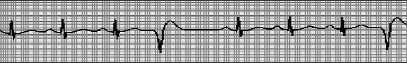
What is this?
V‑tach
How do you identify V‑tach?
shows a ventricular rate of 100–200 bpm, absent P waves, and a wide QRS greater than 0.12 seconds.
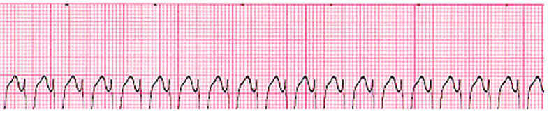
What is this?
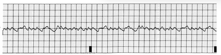
V‑fib (chaotic, asynchronous electrical activity in the ventricles)
How do you identify V‑fib?
P waves, no QRS complexes, a chaotic squiggly baseline, and a ventricular rate (if identifiable) over 300 bpm.
What is this?
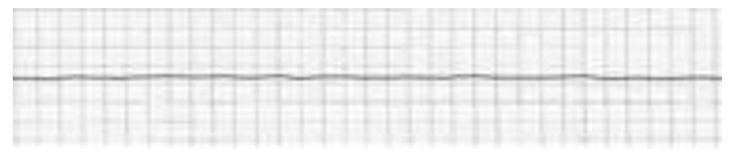
asystole (No electrical activity — flat line.)
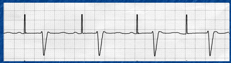
What is this?
ELECTRONIC PACEMAKER RHYTHM
What does an electronic pacemaker do?
It delivers an electrical impulse to the myocardium, causing the cells to depolarize, and it can pace the atria, ventricles, or both.
What is a pacing spike on an ECG?
A thin spike on the ECG tracing that shows electrical current from the pacemaker. After the spike, a P wave or QRS complex will appear.
What is the AV delay?
The time measured from the atrial pacing spike to the ventricular pacing spike, normally 0.12–0.20 seconds.
What is the conduction system considered?
the wiring of the heart
How much fluid normally fills the pericardium?
10–50 mL of pericardial fluid.
What is pericardium filled with?
The pericardium is filled with serous (pericardial) fluid that prevents friction and keeps the lining of the pericardium moist.
Where does the heart lie in the thoracic cavity?
The heart lies in the middle of the thoracic cavity (mediastinum).
What surrounds the heart?
The heart is surrounded by a protective sac called the pericardium.
How big is the human heart?
The heart is slightly larger than a man’s fist.
What are the four chambers of the heart?
The heart has 4 chambers — 2 atria and 2 ventricles.
What are the two types of heart valves?
The two types of heart valves are Atrioventricular (AV) valves and Semilunar (SL) valves.
What are the AV valves?
Tricuspid valve and mitral valve.
Where is the mitral (bicuspid) valve located?
The mitral (bicuspid) valve lies between the left atrium (LA) and the left ventricle (LV).
Where is the tricuspid valve located?
lies between the right atrium (RA) and the right ventricle (RV).
Where is the aortic valve located?
lies between the left ventricle (LV) and the aorta.
Where is the pulmonary (pulmonic) valve located?
lies between the right ventricle (RV) and the pulmonary artery (PA).
What are the semilunar (SL) valves?
Pulmonic valve and aortic valve.
Where is the Semi Lunar valves
between the atria and ventricles
Where is the Thebesian valve located?
is located near the entrance of the coronary sinus.
Where is the Eustachian valve located?
located near the entrance of the inferior vena cava (IVC).
What is the function of heart valves?
To control and maintain blood flow through the heart and body.
What is the Blood flow?
Blood enters the heart from the Superior Vena Cava (SVC), Inferior Vena Cava (IVC), and Coronary Sinus (CS) into the Right atrium(RA). It goes through the tricuspid valve(TV) to the right ventricle(RV), through the pulmonic valve(PV) to the main pulmonary artery(MPA) to the lungs.
From the lungs it returns to the heart through the 4 pulmonary veins into the Left Atrium(LA) through the mitral valve(MV) into the Left Ventricle(LV) out through the Aortic Valve(AOV) to the ascending aorta to the aortic arch to the descending aorta which becomes the abdominal aorta to the body.
What is the cardiac cycle?
One complete contraction and relaxation of the heart.
What is stroke volume?
The amount of blood ejected from a ventricle with each heartbeat.
What is cardiac output?
Amount of blood pumped into aorta each minute by the hear
What is excitability?
The ability of each cardiac cell to respond to an electrical stimulus — the ability to respond to an impulse.
What is Automaticity?
The ability to generate an electrical impulse independently.
What is the QRS duration in a PVC?
has a wide QRS greater than 0.12 seconds with the T wave in the opposite direction.
What does parasympathetic stimulation do to the heart?
Slows heart rate and conduction.
What does sympathetic stimulation do to the heart?
Increases the force of ventricular contraction, heart rate, blood pressure, and cardiac output
What is the absolute refractory period?
Phases 1 & 2 a period when no stimulus, no matter how strong, can excite the cell to produce another action potential (QRS).
What is the relative refractory period?
Phase 3 — the cell can produce an action potential, but only if the stimulus is much stronger than normal (T wave).
What is the nonrefractory period?
Phase 4 — the cell has returned to its resting state and is ready to accept another stimulus (end of T to P).
What is coupling in PVCs?
Two PVCs occurring back‑to‑back.
What is the sarcolemma?
is the plasma membrane of the myocardial cells.
What protein filaments make up myocardial cells?
Actin and myosin.
What are the types of AV blocks?
The AV blocks are 1st‑degree Heart Block, 2nd‑degree Mobitz I (Wenckebach), 2nd‑degree Mobitz II, and 3rd‑degree (complete) heart block
What is the progression of heart blocks?
First-degree → Second-degree Type I → Second-degree Type II → Third-degree (complete heart block).
Where are the coronary arteries located?
located just beyond the cusps of the aortic valve, run across the epicardium, and branch several times to supply the heart during diastole.
What ions provide the electrical charges for cardiac cells?
Sodium and potassium provide the electrical charges for cardiac cells.
What do the horizontal boxes on EKG paper represent?
Horizontal boxes represent time.
What do the vertical boxes on EKG paper represent?
Vertical boxes represent amplitude.
What are the three refractory periods of cardiac cells?
The refractory periods are absolute, relative, and non‑refractory.