DMU 3212 Unit 1
1/74
Earn XP
Description and Tags
Before, during, and after the exam; Sonography & Anatomy
Name | Mastery | Learn | Test | Matching | Spaced | Call with Kai |
|---|
No analytics yet
Send a link to your students to track their progress
75 Terms
Other names for ultrasound (5)
Sonography, sonogram, echocardiogram (echo), doppler, ultrasonography
Example of an ordering practitioner
Physician (doctor), Physician’s Assistant (PA), Nurse Practitioner (NP)
What does an ordering practitioner do?
They see the patient and order the exam .
What does the sonographer do?
Performs and records ultrasound study
What does the Reading Physician do?
They provide final legal interpretation of ultrasound findings. Creates final report (interpretative report).
Examples of a Reading Physician (5)
Radiologist, OB/GYN Physician, Cardiologist, Vascular Surgeon, Sonologist
What is on the Exam Order (US Request Form)? 4 things
Patient ID (Name and DOB), Symptoms/ Reason for exam (ICD 10 code), Type of exam requested, Physician Signature (Physical/ written or electronic)
What can you find on a Patient Chart (Medical Record)?
Patient information (ID and contact info), medical history, physical exam results, symptoms, previous imaging results, lab results
What is an Electronic Medical Record (EMR)?
Digital (computerized) medical record
Meaning of Standard Precautions
Treat every patient as though they may have a bloodborne or infectious disease.
3 Acts of Standard Precautions
Clean/ disinfect ultrasound system between patients, wear appropriate PPE (gloves and masks), handwashing for 30 seconds before and after an exam
Curved Transducer
Abdominal exams, trans abdominal pelvic
Deeper images over a wider area
Curvilinear, curved array
Linear transducer
Vascular and small parts (thyroid, breast, scrotum)
Shallower images
Linear array
Sector Transducer
Cardiac
Deeper with a smaller “footprint”
Phased, phased array
Endocavity Transducer
Transvaginal
In what direction do linear beams travel?
Straight
In what direction do curved beams travel?
Diverge from curve of transducer
In what direction do phased/ sector beams travel?
Diverge significantly from transducer
Steps for beginning the exam
Verify patient identity
Interview the patient
Explain the exam to the patient
Steps for during the exam
Choose appropriate machine presets for your exam
Know your protocol
Know your anatomy and normal sonographic appearance
Obtain images in appropriate scanning planes and label correctly
Protocol
Anatomic images and measurements required for the ordered exam.
Steps for after the exam (Sonographer)
Clean up the ultrasound room
organize your machine
Review images and write up preliminary findings
Submit images to the PACS system
PACS
A computerized method of storing, transmitting, and displaying medical images
DICOM
The international; standard to communicate and manage medical images and data
Steps for after the exam (Reading Physician)
Review preliminary findings and images
Create final report
What may a Final Report include?
Recommend clinical correlation, recommend further imaging, recommend biopsy, potential differential diagnosis
Differential Diagnosis
Other possible cause of finding
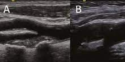
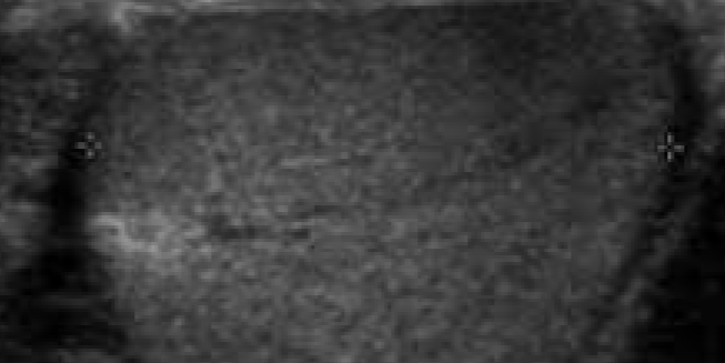
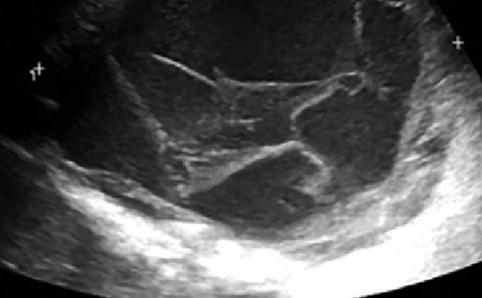
Echo Texture
Sonographic appearance of tissue within the body
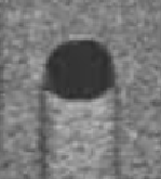
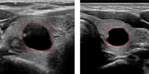
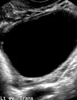
Anechoic
Echo-free appearance, structure will be black, fluid-filled
Isoechoic
The same echogenicity, same shade as another structure

Hypoechoic
Less echogenic, darker than another structure

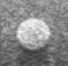
Hyperechoic
More echogenic, brighter than another structure
Gray Scale
Black and white image
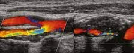
Color Doppler
Presence and direction of blood flow
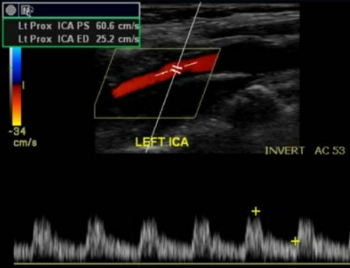
Pulsed Wave Doppler (Spectral)
Speed and direction of blood flow
Continuous Wave (CW) Doppler
Direction and speed of flow, but not location
Cystic
Fluid-filled
Solid
Composed of tissue
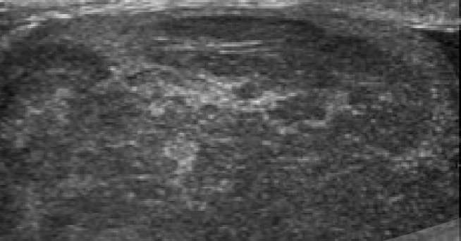
Homogenous
Similar or uniform echo pattern
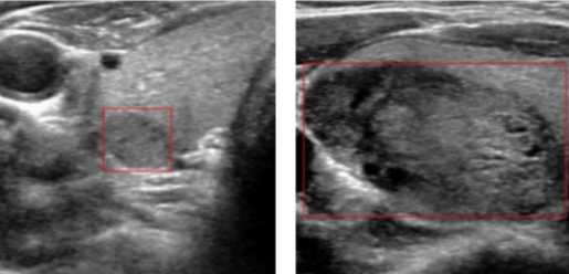
Heterogenous
Irregular or mixed echo pattern
Simple
Uncomplicated, usually referring to cysts
Anechoic, unilocular, thin smooth wall, no blood flow
Complex
Composed of both tissue and fluid
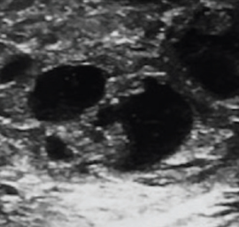
Septations
Thin membranes within a mass
Ipsilateral
On the same side
Contralateral
On the opposite side
NPO
Nothing by mouth
Neoplasm
Any abnormal growth
Benign
Non-cancerous
Malignant
Cancerous
Diffuse Disease
Disease throughout an organ
Superior/ Cephalic
Towards the head
Inferior/ Caudal
Towards the feet
Anterior/ Ventral
Front of body
Posterior/ Dorsal
Back of body
Medial
Towards middle of body
Lateral
Towards edge of body
Proximal
Towards the heart
Distal
Further from the heart
Subcostal
Beneath or below the ribs
Intercostal
Between the ribs
Midline
Vertical line- center of body
Midclavicular
Vertical line- middle of clavicles
Xiphoid Process
Lower end of sternum (breastbone)
Umbilicus
Belly button
Sternal Notch
Top of sternum
Iliac Crest
Top of hip bone
Symphysis Pubis
Joint of left and right pubic bones
Sagittal Plane
Longitudal, long
Divides body into left and right
Transducer indicator notch towards patient’s head
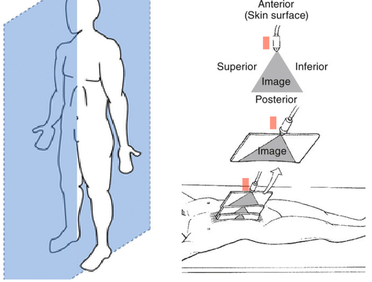
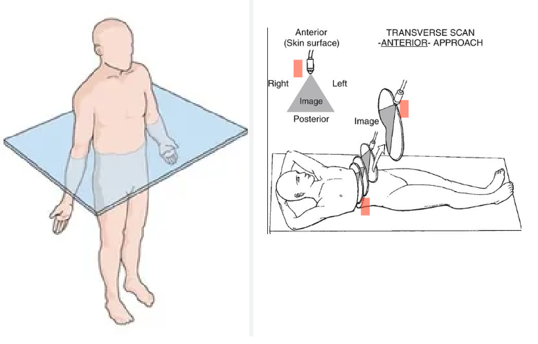
Transverse Plane
Divides body into superior and inferior
Transducer indicator notch towards US machine
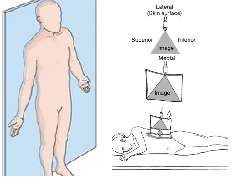
Coronal Plane
Divides body anterior to posterior
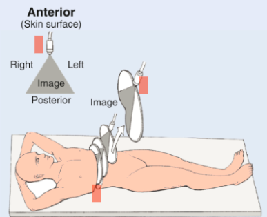
Transducer Indicator
Indicator points towards patient’s head in sagittal or towards the machine in transverse
The indicator will correspond with the left side of your screen
If indicator is to patient’s anatomic right, right side anatomy is seen on the left of the screen in transverse
Supine
Laying on back
Indicator towards machine (patient’s right)
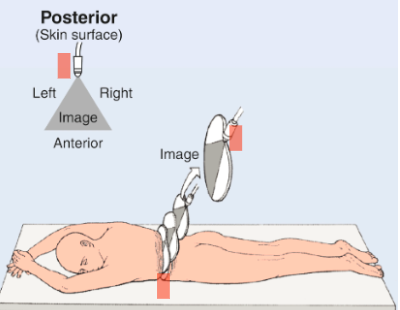
Prone
Laying on belly (not frequently used for US)
Indicator towards machine (patient’s left)
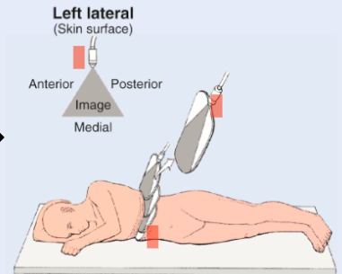
Left Lateral Decubitus (LLD)
Patient lays on left side
Enables scanning of right lateral surface
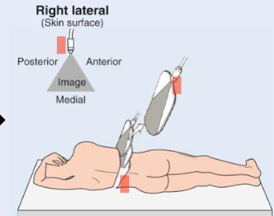
Right Lateral Decubitus (RLD)
Patient lays on right side
Enables scanning of left lateral surface