16. Surgery of the Ear
1/52
There's no tags or description
Looks like no tags are added yet.
Name | Mastery | Learn | Test | Matching | Spaced | Call with Kai |
|---|
No analytics yet
Send a link to your students to track their progress
53 Terms
inflammation of the vertical or horizontal ear canal or both
define otitis externa
inflammation of the tympanic cavity and membrane
define otitis media
inflammation of the inner ear that typically causes vestibular disease in dogs
otitis interna
extension of infection into the petrosal bone from otitis media
otitis interna is typically caused by....
age-related hearing loss
define presbycusis
diminished hearing
what can total ear canal ablation (TECA) result in which should be mentioned to the owners to understand their expectations for the procedure
reduces owner dissatisfaction associated with any perceived hearing loss after sx.
why should we be sure the owner is aware of the dog's hearing deficits before sx.
painful
ear disease and the surgical interventions that we use to treat them are _____
should be fully integrated into every phase of diagnosis, treatment, and recovery
in terms of pain management during ear procedures...
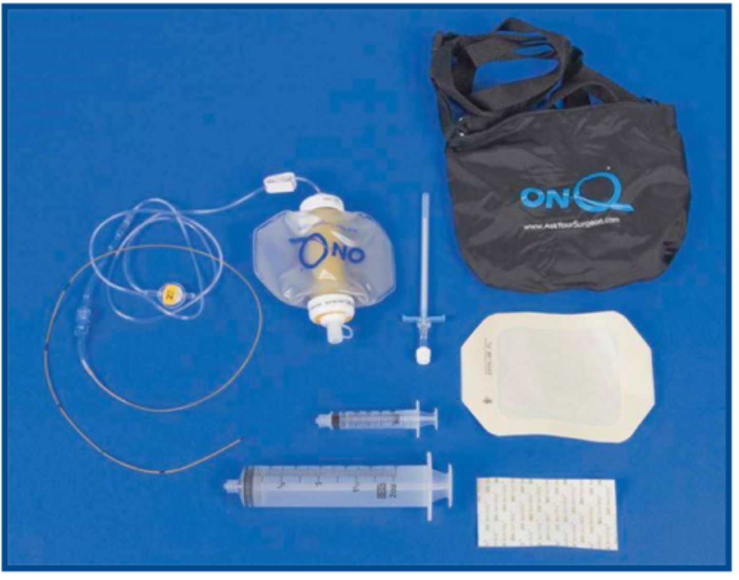
CRI delivery catheter kit for local anesthetic postoperative
what is shown here
find a pain scale that the staff can utilize quickly, easily, and consistently
no pain scale is perfect so it is important for you to....
guide analgesic therapy not deny it
what is the role of a pain scale
provide timely and adequate pain management
in terms of analgesic therapy, it can be helpful to...
do not spend too much time convincing yourself a patient is in pain just treat early
in terms of pain scales...
sicker
____ patients may need additional pain monitoring
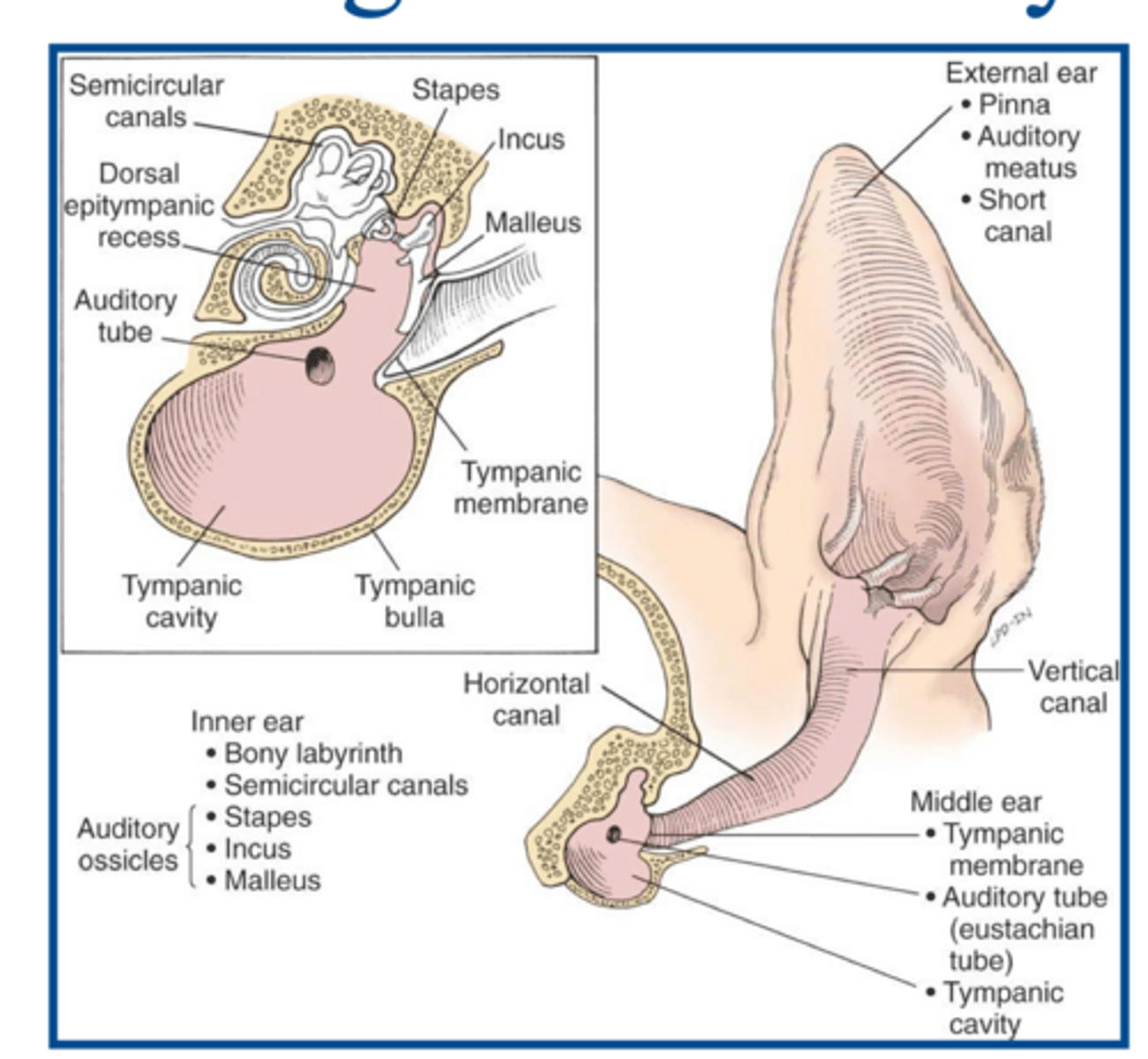
1. inner ear: membranous and bony labyrinth for hearing and balance
2. middle ear: tympanic cavity and auditory tube
3. external: auditory meatus and short canal
what are the three primary parts of the ear

tympanic membrane
what structure separates the middle and external ear canal

opening of horizontal canal into middle ear
what is the external acoustic meatus
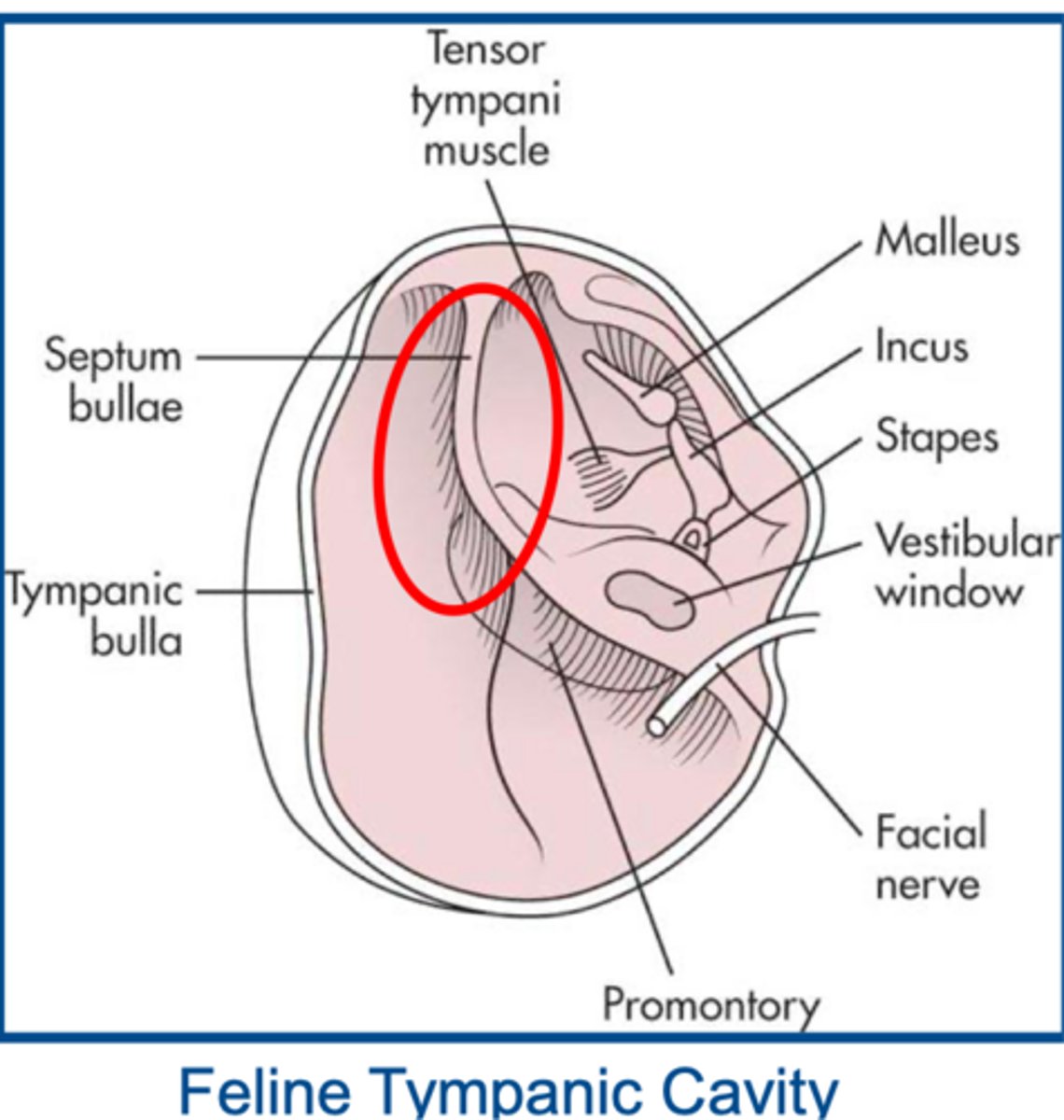
divided into two parts by thin bony septum...will need to be perforated to drain the ear
significance of the feline tympanic cavity
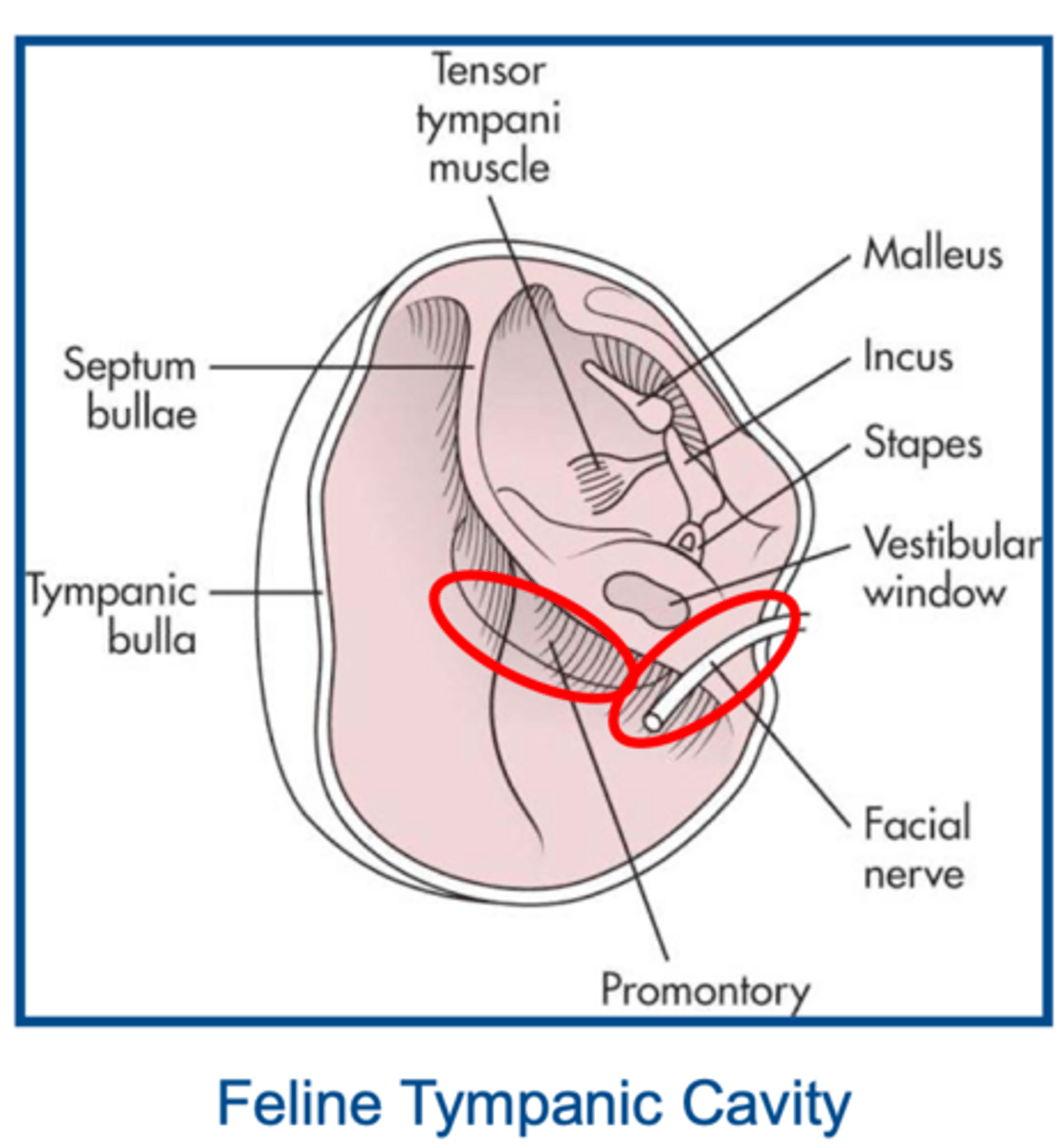
vulnerable location leading to the facial nerves often being traumatized during sx. causing Horner's syndrome
significance of location of nerves within the tympanic cavity
1. drooping of the eyelid on affected side-ptosis
2. pupil of affected eye will be constricted/miosis
3. affected eye appears sunken-enophthalmos
4. prominent third eyelid
most common C/s of Horner's Syndrome
1. diminished palpebral reflex
2. widened palpebral fissure
3. drooping of the ear and lip
4. excessive drooling
5. blepharospasm
6. elevation and wrinkling of the lip
7. caudal displacement of the labial commissure
8. elevation of the ear on the affected side
most common C/s of facial nerve paralysis
very common to happen but typically resolves within weeks-months
prognosis of facial nerve paralysis and horner syndrome in TECA-LBO
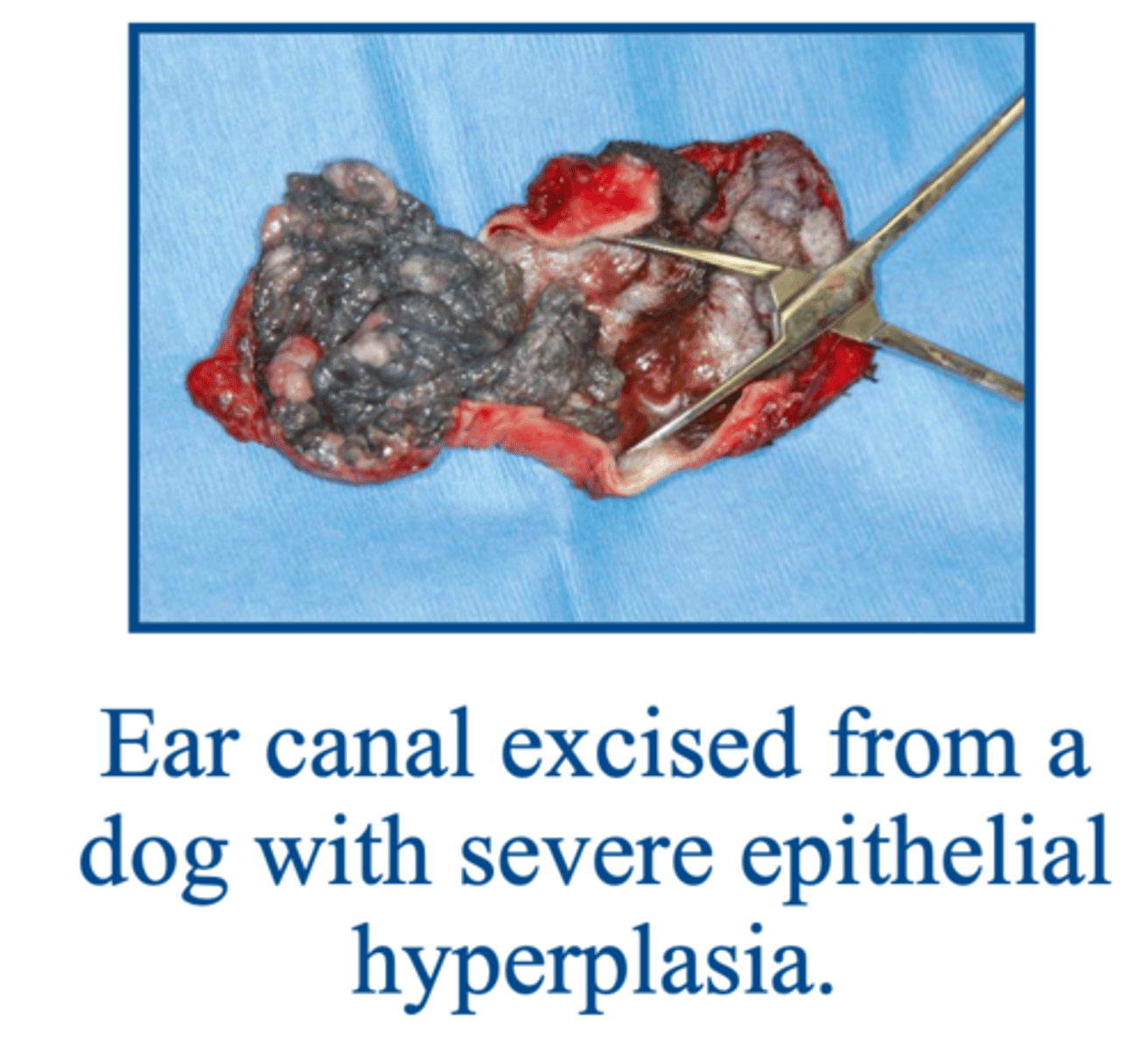
1. minimal hyperplasia of the ear canal epithelium
2. small neoplastic lesions of the lateral aspect of the vertical canal
indications for lateral ear canal resection
low
owner satisfaction for lateral ear canal resection in chronic otitis externa dogs is often ___
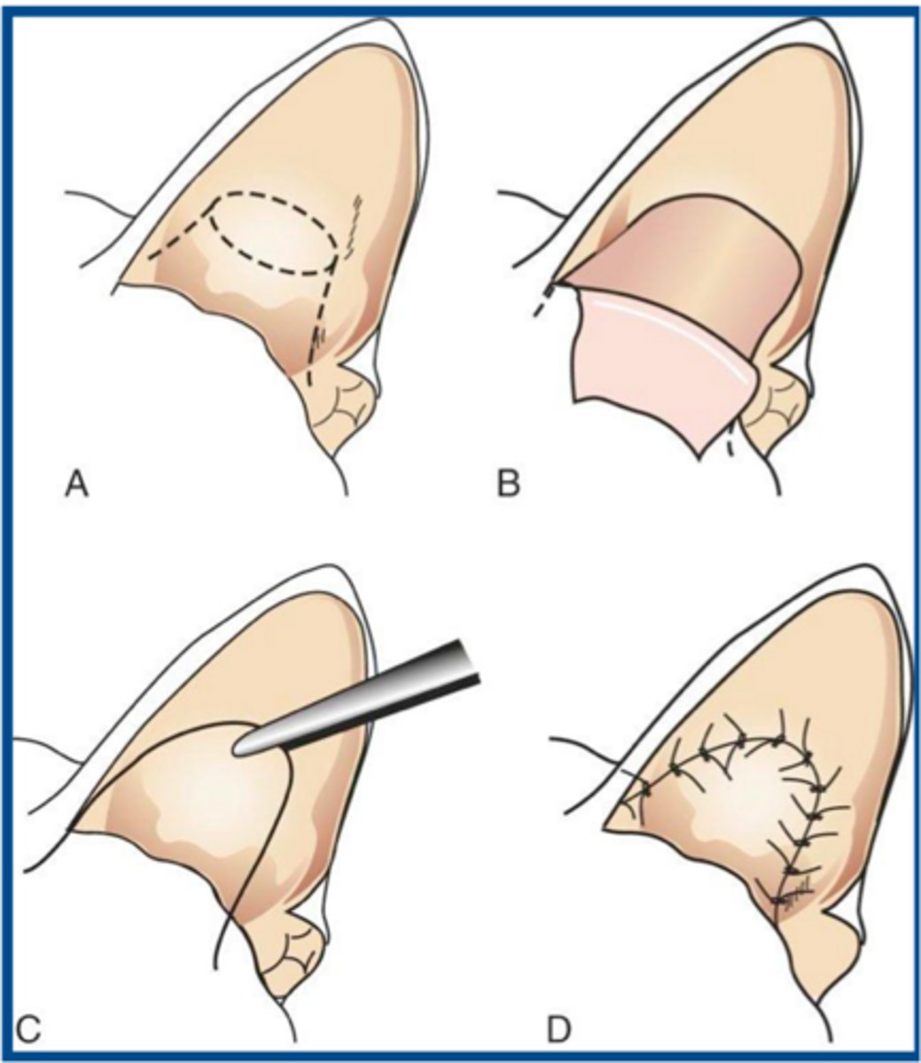
zep procedure (lateral ear canal resection) used to restrict hair growth at horizontal canal opening when there are small cancers found early on
what procedure is shown here and when is it typically used
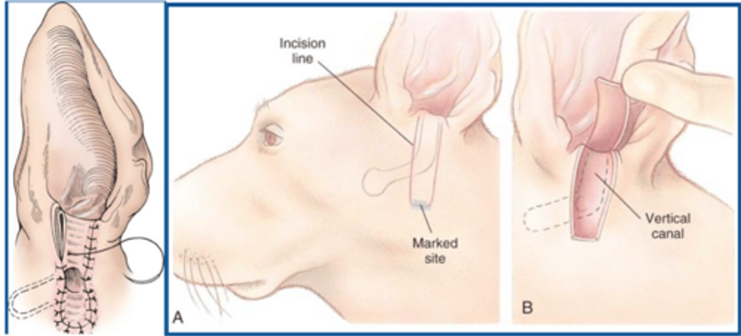
it is not a cure and medical management will be needed for the remainder of the animals life
when doing a lateral ear canal resection, it is important for the owner to understand that....

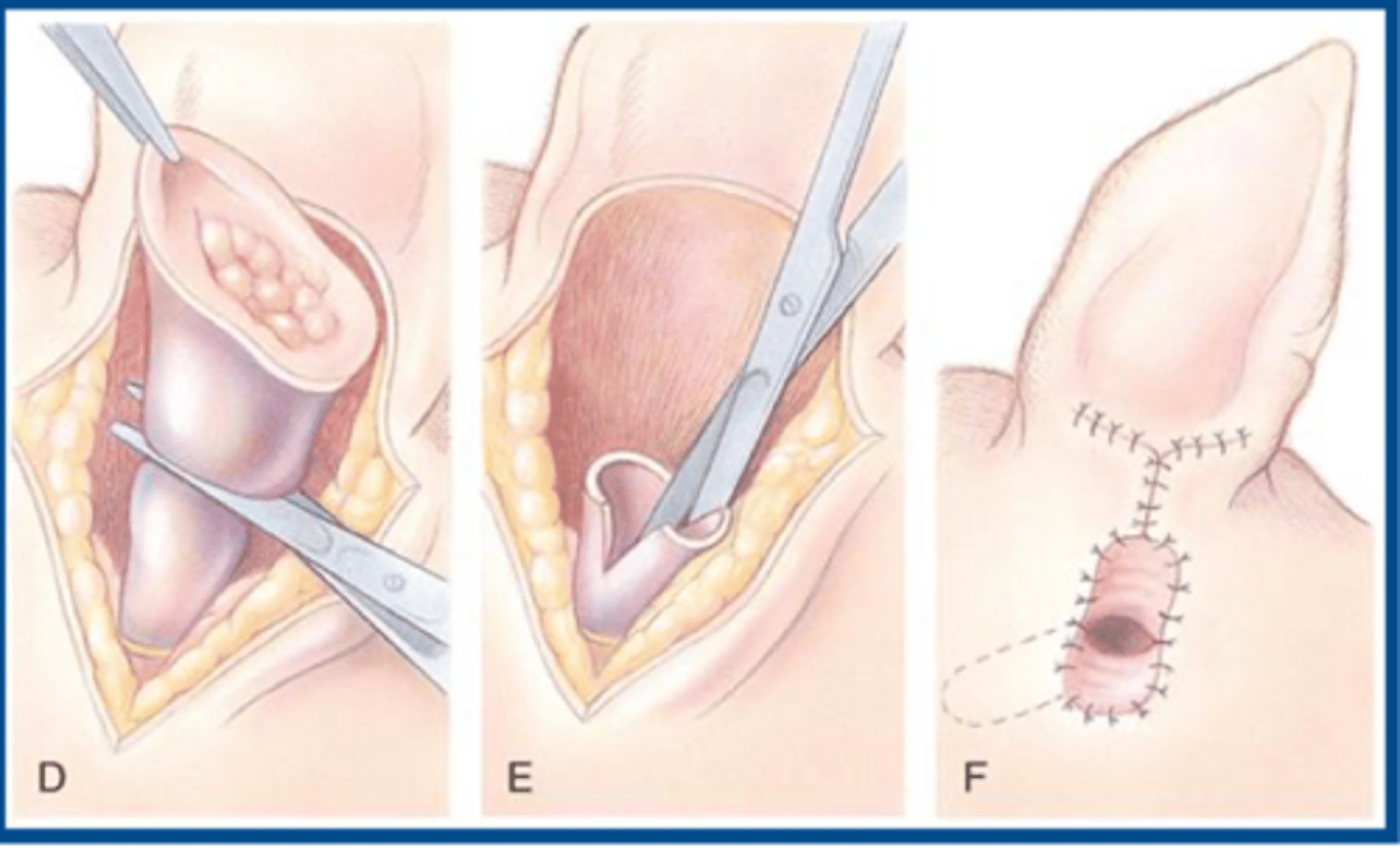
entire vertical canal is diseased but the horizontal canal is normal
when might the vertical ear canal ablation be performed

1. neoplasia is confined to vertical canal
2. some animals with chronic otitis externa
when might the vertical ear canal ablation be the technique of choice
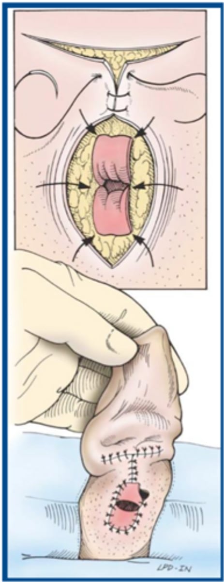
vertical ear canal ablation
does a vertical or lateral ear canal resection provide better cosmetic appearance
if you do not remove the avenue for drainage of exudative material by performing a TECA without treating otitis media...it is disastrous
why must a bulla osteotomy (LBO) always be performed in conjunction with a TECA for otitis externa and media
potential for serious complications due to nerve locations and vessels
why should TECA-LBO not be performed on animals with mild disease or by surgeons unfamiliar with ear anatomy
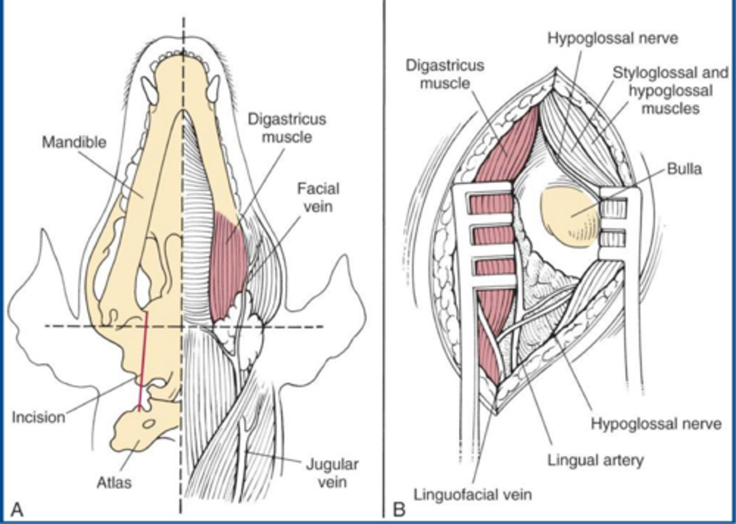
modified TECA-LBO in a cat
what procedure is shown here
lateral bulla osteotomy
what procedure is shown here
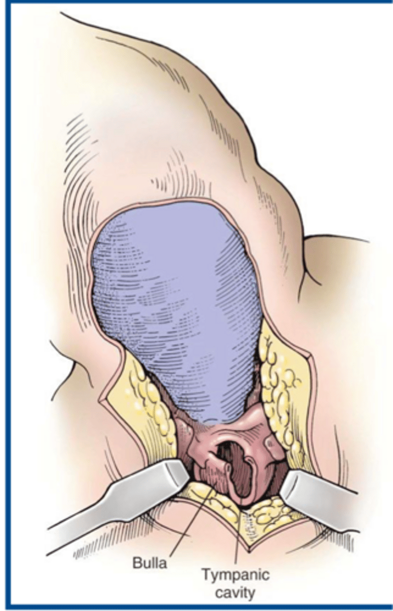
ideal when middle ear neoplasia is suspected in cats w/ nasopharyngeal polyps...allow both bullae to be opened without repositioning the animal
significance of a ventral bulla osteotomy
1. excessive swelling can impair respiration
2. need to note presence of abnormalities prior to sx. to avoid them being considered as surgical complications
3. Horner's syndrome and facial n. paralysis common in cats but typically transitory
in terms of complications associated with ventral bulla osteotomy
1. superficial wound infection
2. facial nerve paralysis
3. vestibular dysfunction
4. deafness
5. avascular necrosis of the skin of the pinna
6. chronic fistulations or abscessation
7. intraoperative arterial hemorrhage which may be life threatening
complications associated with TECA-LBO
1. usually resolves within a few weeks of sx
2. reported in 56% of cats aft TECA
3. permanent in approximately 1/4 of cats aft TECA
stats associated with facial nerve paralysis
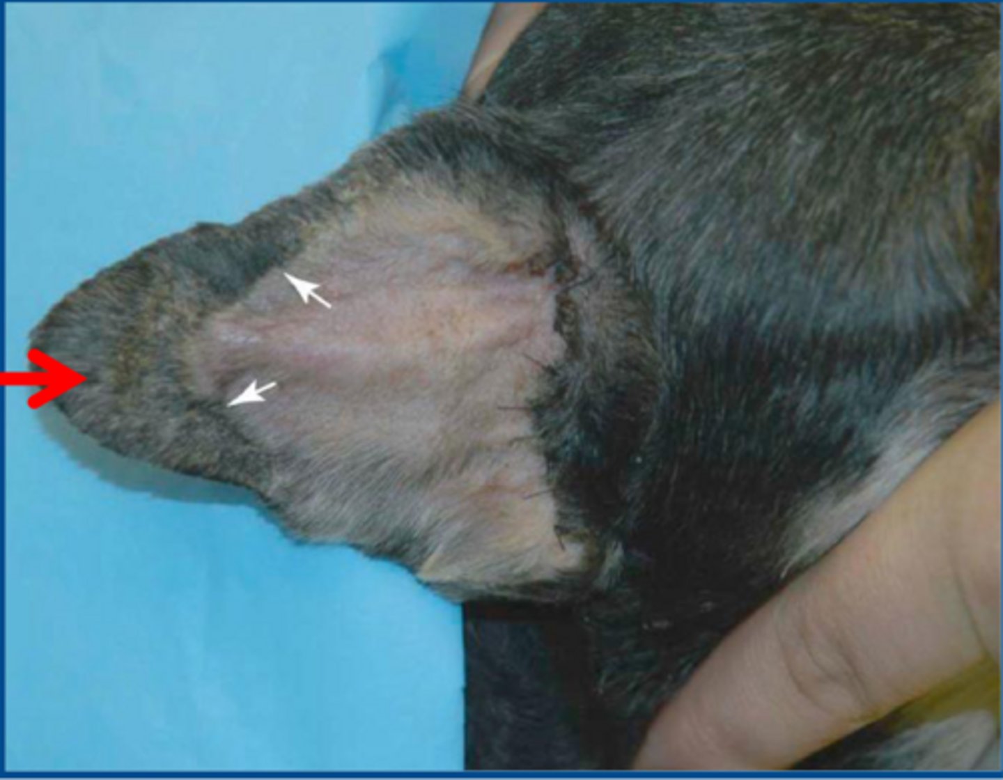
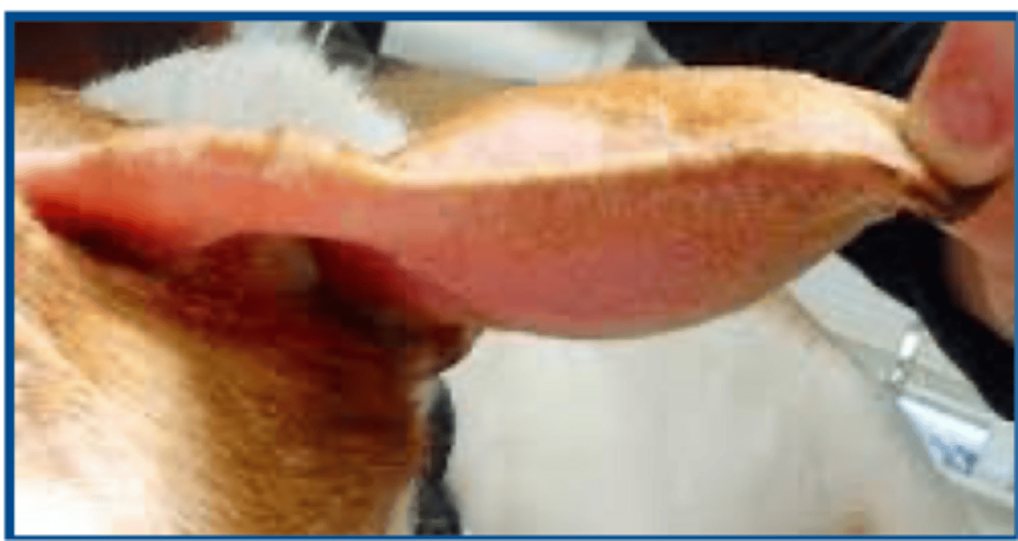
aural hematoma-collection of blood in cartilage plate of the ear secondary typically to shaking and scratching
what condition is shown here
otodectes cynotis
aural hematomas in cats are usually secondary to....
underlying disease must be ID and tx.
how do we reduce likelihood of aural hematoma recurrence
1. incision tissue overlying the hematoma
2. evacuating blood clots and fibrin
3. holding cartilage in apposition until scar tissue can form
most common tx. of aural hematomas
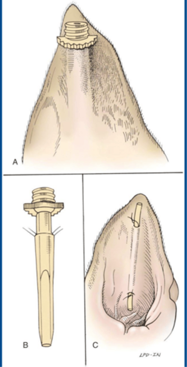
placement of drain or cannula to provide drainage for several weeks during healing
what is an alternative method for sx. tx. of aural hematoma...though not really ideal unless owners can keep up with it
1. sutures placed vertically and offset to maintain blood supply-->parallel to major vessles
2. excision left open to drain for up to 3wks...do NOT suture incision closed
3. "s" shape incision on concave surface of ear from end-to-end of the hematoma
4. leave no pockets to collect fluid
5. do NOT ligate visible branches of great auricular aa.
characteristics of aural hematoma tx.
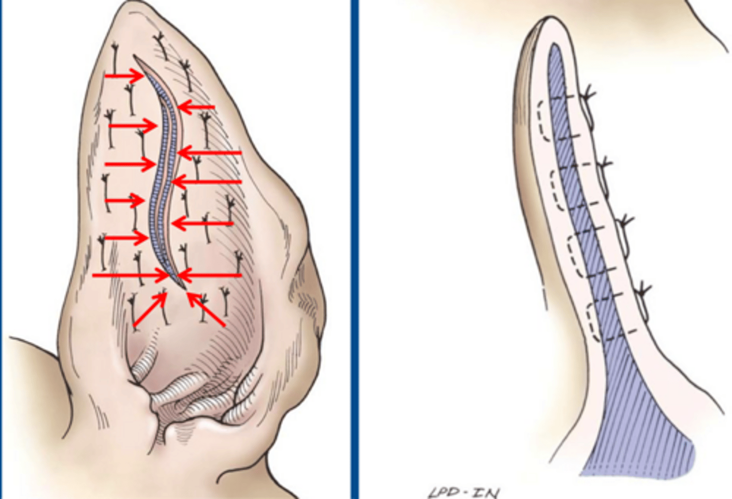
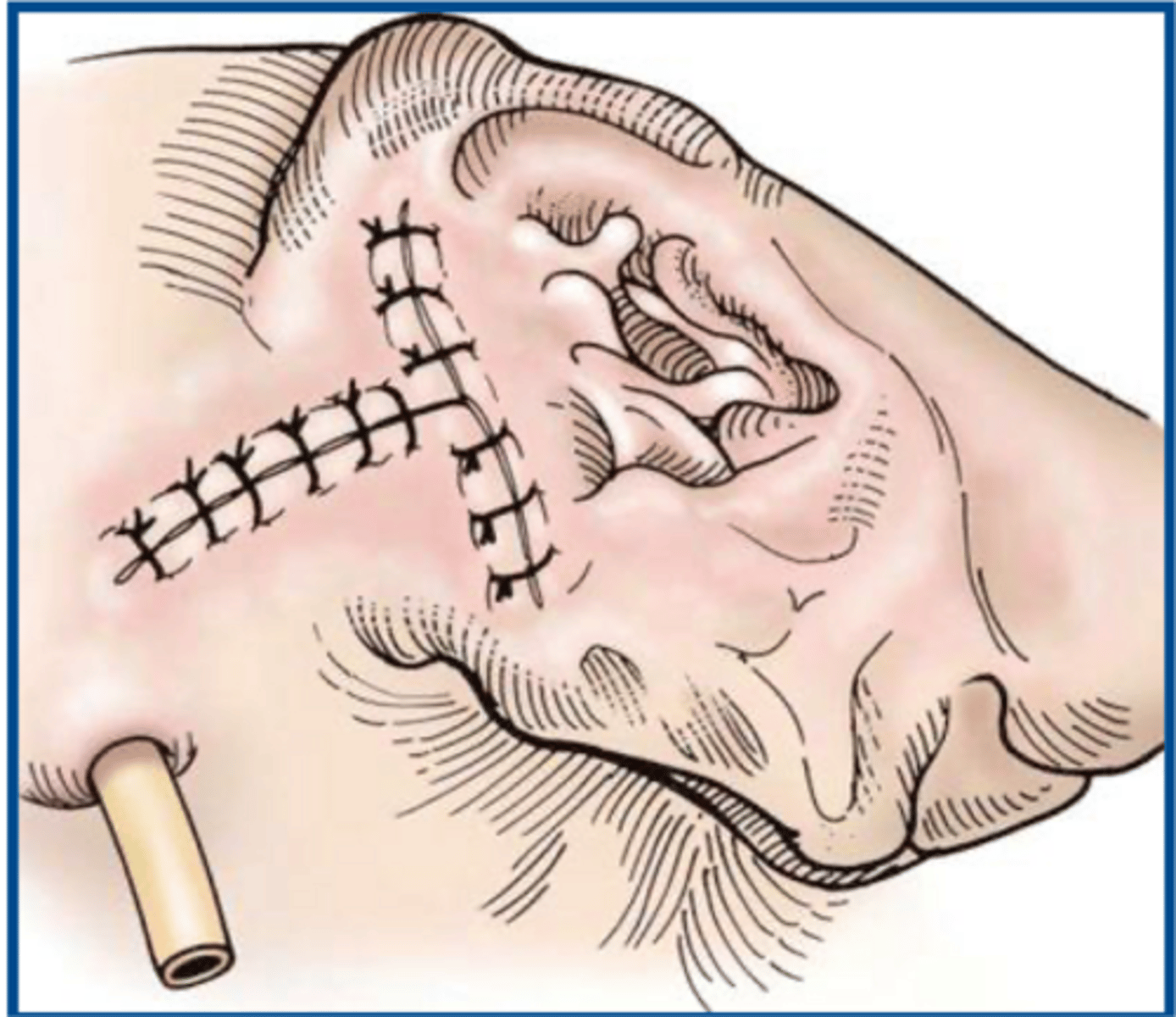
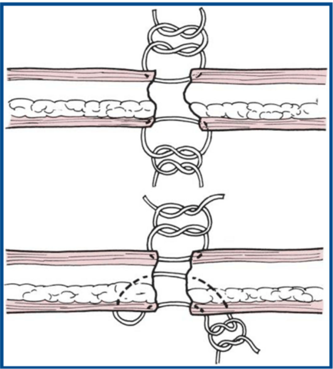
suture placement for repair of pinna lacerations
what is shown here
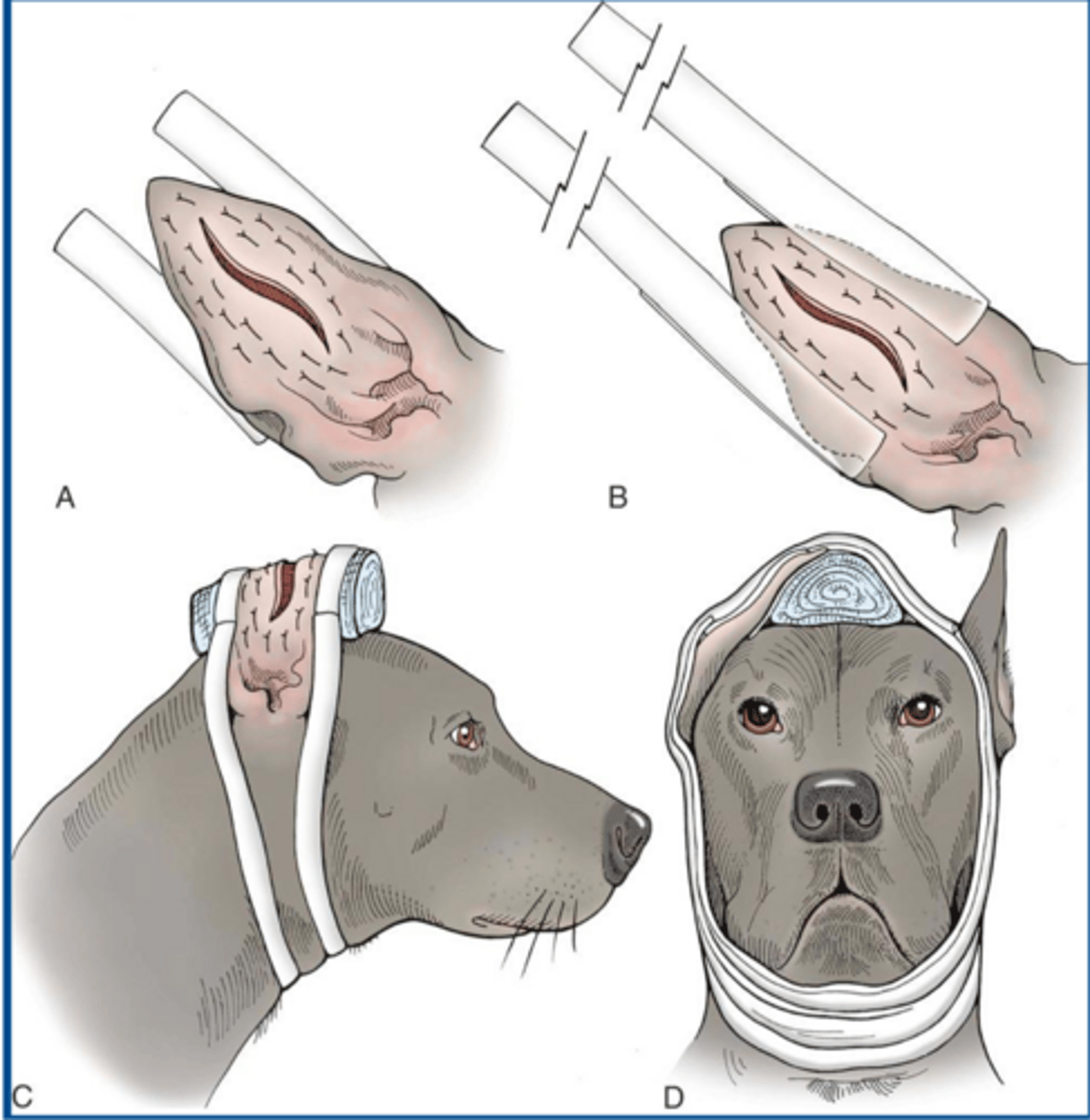
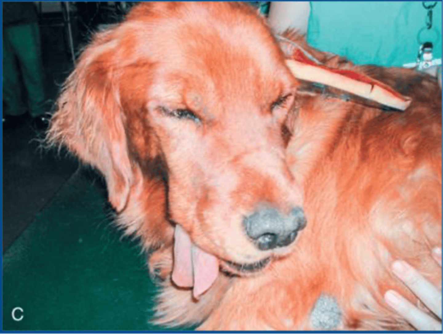
bandaging the ear after a surgical procedure
what procedure is shown here
aural hematoma pad placement
what procedure is shown here
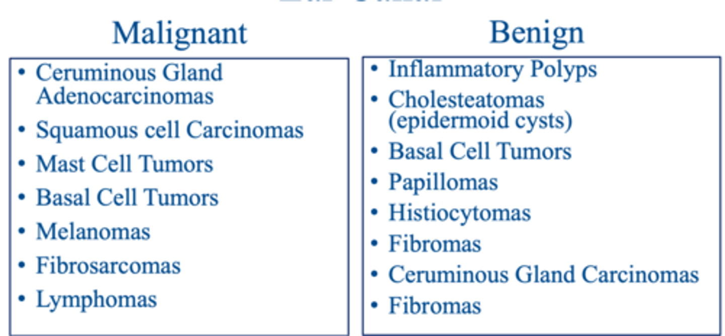
1. uncommon in dogs and cats...but more aggressive in cats
2. benign or malignant
3. most common are from ceruminous glands
4. often associated with otitis externa, media, interna
neoplasia of the pinna and external ear canal...
malignant vs. benign neoplasia of the pinna and external ear canal
what is shown here
achieving wide margins to prevent local recurrence
what is the most important aspect of sx. of ear neoplasms
adjunctive therapy like radiation should be considered
if aggressive sx. therapy of ear neoplasms cannot provide clean margins...
cosmetic defects due to wide margins of normal tissue
what should owners be warned of prior to excision of malignant ear tumors
stopped on slide 78...anything beyond this was not on the objectives