femoral canal
1/29
There's no tags or description
Looks like no tags are added yet.
Name | Mastery | Learn | Test | Matching | Spaced | Call with Kai |
|---|
No analytics yet
Send a link to your students to track their progress
30 Terms
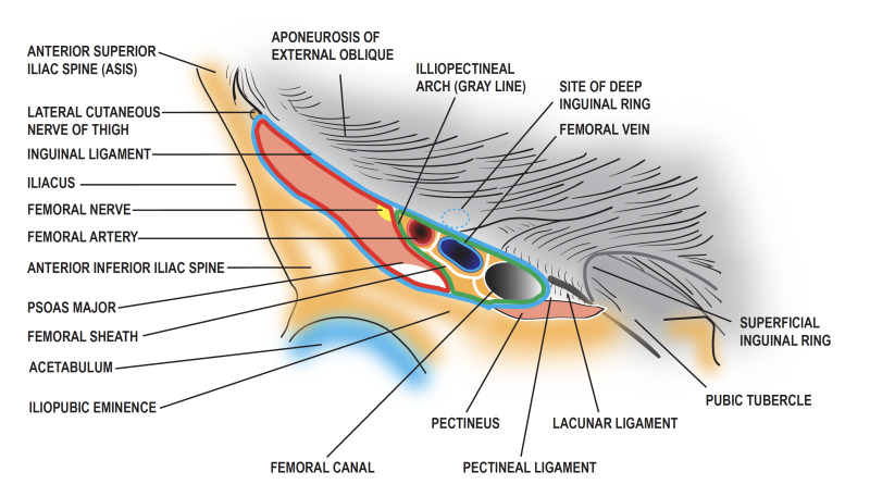
-Femoral Canal – Overview
The femoral canal is the medial compartment of the femoral sheath.
Unlike the other compartments, it does not transmit major vessels.
It is filled with loose fibrofatty tissue, lymphatics, and a lymph node (Cloquet’s node).
Its main function is to allow expansion of the femoral vein when venous return increases.
Femoral Ring.
The femoral ring is the upper opening (abdominal end) of the femoral canal.
It is:
About ½ inch wide
Oval-shaped
It is normally closed by the femoral septum, which is made of extraperitoneal fat.
Boundaries of the Femoral Ring
Anteriorly: Inguinal ligament
Posteriorly: Pectineal line + pectineus muscle and its fascia
Laterally: Femoral vein
Medially: Lacunar ligament (sharp, concave edge
Mnemonic to Remember Femoral Ring Boundaries
Think: “LIP-L” (like lips around the ring)
L → Lateral → Femoral vein
I → Inferior (posterior in anatomy position here) → Pectineal line + pectineus
P → Proximal (anterior) → Inguinal ligament
L → Medial → Lacunar ligament
👉 Or a simpler phrase:
“V-I-P-L” (Vein, Inguinal, Pectineal, Lacunar)
Move clockwise from lateral:
Vein → Inguinal → Pectineal → Lacunar’
What are the boundaries of the femoral ring?
:
Anterior: Inguinal ligament
Posterior: Pectineal line + pectineus muscle & fascia
Lateral: Femoral vein.
Medial: Lacunar ligament
Extra points:
Femoral ring = upper opening of femoral canal
Closed by femoral septum
Site of femoral hernia (high risk of strangulation)
(Anterior)
Inguinal Ligament
───────
Medial Lateral
Lacunar Lig. Femoral Vein
│ │
│ ○ │ ← Femoral ring
│ │
Pectineal Line
+ Pectineus Muscle
(Posterior).
.Clinical Hook (helps memory stick)
The lacunar ligament (medial) is sharp →
👉 this is why strangulation of femoral hernia is common
.🧠 Functions of Femoral Canal
👉 Think: “V-L” (VAllows expansion of femoral vein
.Allows expansion of femoral vein
Acts as a dead space when venous return increases
Passage for lymphatics
Drains lymph from lower limb → external iliac lymph nodes
Applied Anatomy (Femoral Hernia)’
The femoral ring is a weak point → site of femoral hernia
A loop of intestine may protrude into femoral canal
📌Why more common in females?
Think: “Wide + Small + Pressure”
Wider pelvis → larger femoral canal
Smaller femoral vessels → more space
Pregnancy → ↑ intra-abdominal pressure
I🚨 Strangulated Femoral Hernia
‘Treated by cutting the lacunar ligament
⚠ Must be careful of:
Accessory obturator artery (can be injured → bleeding)
Direction of Femoral Hernia (VERY IMPORTANT)
.👉 Think: “Down → Forward → Up & Lateral”
Downward → through femoral canal
Forward → through saphenous opening
Upward & lateral → along superficial vessels
.Functions of femoral canal + applied anatomy of femoral hernia
Functions:
Allows expansion of femoral vein (dead space)
Transmits lymphatics to external iliac nodes
Femoral hernia:
Occurs through femoral ring (weak point)
More common in females (wide pelvis, small vessels, pregnancy)
Direction:
Downward → Forward → Upward & lateral
Clinical:
Strangulation common
Lacunar ligament is cut (beware accessory obturator artery)
Q: What is the main function of the femoral canal?.
A: Provides space for expansion of the femoral vein.
Q: What structures pass through the femoral canal?.
‘A: Lymphatics from lower limb to external iliac lymph nodes.
‘Q: Why is the femoral canal called a “dead space”?
A: Because it allows expansion of the femoral vein.
Q: Through which structure does a femoral hernia occur?
A: Femoral ring.
IQ: Why is the femoral ring clinically important?
A: It is a weak point prone to herniation.
Q: Why are femoral hernias more common in females?
A: Wider pelvis, smaller femoral vessels, increased intra-abdominal pressure (pregnancy).
Q: What is the direction of a femoral hernia (step 1)?
A: Downward through femoral canal.
Q: What is the direction of a femoral hernia (step 2)?
A: Forward through saphenous opening.
Q: What is the direction of a femoral hernia (step 3)?
A: Upward and laterally along superficial vessels.
Q: What structure is cut to relieve strangulated femoral hernia?
A: Lacunar ligament.
Q: Which artery is at risk when cutting the lacunar ligament?
A: Accessory obturator artery.
Q: Give a quick summary of femoral canal function and hernia direction.
.
A: Vein expansion + lymphatics; hernia goes Down → Forward → Up & lateral.
Direction of Enlarging Femoral Hernia
1⃣
Think of it as a three-step path:
Downward → through the femoral canal
Forward → bulges through the saphenous opening
Upward & laterally → along the superficial epigastric and superficial circumflex iliac vessels
Mnemonic: “Down → Forward → Up & Lateral” (DFUL)
2⃣ Coverings of Femoral Hernia (From Inside Out)
Peritoneum of hernial sac
Femoral septum (extraperitoneal fat)
Anterior wall of femoral sheath
Cribriform fascia
Superficial fascia
Tip: Think “P-F-A-C-S” (Peritoneum → Femoral septum → Anterior wall → Cribriform fascia → Superficial fascia)
3⃣ Reduction of Femoral Hernia
Position thigh: slightly flexed & medially rotated → relaxes fascia and ligaments
Reduce hernia opposite the path of hernial sac (reverse DFUL)
Mnemonic: “Flex, Rotate, Reverse the Flow”
Q: What is the first direction of an enlarging femoral hernia?.
A: Downward through the femoral canal.
Q: What is the second direction of an enlarging femoral hernia?
A: Forward, bulging through the saphenous opening.
Q: What is the third direction of an enlarging femoral hernia?
A: Upward and laterally along superficial epigastric and superficial circumflex iliac vessels.
Q: List the coverings of a femoral hernia from inside outward.
A: Peritoneum → Femoral septum → Anterior wall of femoral sheath → Cribriform fascia → Superficial fascia.
Q: How is a femoral hernia reduced surgically?
A: Thigh slightly flexed & medially rotated, then hernia pushed back opposite to its course.