Biology 223 - Lab 4: Axial Muscles
1/68
There's no tags or description
Looks like no tags are added yet.
Name | Mastery | Learn | Test | Matching | Spaced | Call with Kai |
|---|
No analytics yet
Send a link to your students to track their progress
69 Terms
Reflect
The act of cutting a muscle and pulling it away to view deeper muscles.
Origin
End of a muscle that is fixed during contraction. (usually proximal for appendicular muscles)
Insertion
End of a muscle that undergoes movement during contraction ( usually distal for appendicular muscles).
Innervation
refers to nerve penetration of muscles
Agonist
The muscle that is responsible for performing the bulk of movement.
Antagonist
The muscle opposing the action of the agonist.
Synergist
The muscle that assists the agonist muscle perform particular movements.
Flexion
Type of movement that reduces the angle of a joint.
Extension
Type of movement that increases the angle of a joint.
Adduction
Movement towards the longitudinal axis of the body.
Abduction
Movement away from the longitudinal axis of the body.
Pronation
Rotating the forearm so that palms face posterior.
Supination
Rotating the forearm so that palms face anterior.
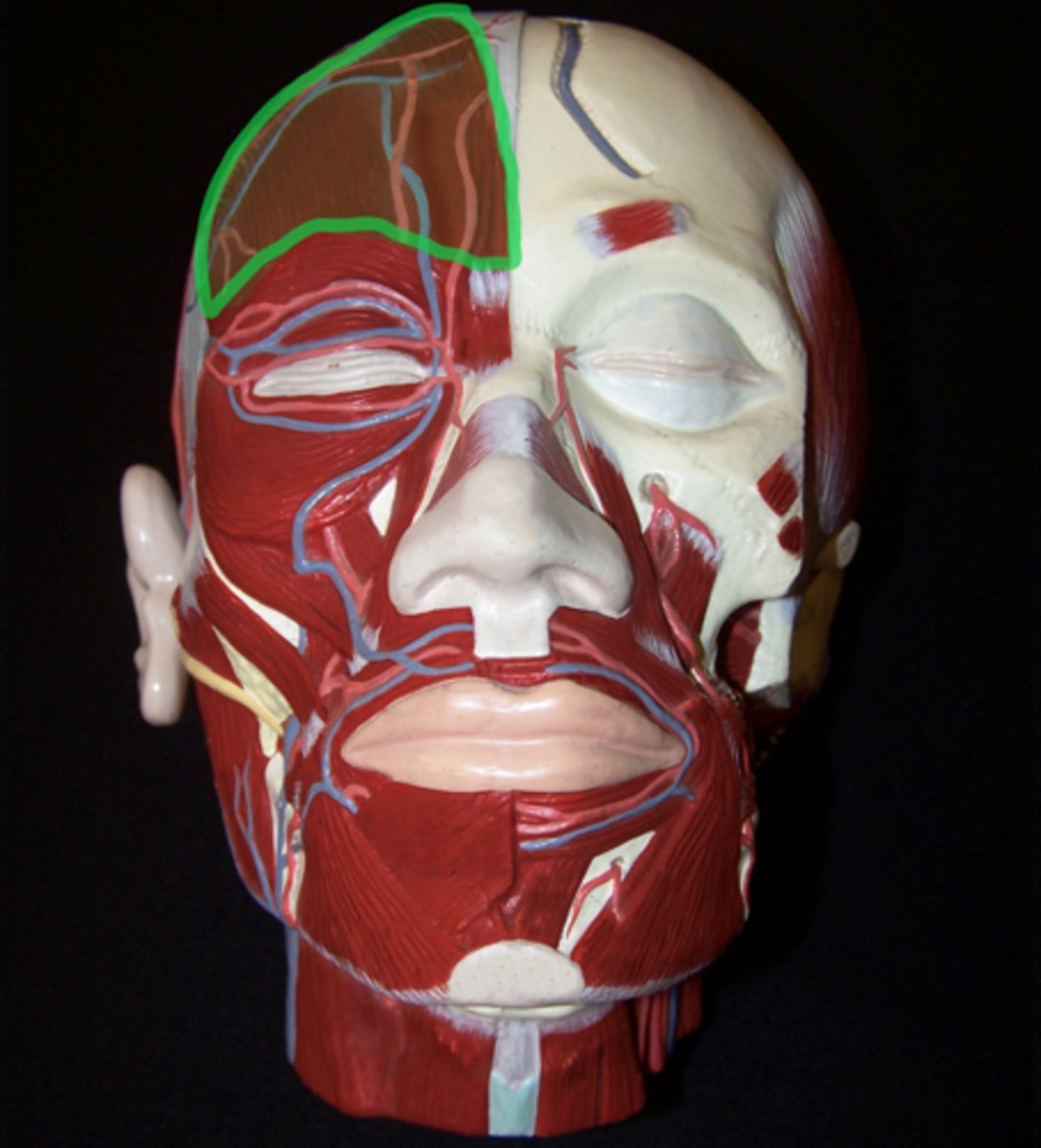
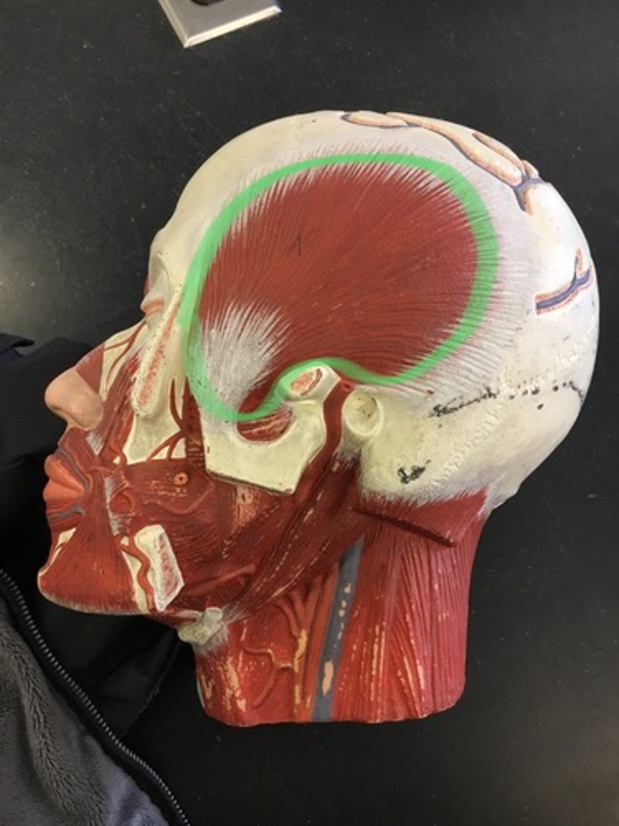
Frontal belly of occipitofrontalis
Superficial side, right on the forehead.
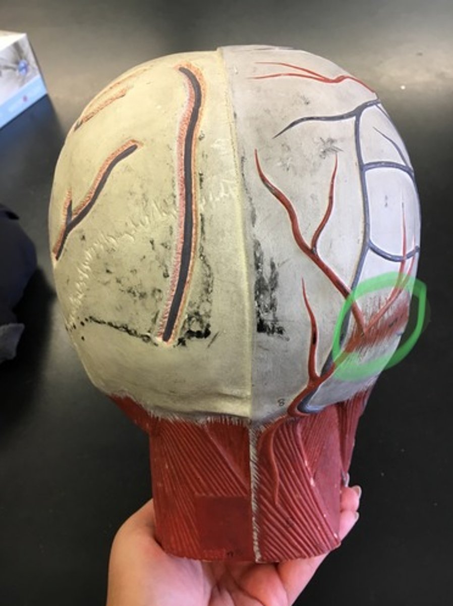
Occipital belly of occipitofrontalis
Superficial side, small, in the back
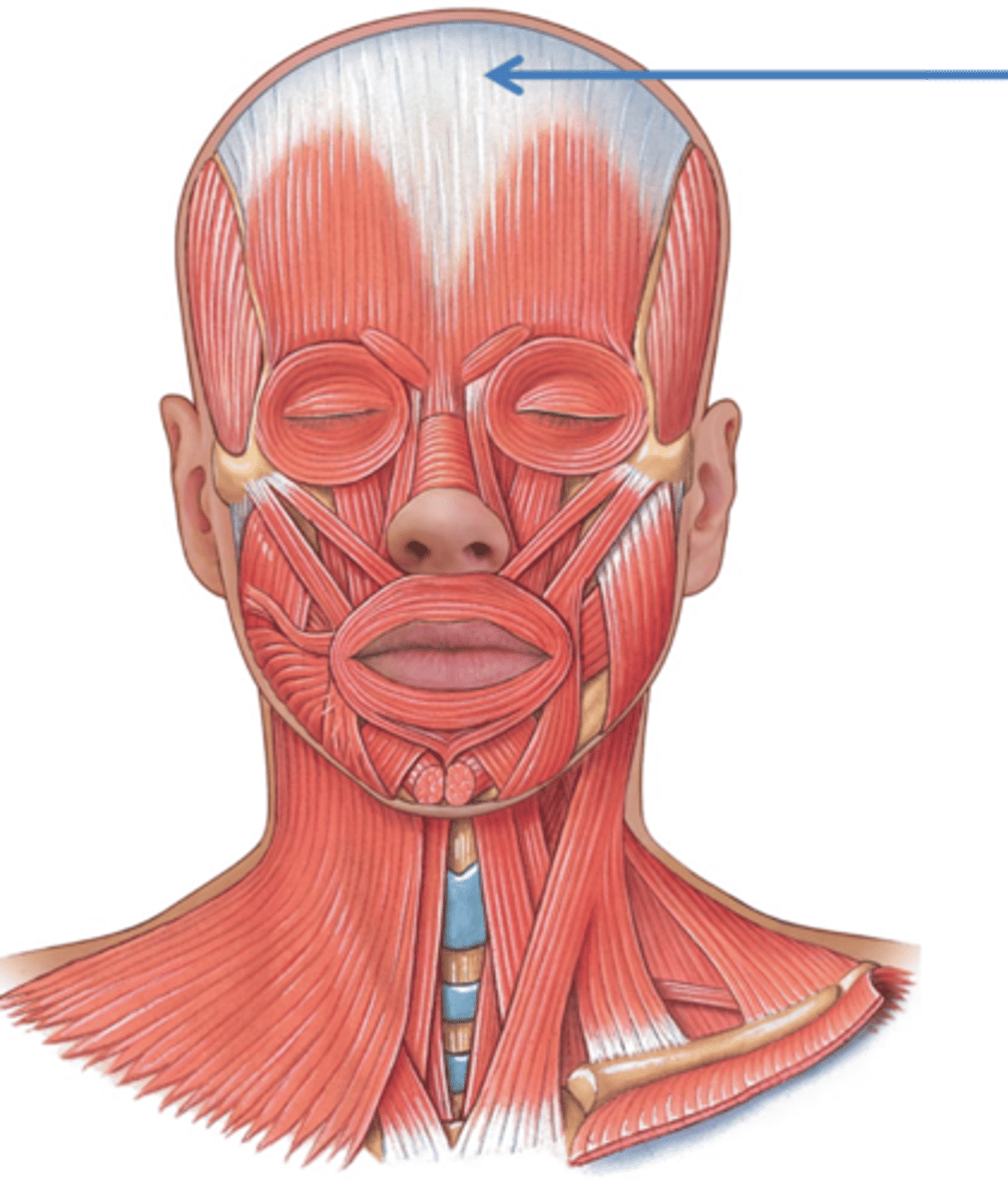
Epicranial aponeurosis
Gray matter, connects all occipitofrontalis
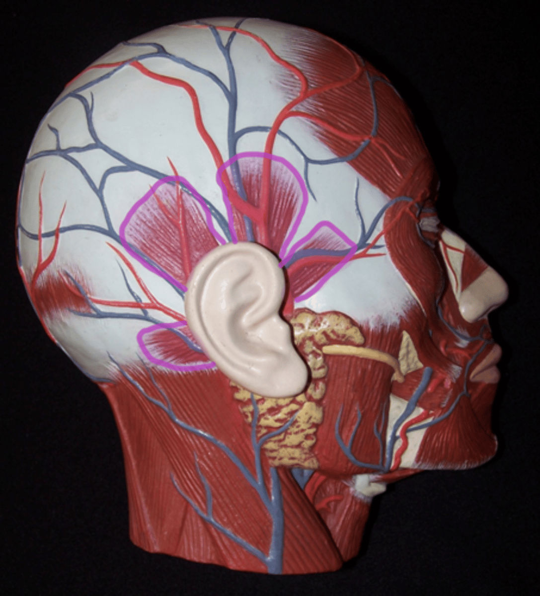
Temporoparietalis
Windmill-like, by ear, above temporalis
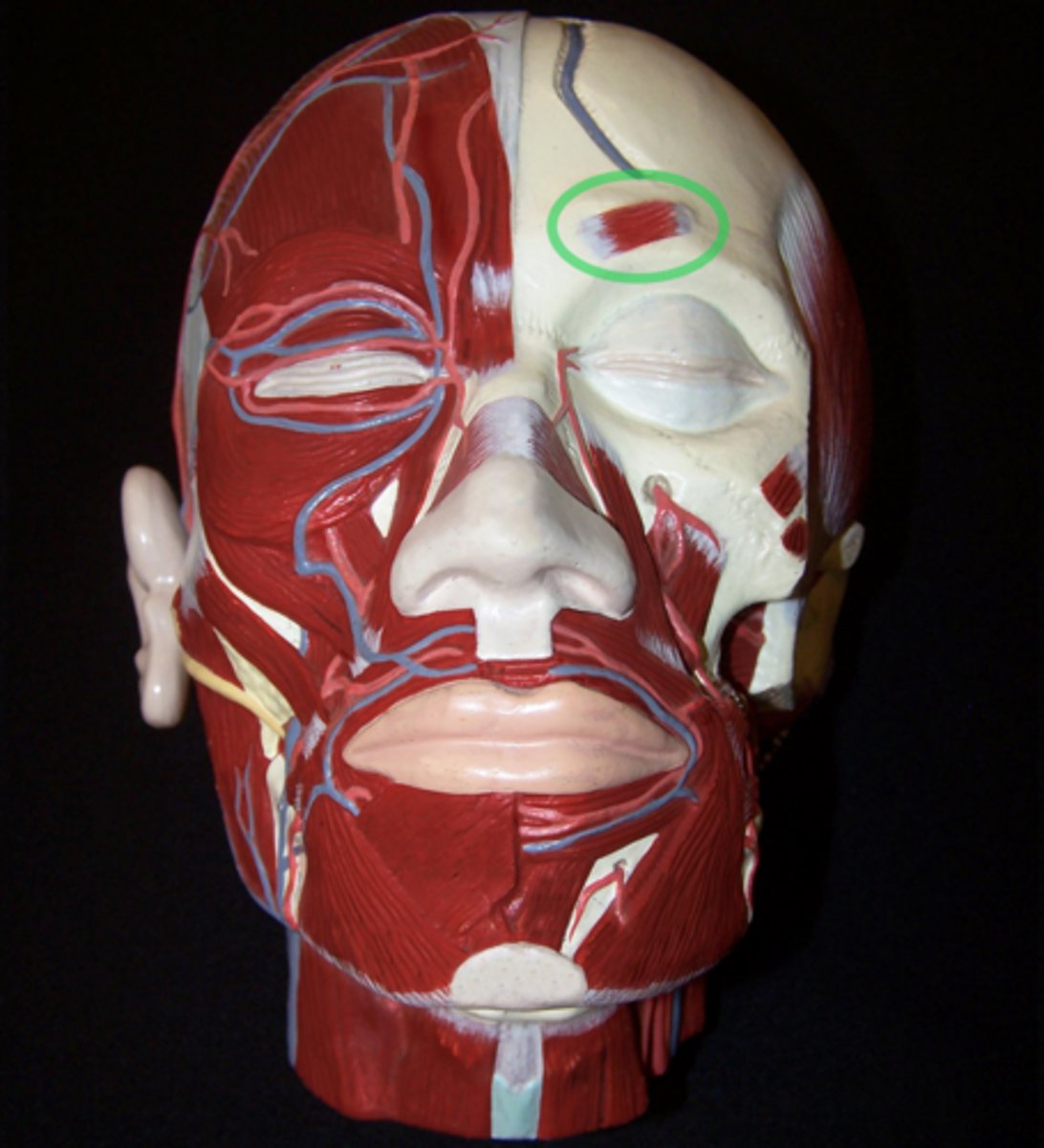
Corrugator supercilii
Deep side; where eyebrow would go. Eyebrow muscle under frontal belly of occipitofrontalis.
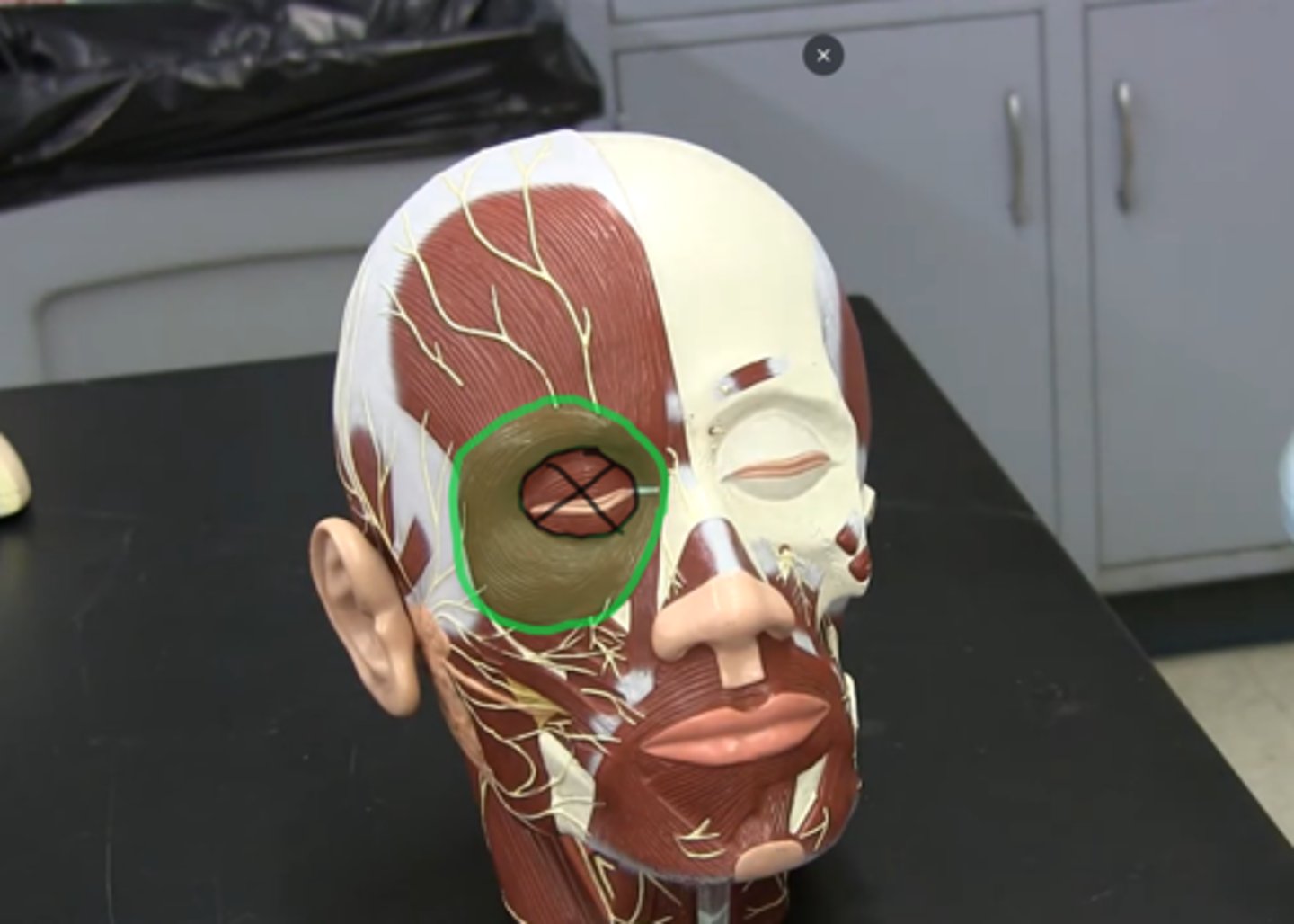
Orbicularis oculi
All around the eye; does not include eyelid.
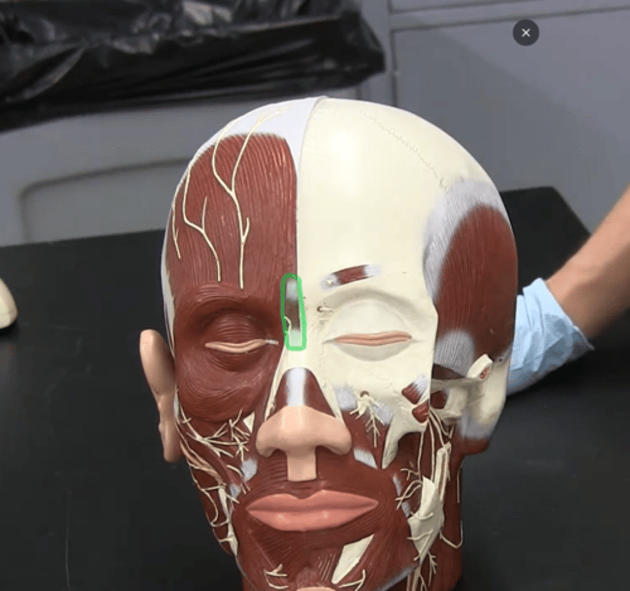
Procerus
Vertical striations running between orbicularis oculi.
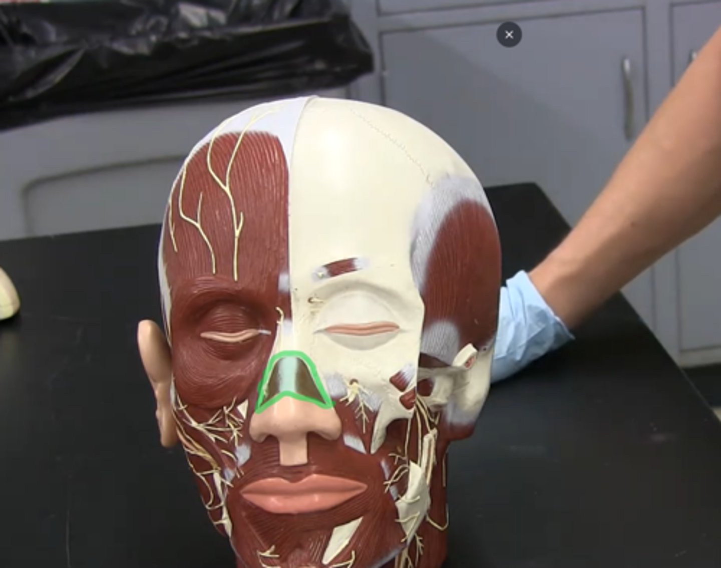
Nasalis
Horizontal striations across bridge of the nose.
Zygomaticus major
(Bottom) a slender band of muscle on each side of the face that arises from the zygomatic bone, inserts into the orbicularis oris and skin at the corner of the mouth, and acts to pull the corner of the mouth upward and backward when smiling or laughing.
*The structure is clipped on this model so you can only see s small piece of it*
Zygomaticus minor
(Top) a muscle of facial expression. It originates from zygomatic bone and continues with orbicularis oculi on the lateral face of the levator labii superioris and then inserts into the outer part of the upper lip.
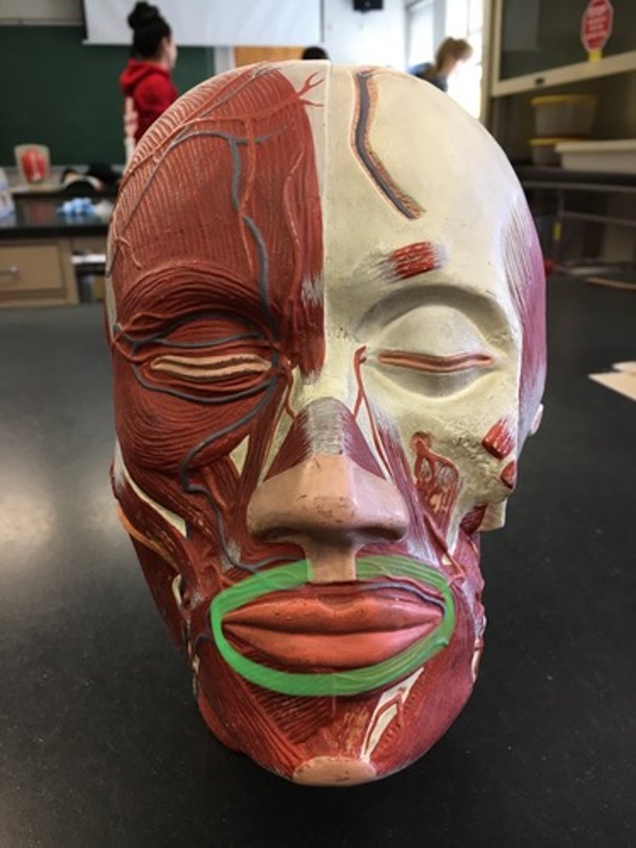
Orbicularis oris
Around the mouth.
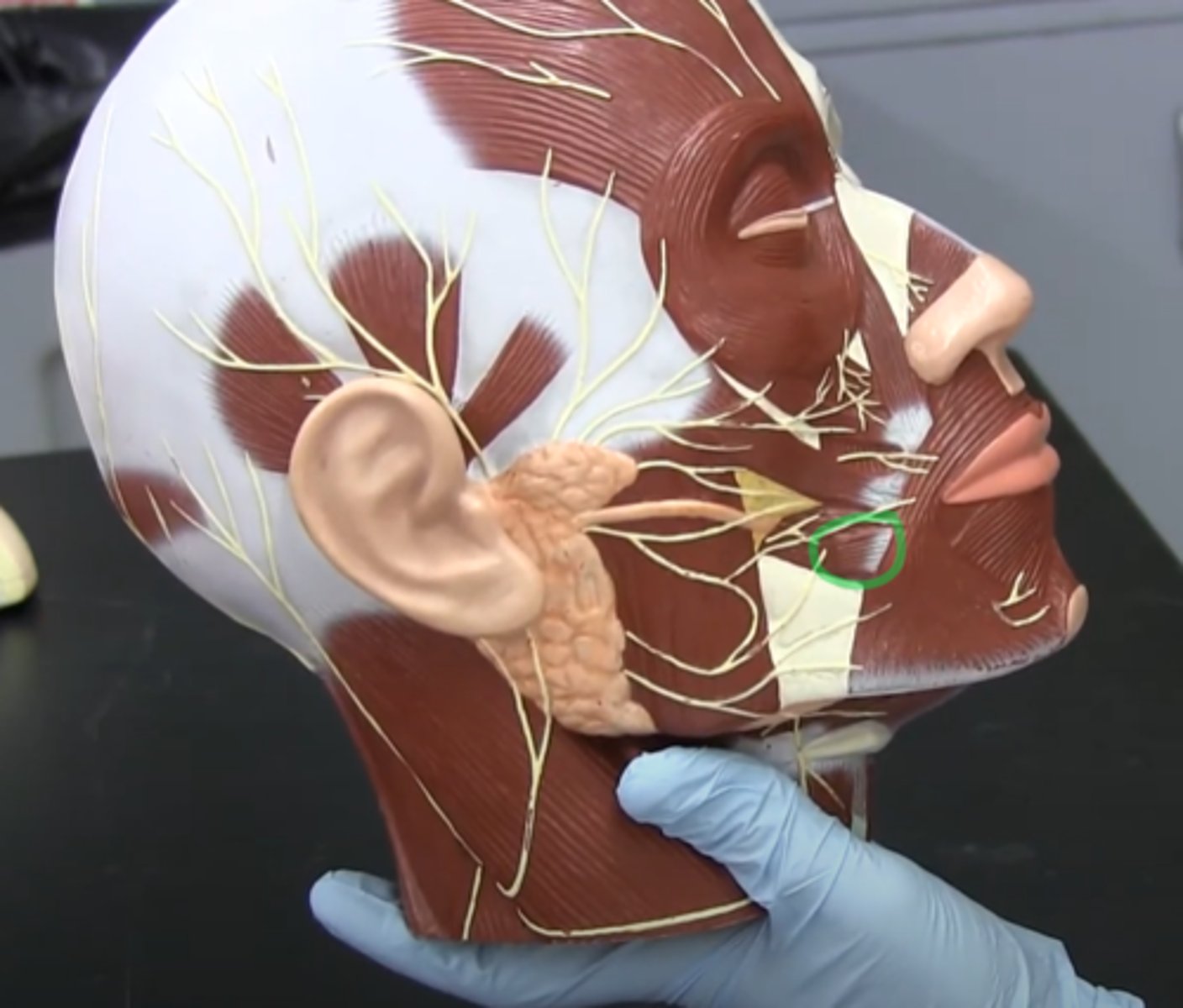
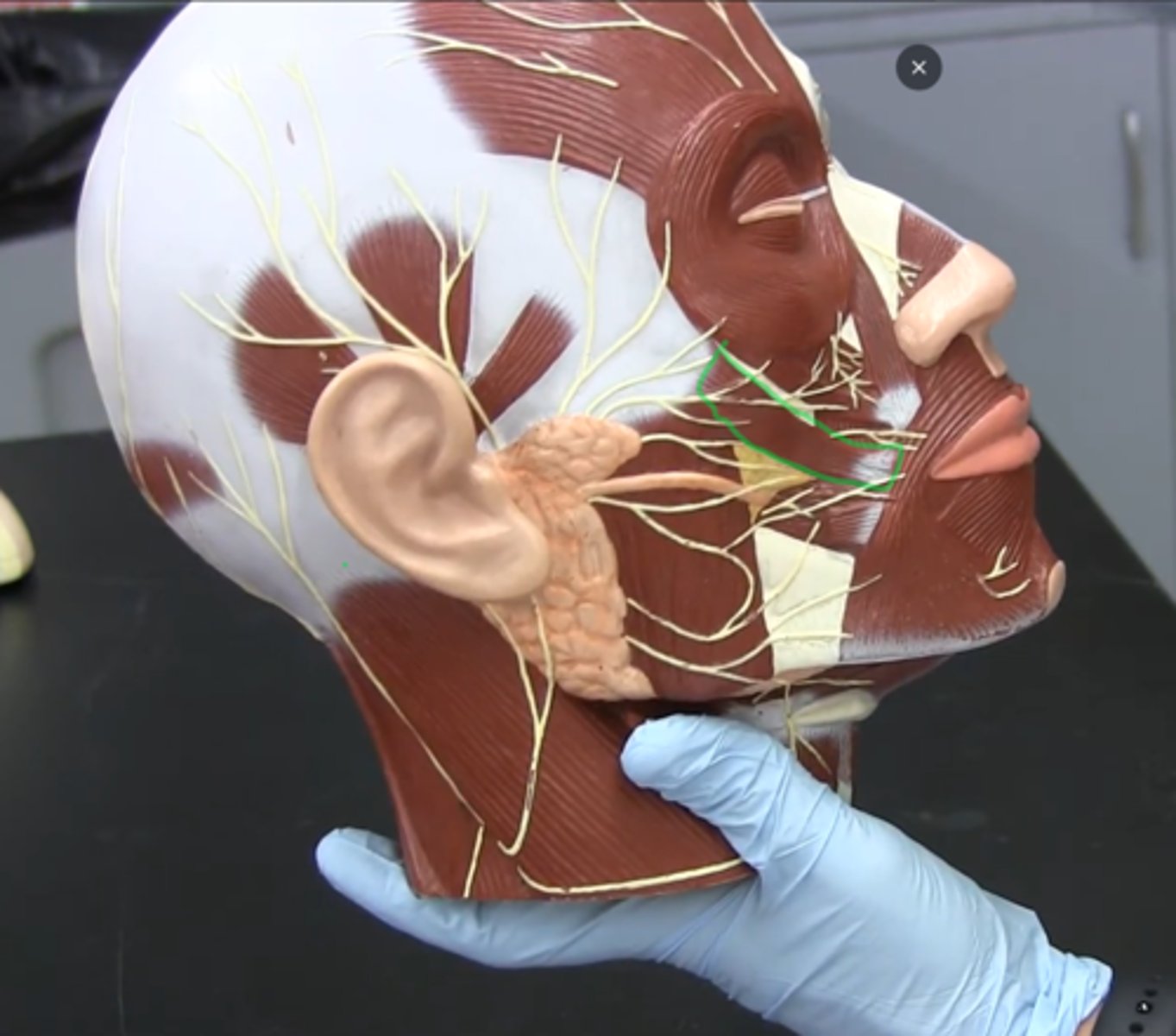
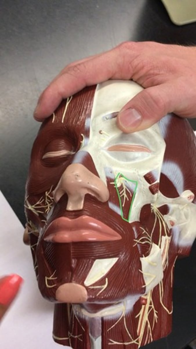
Levator labii superioris
Cheek, underneath eye (deep side) yellow nerve on it.
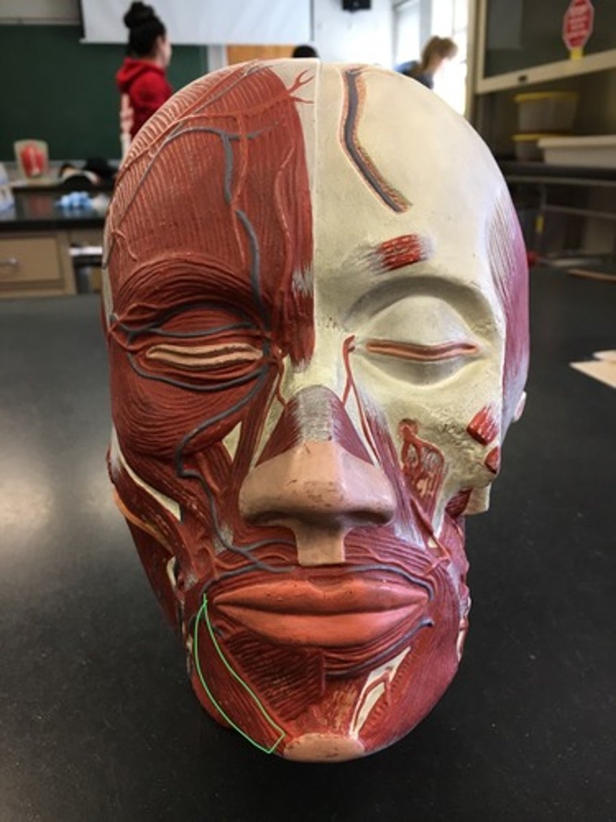
Depressor anguli oris (superficial)
Triangular muscle, helps frowning (lateral)
Depressor labii inferioris (superficial)
Facial muscle that helps lower the bottom lip; Outer surface of mandible along oblique line (medial)

Mentalis
where the chin is; right where mental protuberance is (skin patch on model)

Buccinator
(Deep side) muscle with a big orange dot on it on the model.
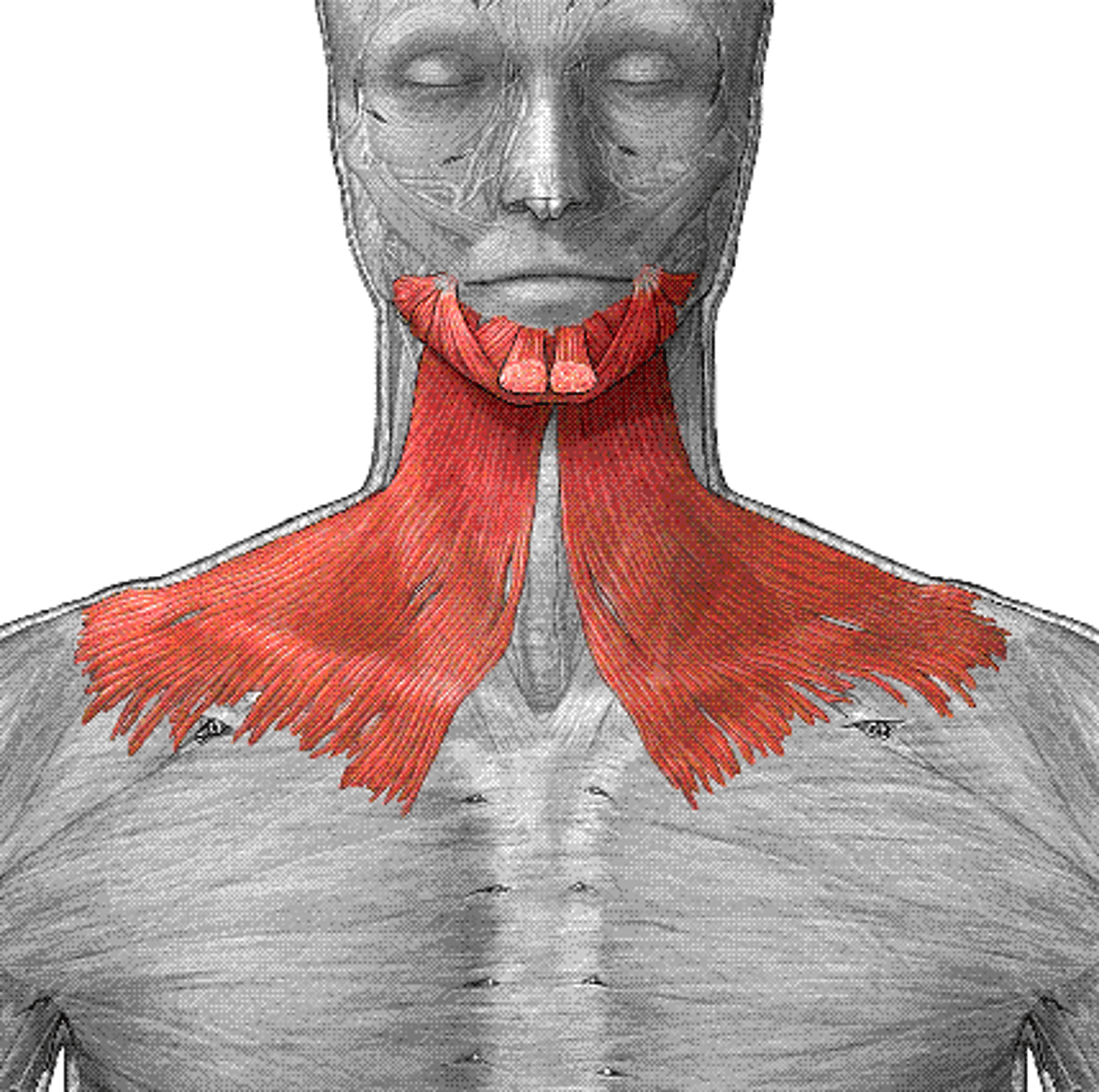
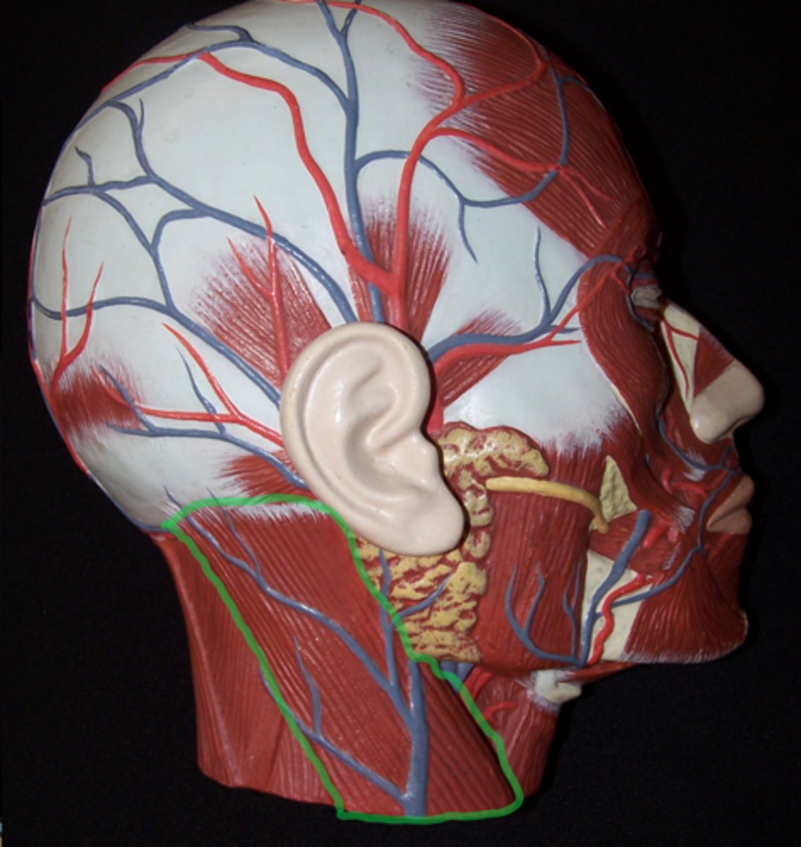
Platysma
Sheet muscle covering all neck muscles.
*No model for this structure, will either be shown on diagram for cadaver for practical*
Temporalis
Deep side, where temporal bone is.
Origin: Temporal Bone
Insertion: Coronoid Process of Mandible
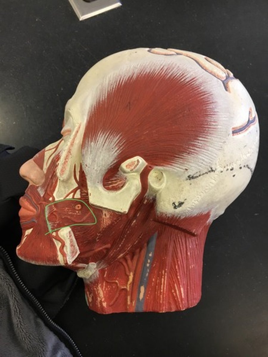
Masseter
(Superficial side) helps chew, vertical striations under buccinator.
Origin: Zygomatic Arch
Insertion: Ramus of the Mandible
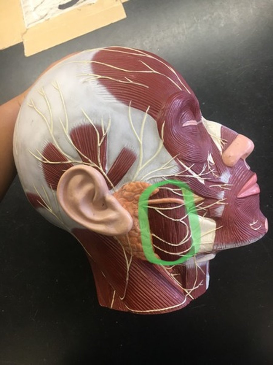
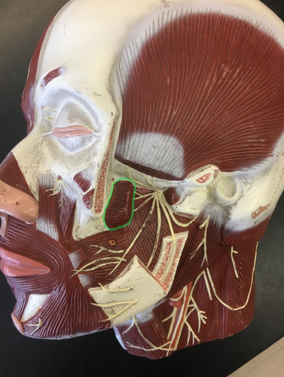
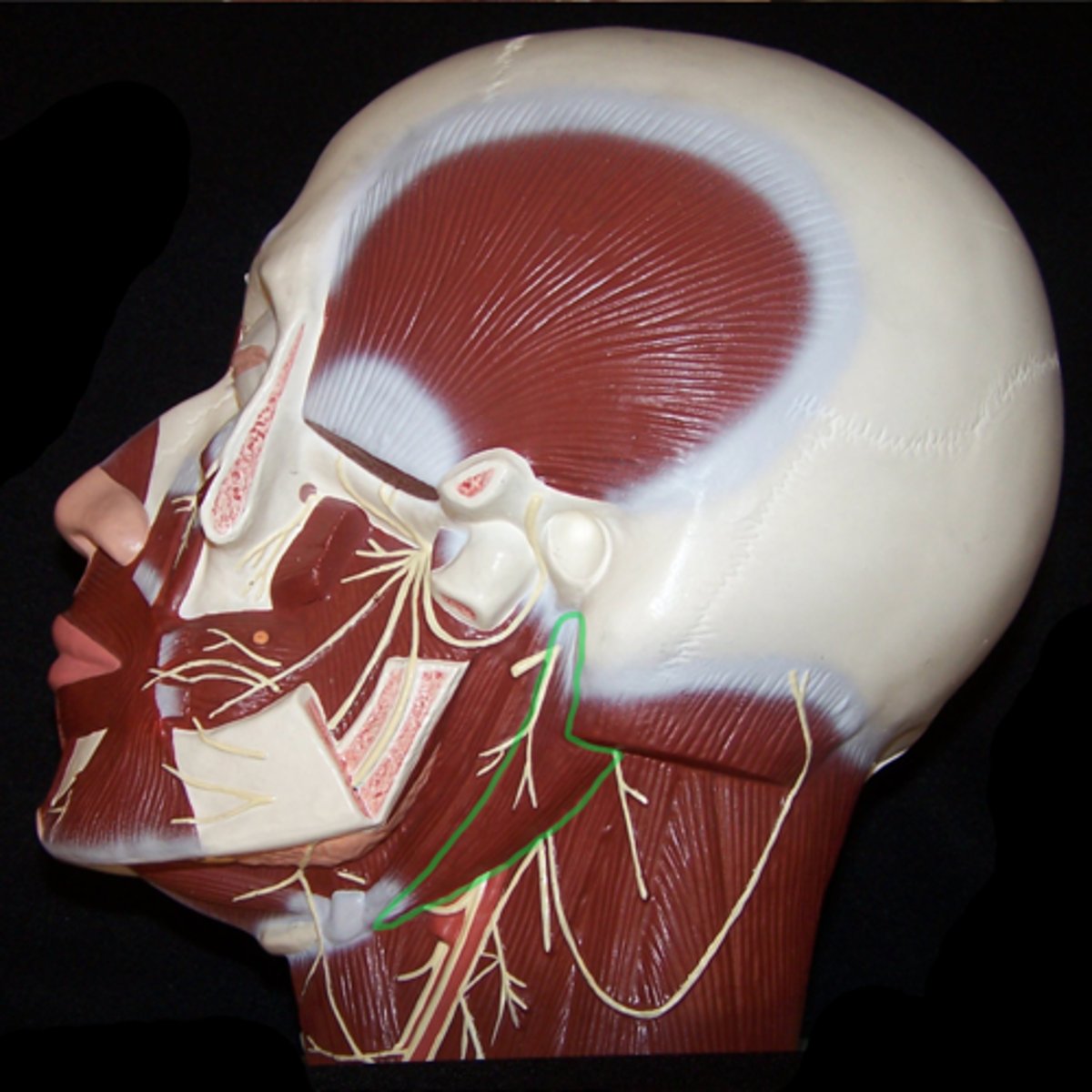
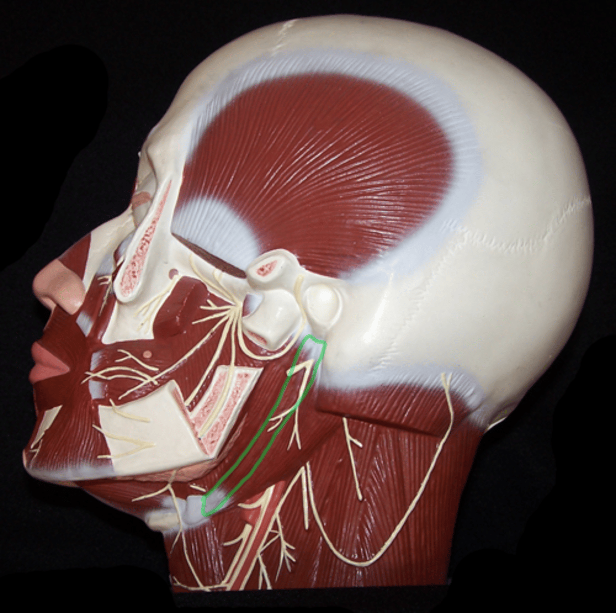
Medial pterygoid (deep side)
(bottom) vertical striations
Origin: Pterygoid Process
Insertion: Ramus of the Mandible
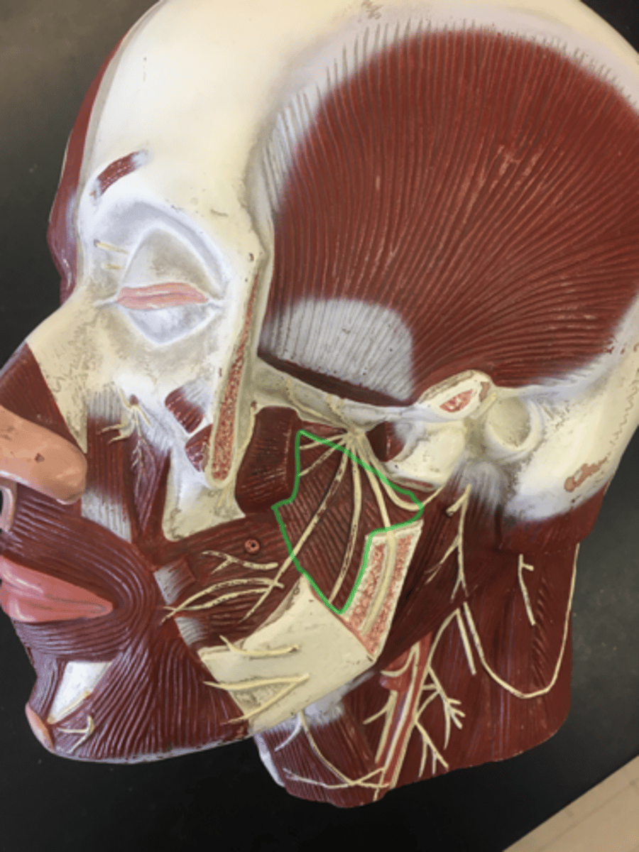
Lateral pterygoid (deep side)
(top) sits, above medial pterygoid, horizontal striations.
Origin: Pterygoid Process
Insertion: Ramus of the Mandible
Digastric, anterior belly
From hyoid to mental protuberance.
Origin: Mental Protuberance
Insertion: Hyoid Bone
Digastric, posterior belly
(lateral) from hyoid to mastoid process; back/below.
Origin: Mastoid Process
Insertion: Hyoid Bone
Stylohyoid (deep side)
Anterior to digastric posterior belly.
Origin: Styloid Process of Temporal Bone
Insertion: Hyoid Bone
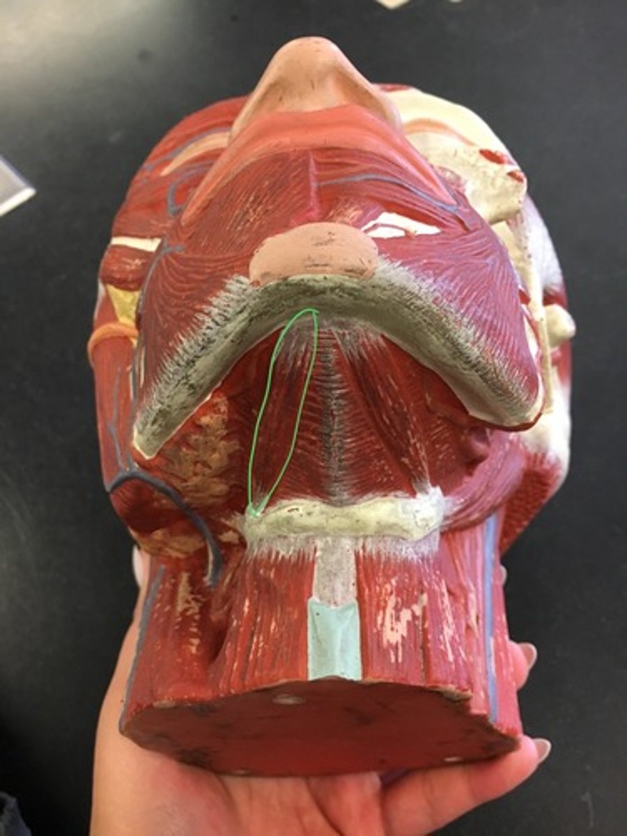
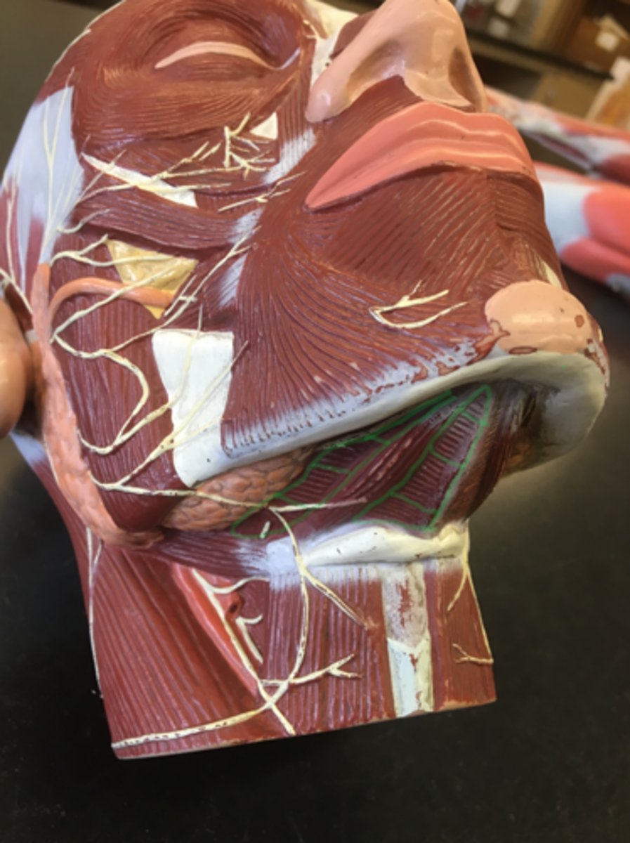
Mylohyoid
Sheet muscle below anterior belly; has horizontal striations
Origin: Body of Mandible
Insertion: Hyoid Bone
Sternohyoid
(medial) running down the hyoid.
Origin: Manubrium and Clavicle
Insertion: Hyoid Bone
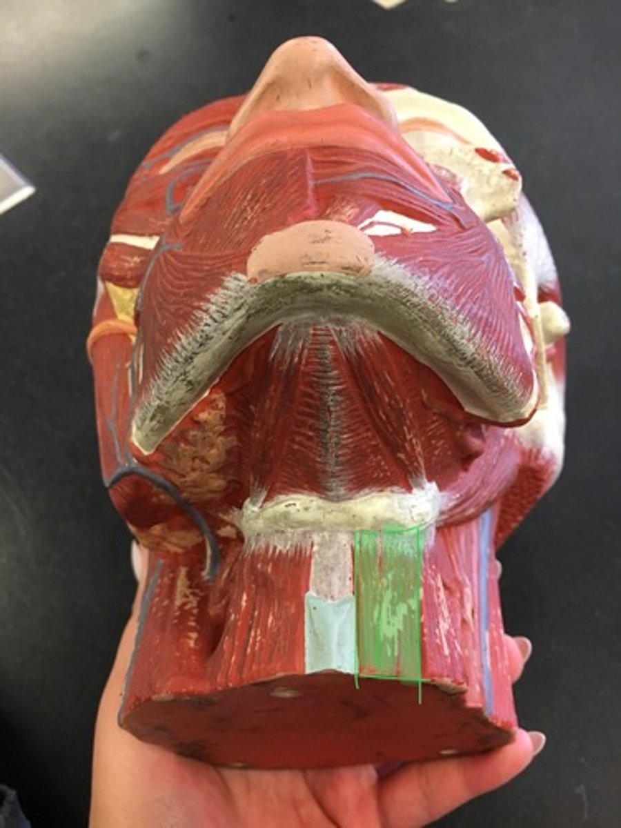
Omohyoid
(lateral) runs down the hyoid, "goes away to Omaha"
Origin: Superior Border of Scapula
Insertion: Hyoid bone

Sternocleidomastoid (SCM)
Huge muscle on side of neck (superficial side).
Origin: Manubrium and Clavicle
Insertion: Mastoid Process
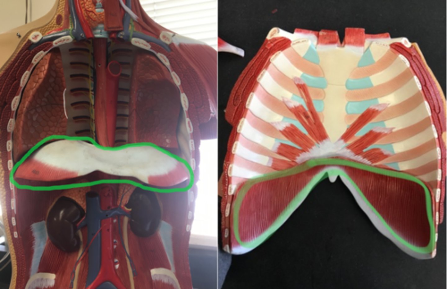
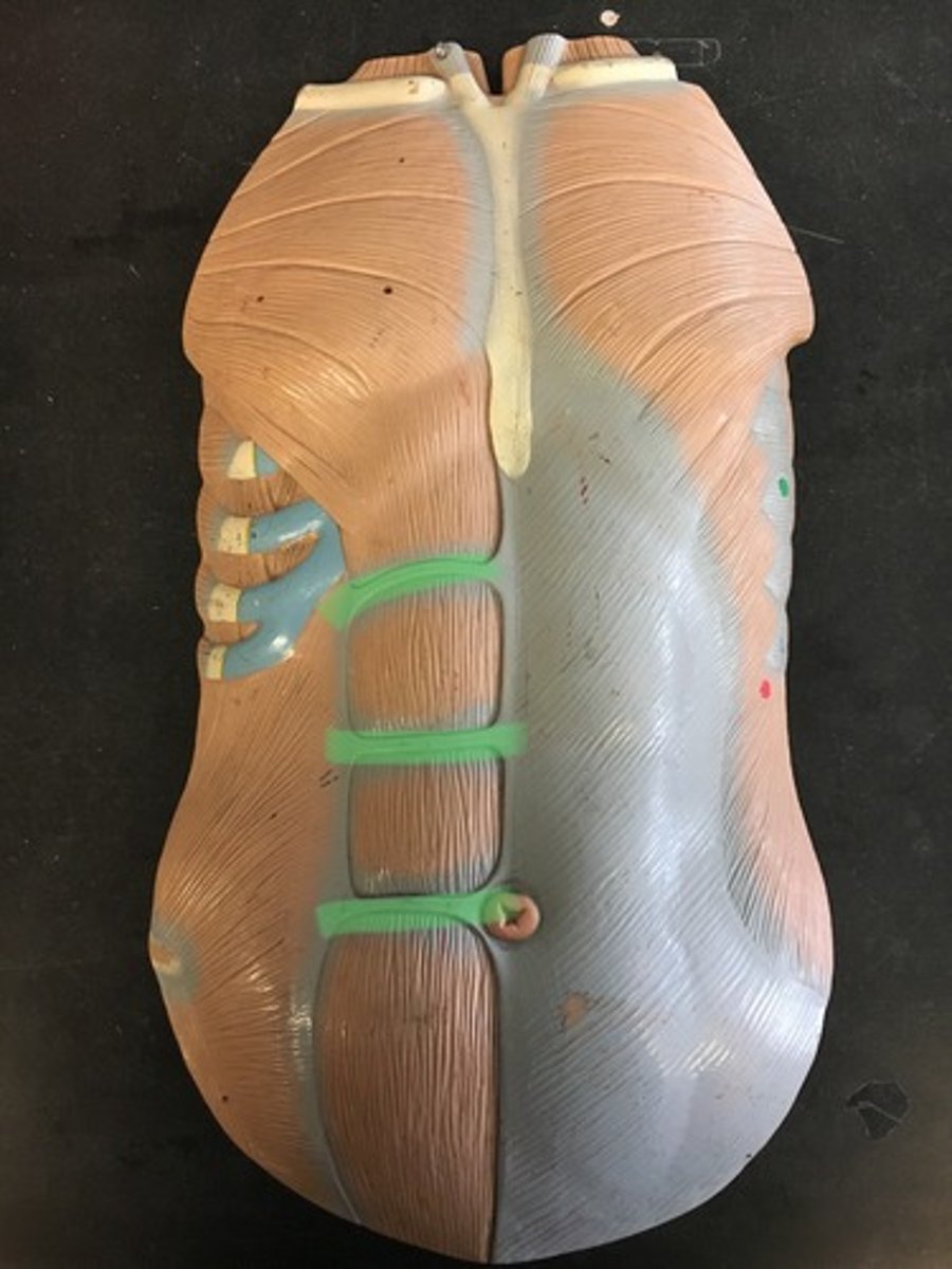
Diaphragm
Under chest cavity, on model all of red portion on the bottom sheet muscle, vertical striations.
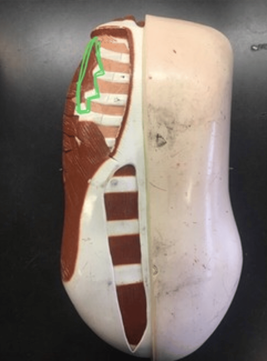
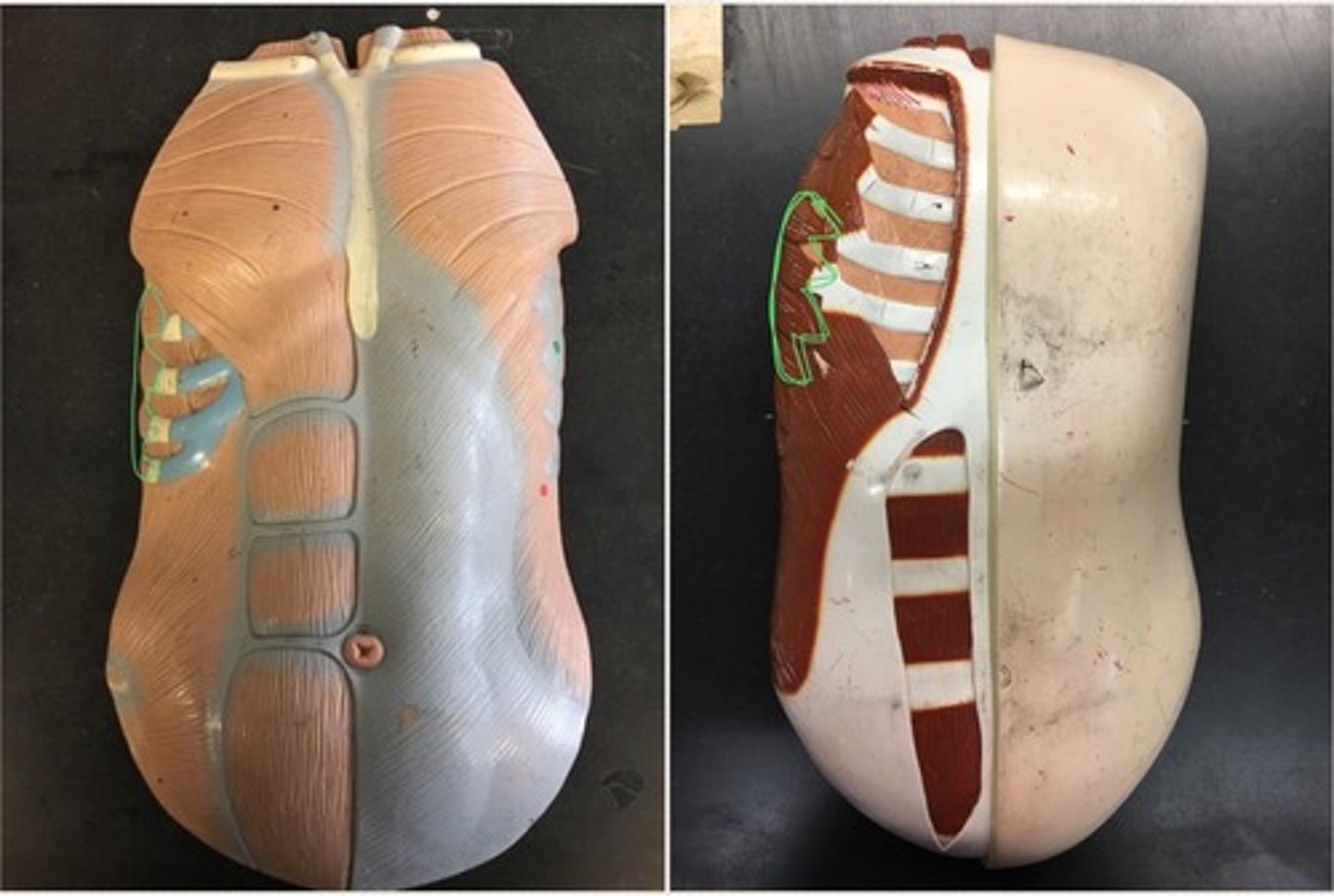
External intercostals (lateral; in between ribs)
More to the side; downward.
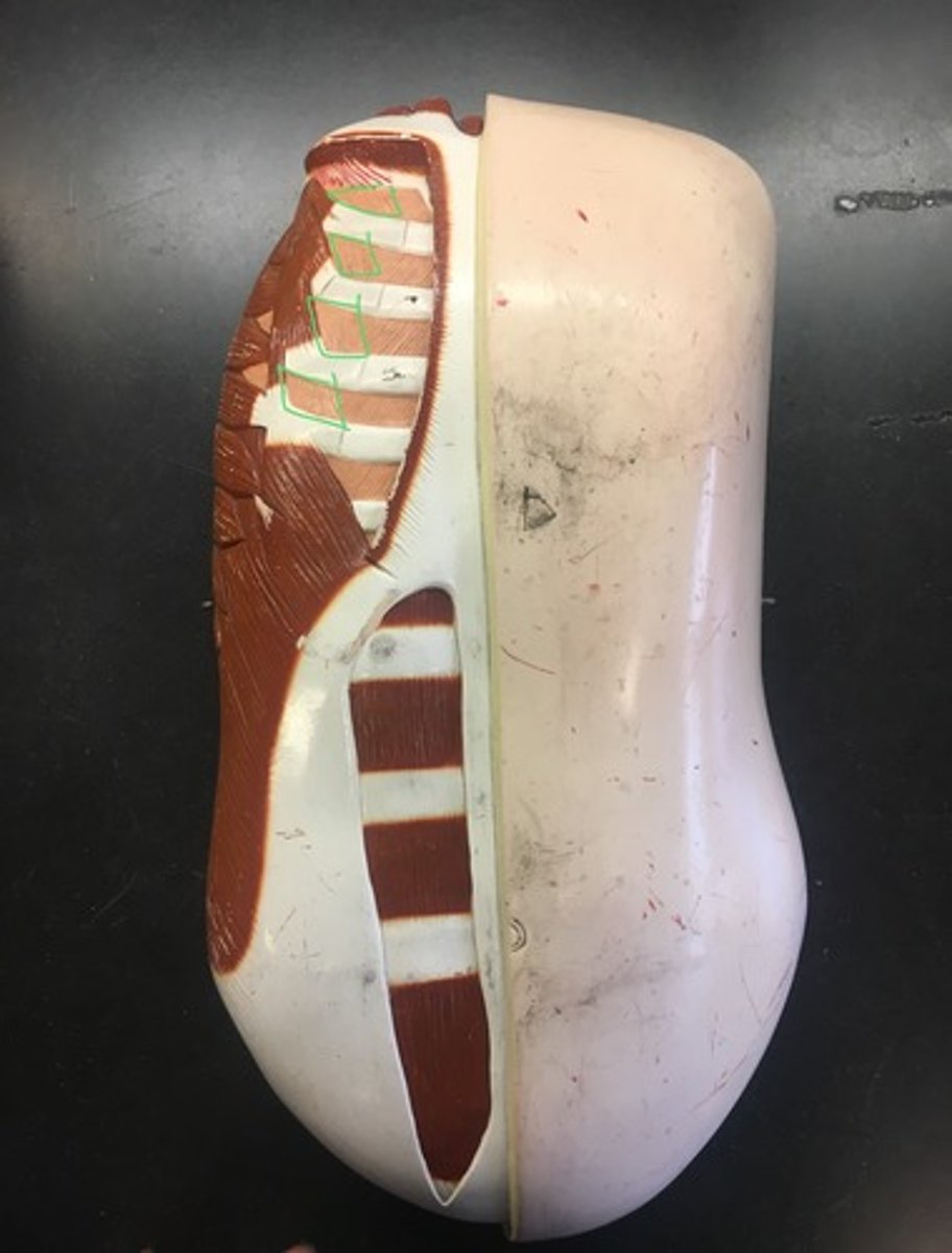
Internal intercostals (medial; in between ribs)
diagonal striations upward.

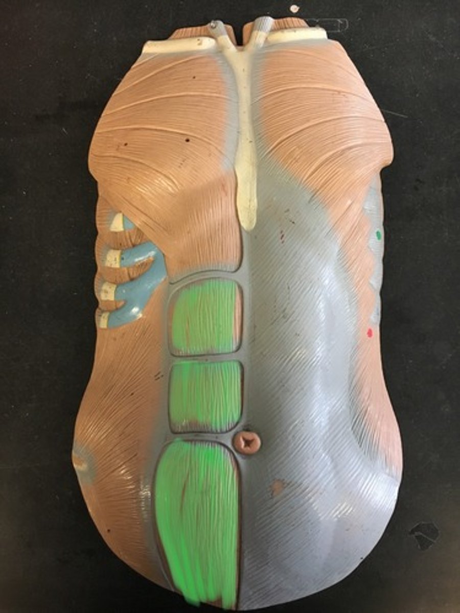
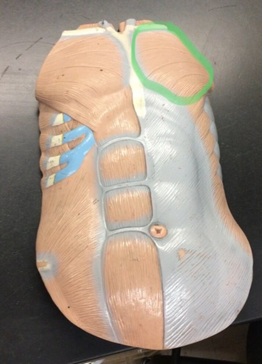
Rectus abdominis
a paired muscle running vertically on each side of the anterior wall of the human abdomen
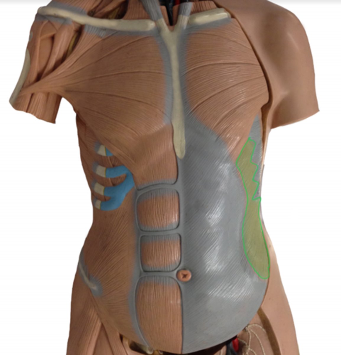
External obliques
downward striations; greyish part on model
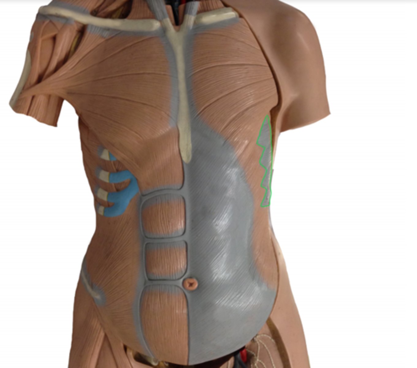
Internal obliques
below external obliques; upward striations; one layer down external oblique.
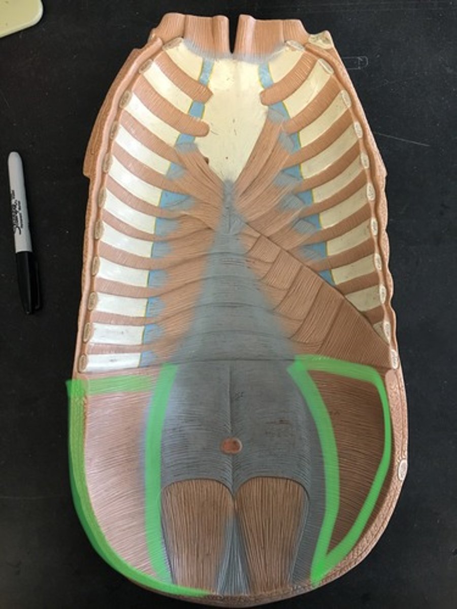
Transverse abdominis (inside view on model)
Below external oblique; horizontal striations.
Linea alba
fibrous structure that runs down the midline of the abdomen in humans and other vertebrates.
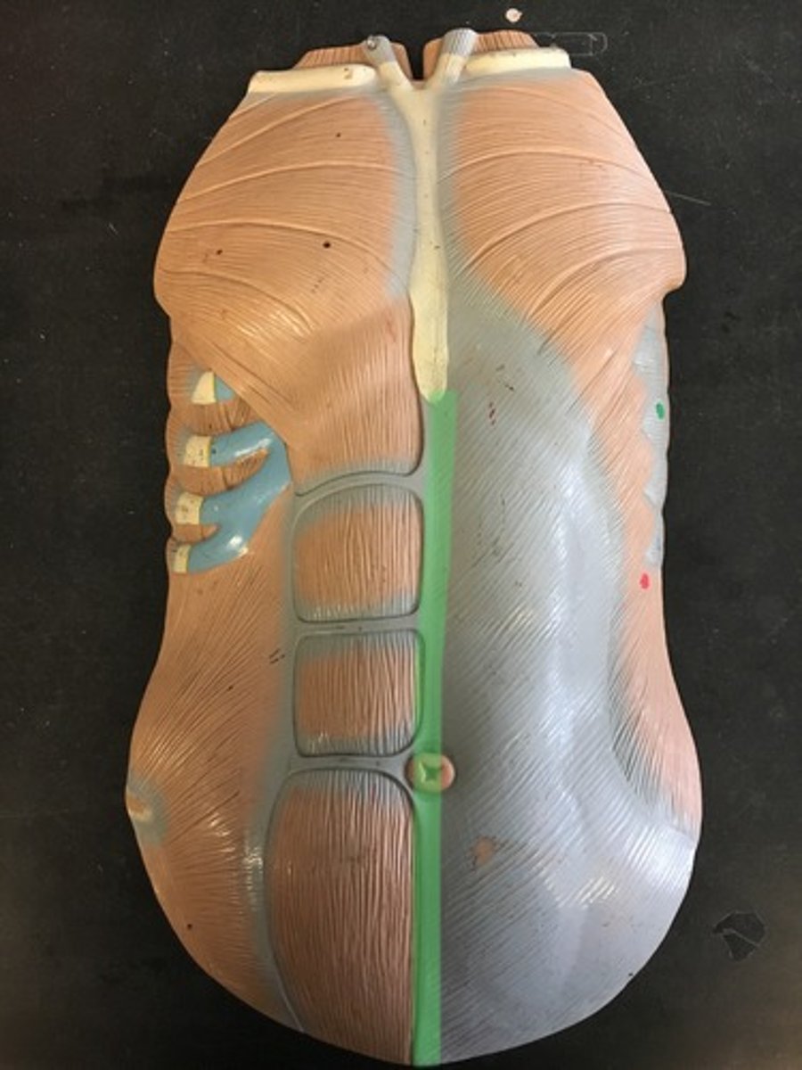
Tendinous inscriptions
separates 6 pack horizontally.
Pectoralis major
Chest muscle
Pectoralis minor
under pectoralis major; individually on ribs
Serratus anterior
muscle on your side; looks like bear claw, (grayish on model); also connects to ribs
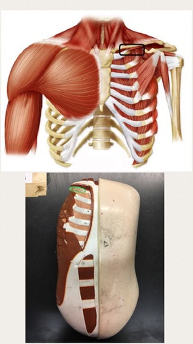
Subclavius
Tiny muscle under clavicle
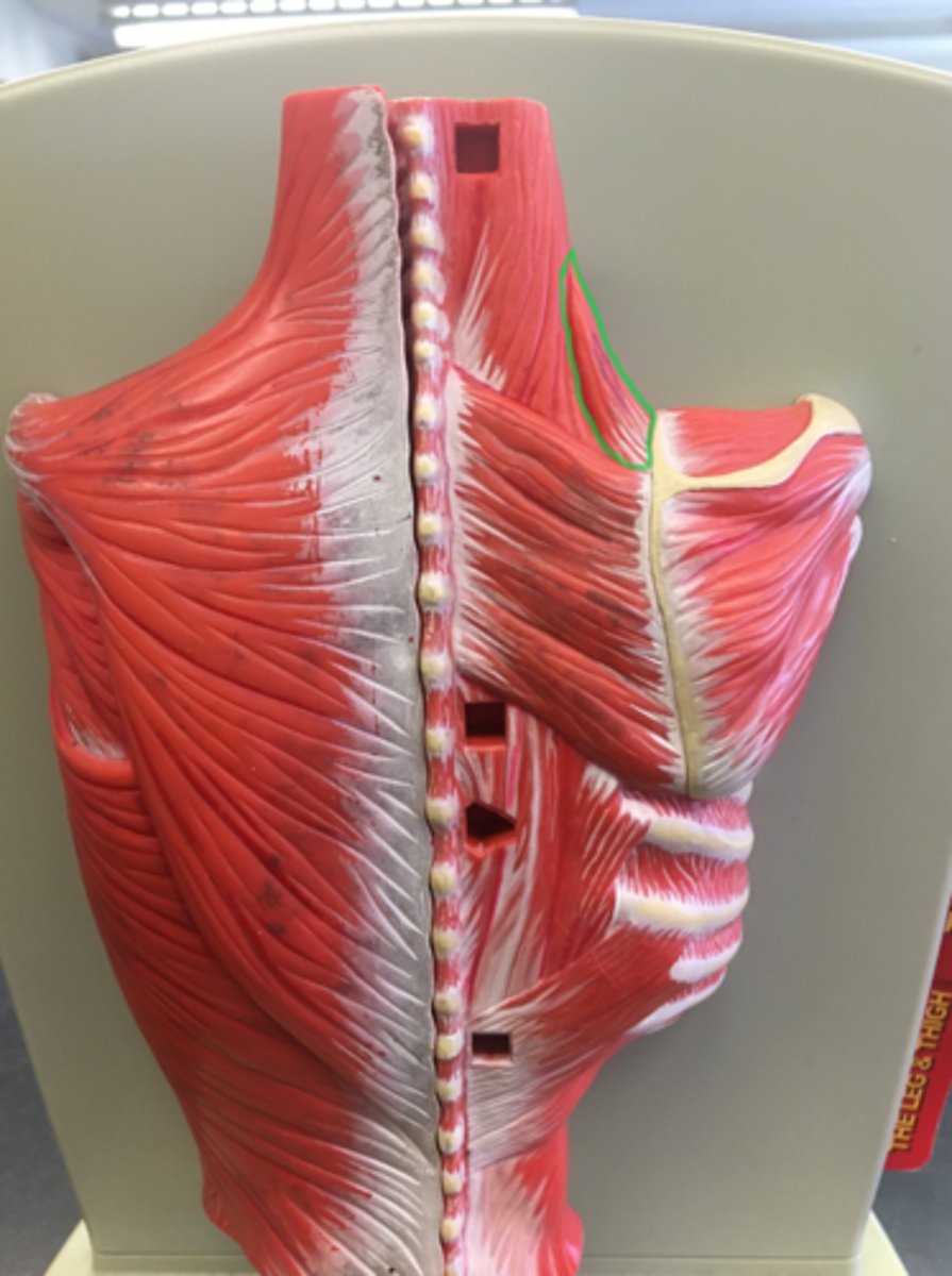
Levator scapulae
on the back; side of neck; small and triangular, by scapuli, small triangle on model
Rhomboid major (bottom)
bigger; muscle on the back that connects the scapula with the vertebrae of the spinal column.
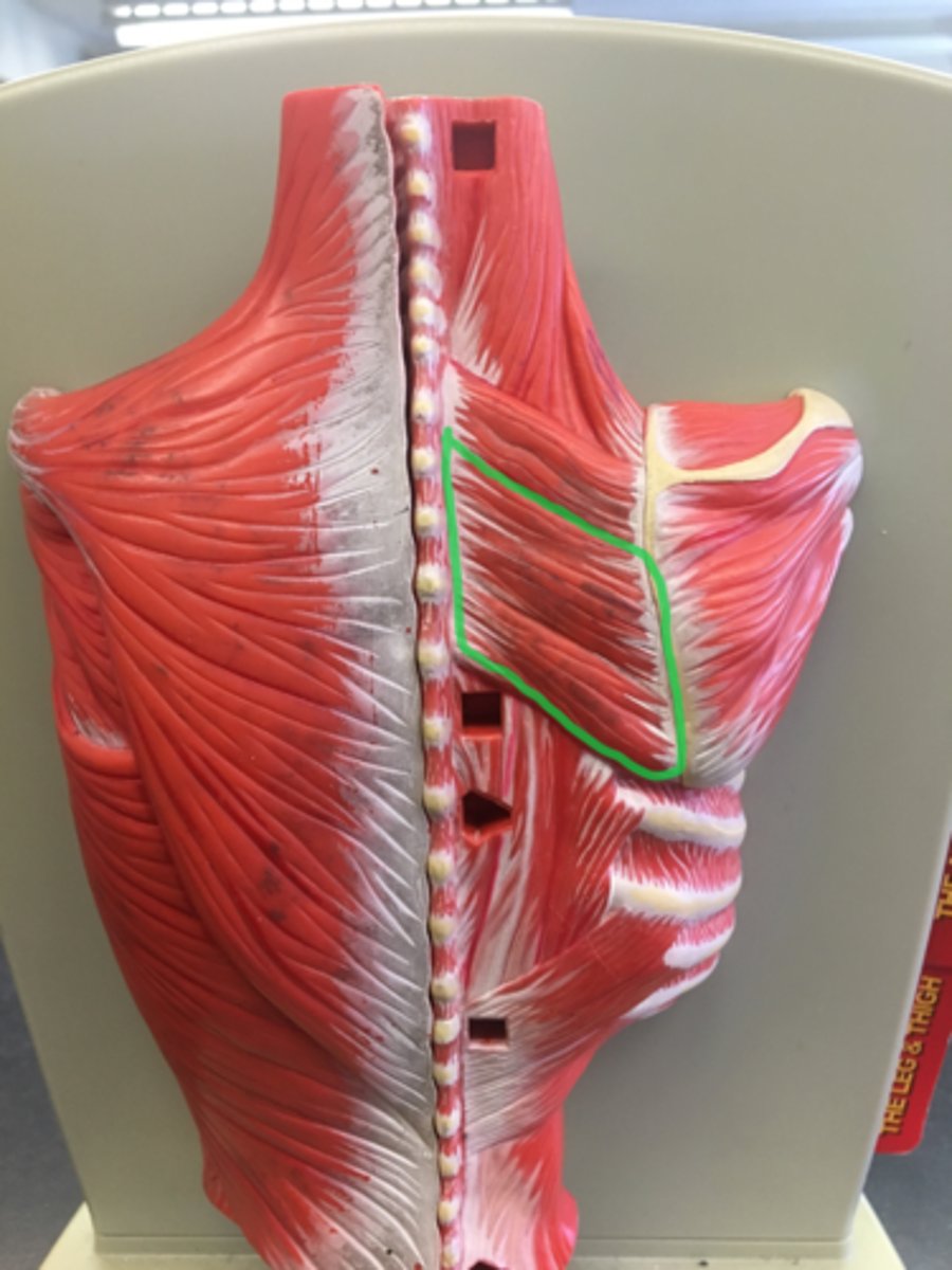
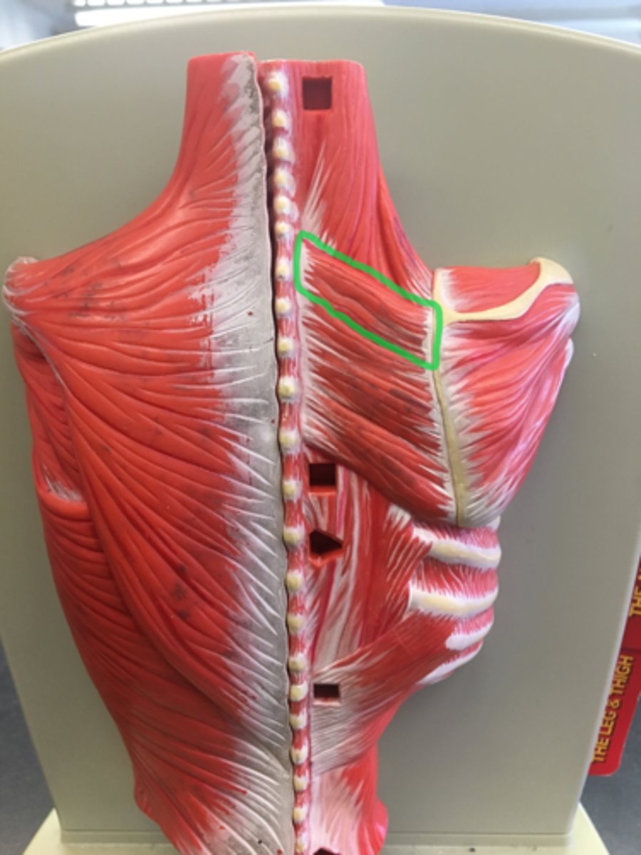
Rhomboid minor (top)
smaller; muscle on the back that connects the scapula with the vertebrae of the spinal column.
Trapezius
Big muscle that looks like kite on back
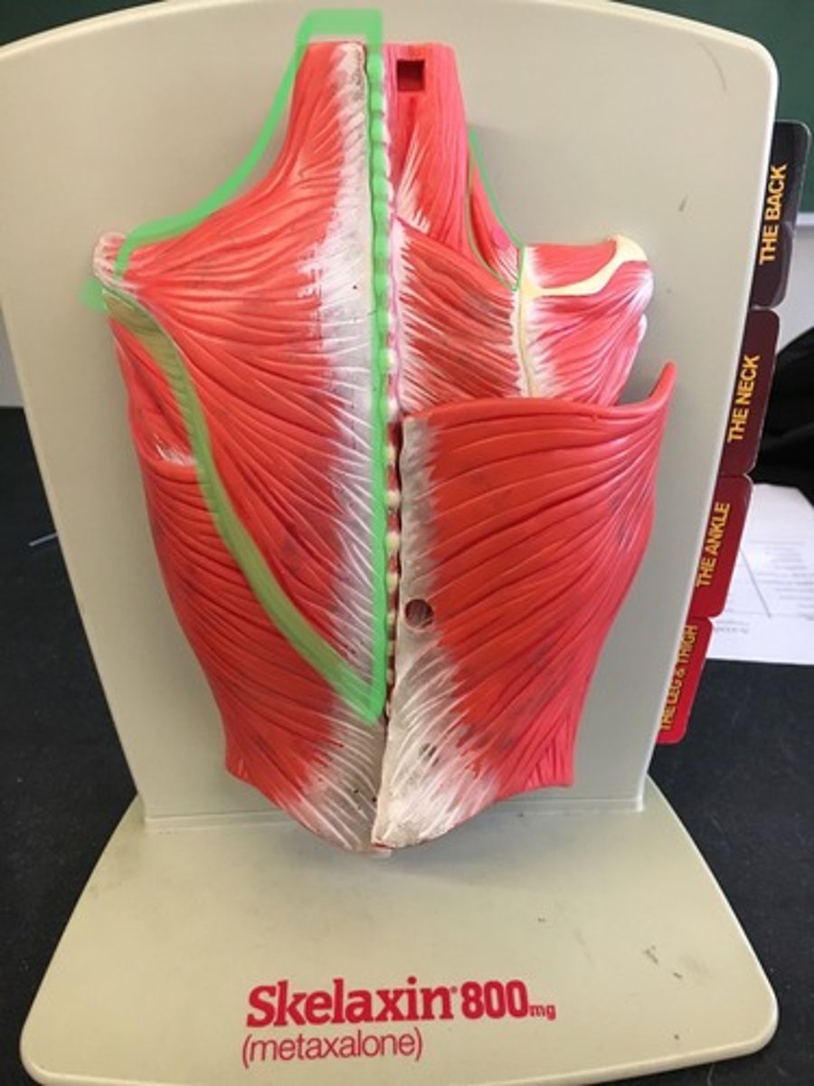
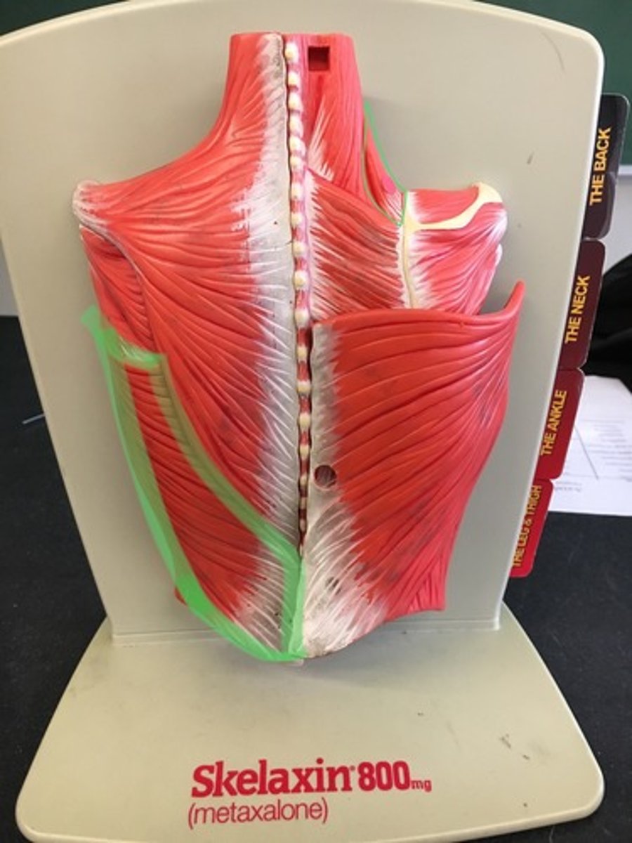
Latissimus dorsi
lower back; sheet muscle.
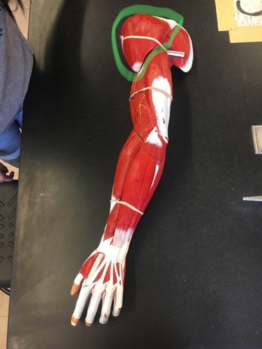
Deltoid
Rounded, triangular muscle located on the uppermost part of the arm and the top of the shoulder.
Origin: Clavicle & Spine of Scapula
Insertion: Deltoid Tuberosity
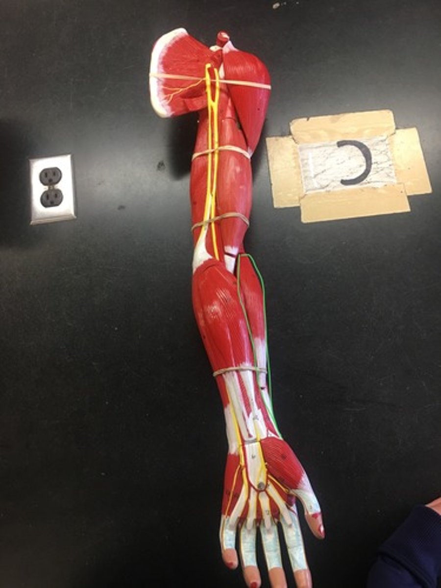
Biceps brachii
Biceps brachii, short head:
Origin: Coracoid Process
Insertion: Radial Tuberosity
Biceps brachii, long head:
Origin: Glenoid Cavity
Insertion: Radial Tuberosity
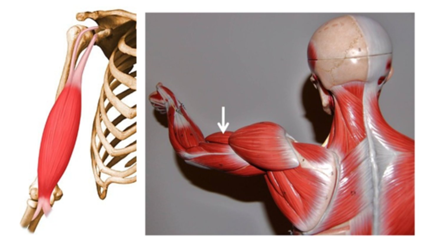
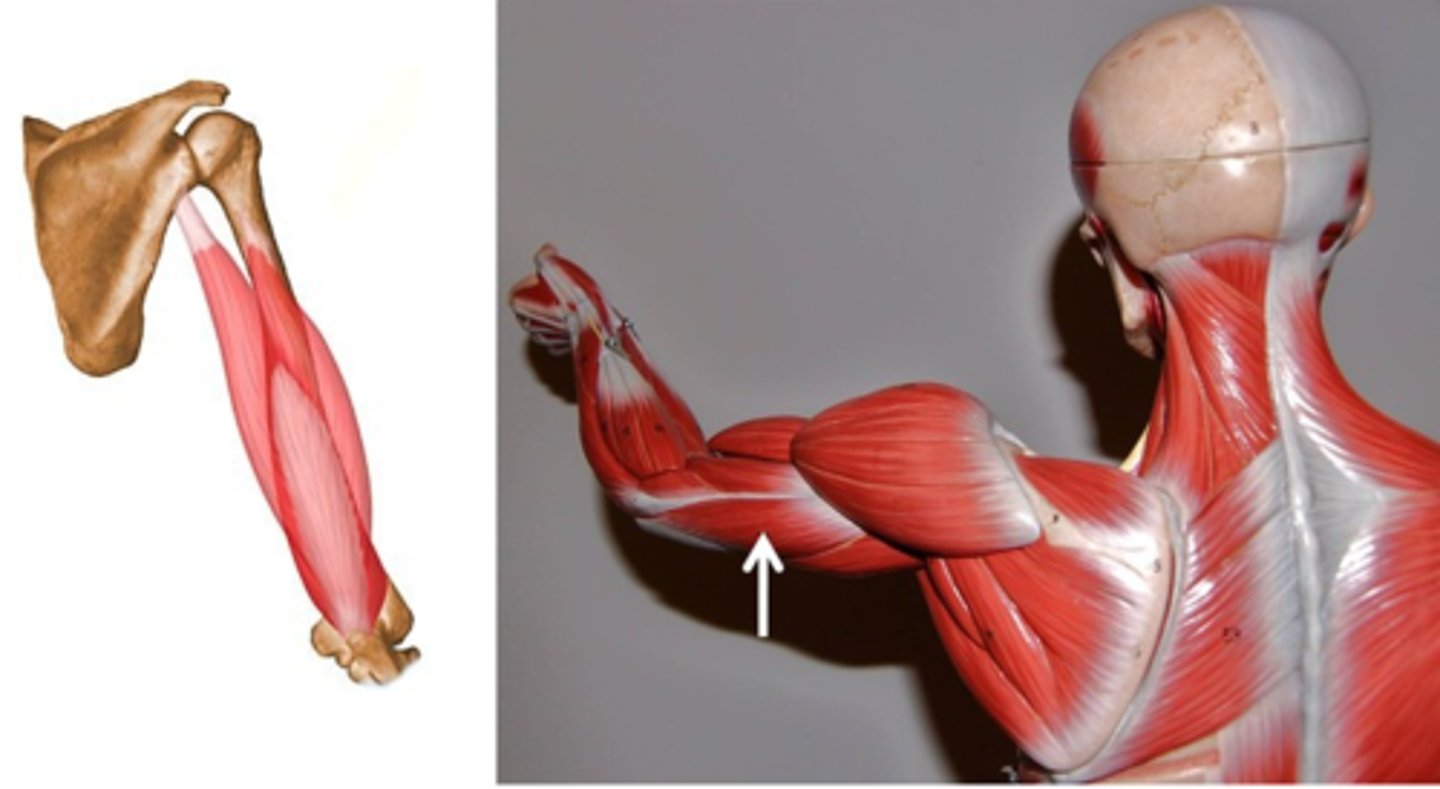
triceps brachii
Triceps brachii, lateral head:
Origin: Proximal Humerus
Insertion: Olecranon
Triceps brachii, long head
Origin: Scapula
Insertion: Olecranon
Triceps brachii, medial head
Origin: Proximal Humerus
Insertion: Olecranon
Brachioradialis
Origin: Lateral Epicondyle of Humerus
Insertion: Styloid Process of Radius
Rectus femoris
Origin: Anterior Inferior Iliac Spine
Insertion: Tibial Tuberosity
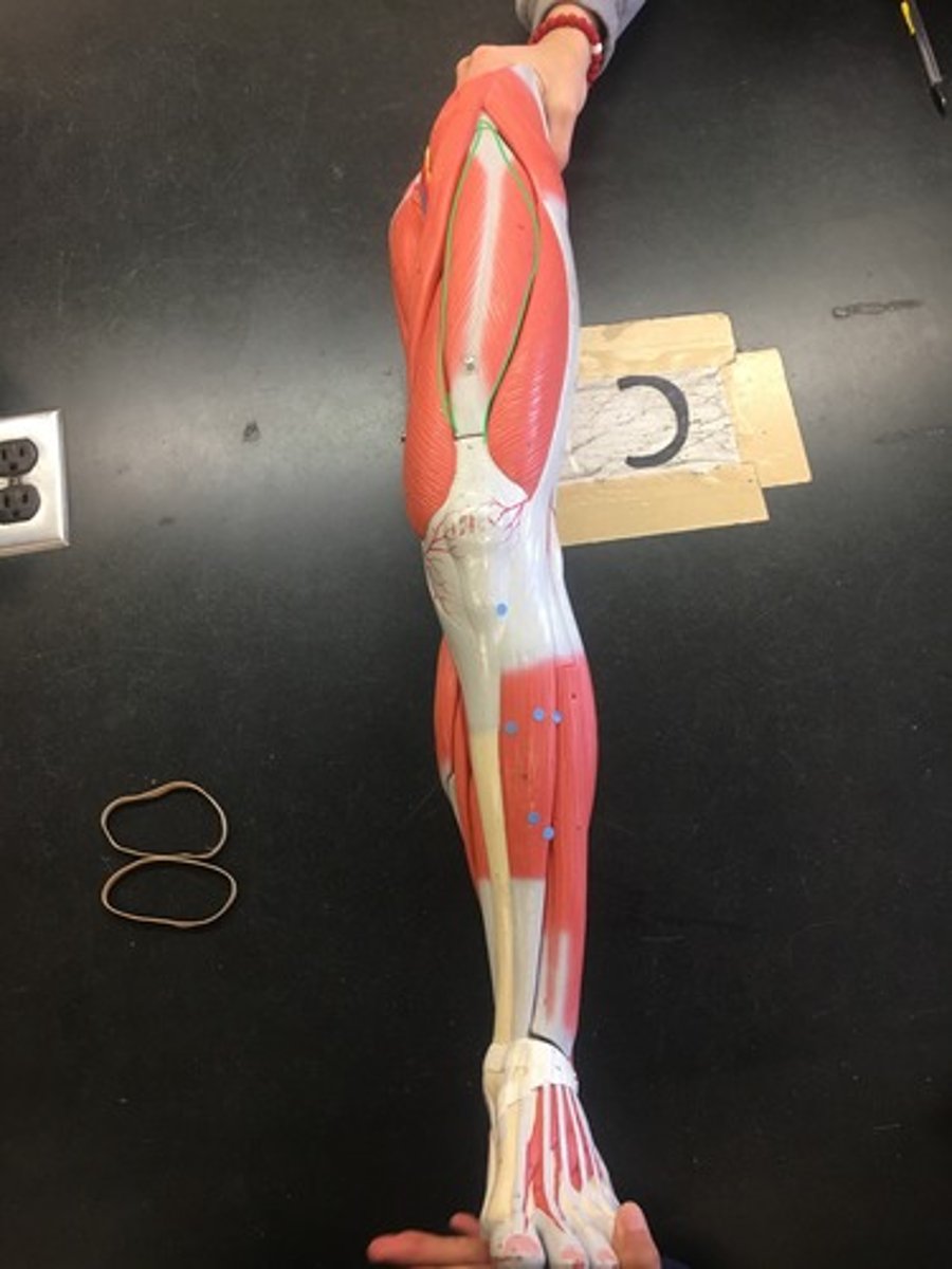
Sartorius
Origin: Anterior Superior Iliac Spine
Insertion: Medial Condyle of Tibia

Gracilis
Origin: Inferior Pubic Ramus
Insertion: Medial Condyle of Tibia
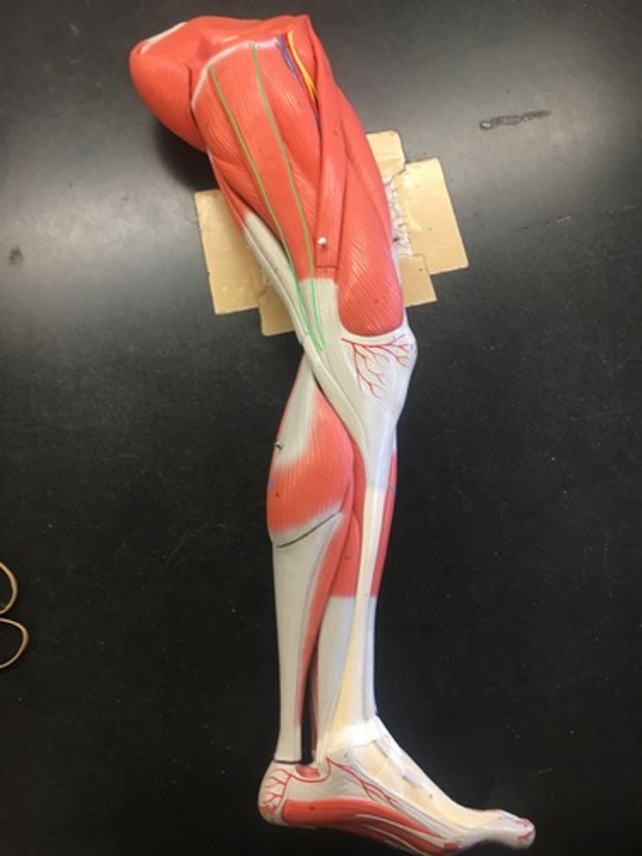
Gluteus maximus
Origin: ilium & sacrum
Insertion: Iliotibial Tract & Proximal Femur
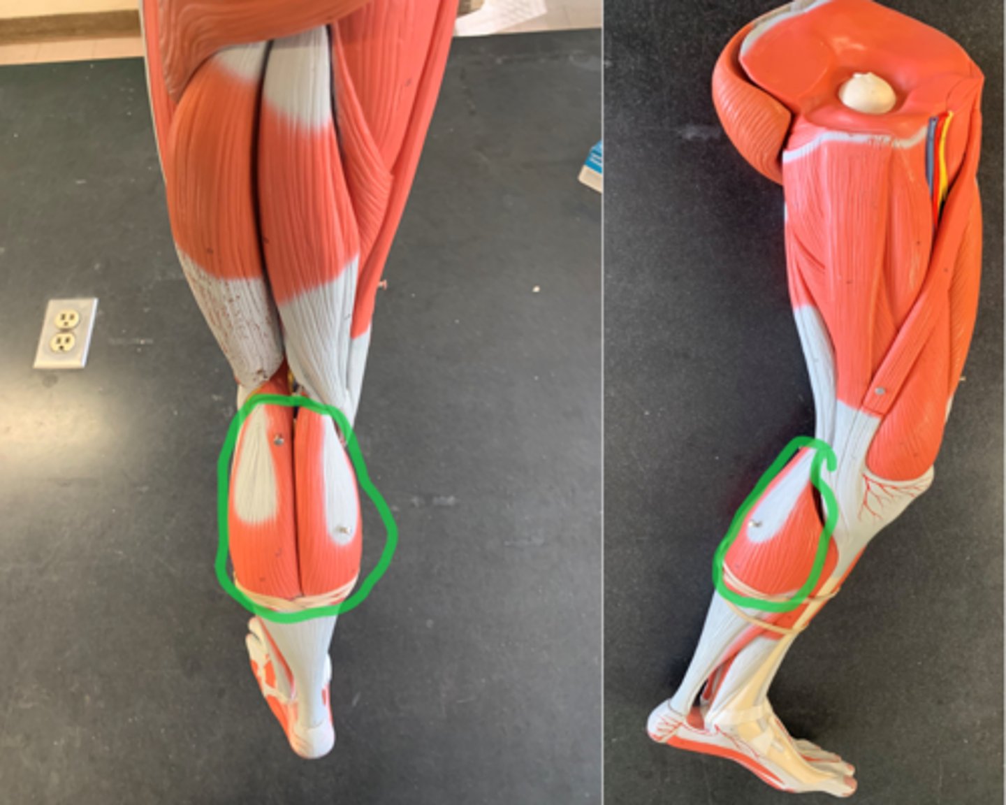
Gastrocnemius
Gastrocnemius, medial head:
Origin: Medial Condyle of Femur
Insertion: Calcaneus
Gastrocnemius, lateral head
Origin: Lateral Condyle of Femur
Insertion: Calcaneus
Tibialis anterior
Origin: Lateral condyle of tibia
Insertion: Metatarsal I
