BIMM 121_Entire Course Material_Grossman
 Call Kai
 Learn
Learn Practice Test
 Spaced Repetition
 Match
Match Flashcards
 Knowt Play
1/174

There's no tags or description
Looks like no tags are added yet.
Name | Mastery | Learn | Test | Matching | Spaced | Call with Kai |
|---|
No analytics yet
Send a link to your students to track their progress
175 Terms
Prokaryotic vs Eukaryotic Cells
(Lecture #1 Content)
-Prokaryotic: smaller, lack nucleus and organelles
-Eukaryotic: larger, have a nucleus and organelles
-Both have DNA, proteins/ATP, and a plasma membrane.
General Types of Media
(Lecture #1 Content)
-Rich: lots of nutrients
-Minimal: limited nutrients
-Undefined: exact composition unknown
-Defined: exact composition known
-Selective: only certain microbes grow
-Differential: microbes look different based on properties
King’s B & TSA agar
(Lecture #1 Content)
-Rich & undefined (digests = exact composition unknown)
MacConkey agar
(Lecture #1 Content)
-Selective: bile salts + crystal violet inhibit gram-positive and allow gram-negative growth.
-Differential: lactose + neutral red pH turns pink when lactose is fermented (acid produced)
-Rich & undefined (digests = exact composition unknown)
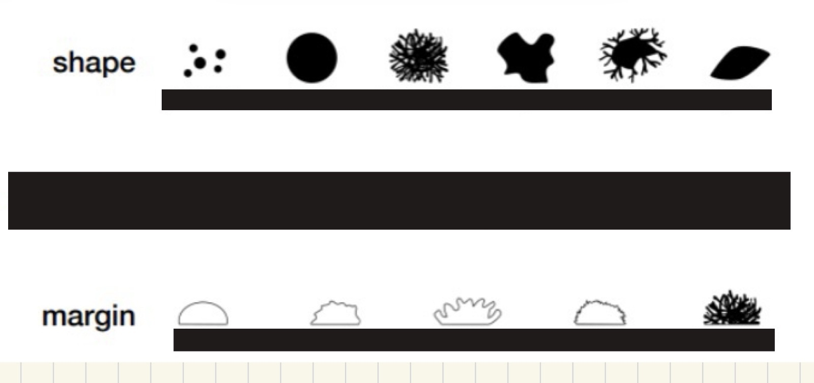
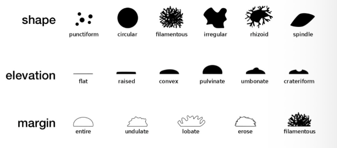
Colony morphology:
Describes appearance and characteristics of a colony
-For shape, know circular, filamentous, and irregular
-Don’t need to know any elevation
-For Margin, know entire and filamentous
(Lecture #2 Content)
T-streak:
(Lecture #2 Content)
separate bacteria into isolated colonies on an agar plate.
Respiration & Aerobic vs Anaerobic respiration
(Lecture #2 Content)
(Study)
-Respiration: glucose —> 2 pyruvate —> acetyl-CoA (Uses glycolysis: glucose → pyruvate)
-Aerobic respiration: uses ETC and O₂ as final electron acceptor.
-Anaerobic respiration: uses ETC and non-O₂ as final electron acceptor.
Microbial physiology
(Lecture #2 Content)
-Obligate aerobe: O₂ dependent
-Obligate anaerobe: O₂ independent
-Facultative anaerobe: grow with or without O₂ but prefer O₂
-Microaerophile: require low O₂, but poisoned by too much
-Aerotolerant: do not use O₂ but tolerate it
Fermentation & Lactic vs Ethanol Fermentation:
(Lecture #2 Content)
(Study)
-Fermentation: No ETC. Electrons go to organic molecule (ex: pyruvate) to regenerate NAD⁺. Use glycolysis (glucose → pyruvate)
-Types:
1. Lactic acid fermentation: Glucose → 2 pyruvate → 2 lactic acids. Ex: milk to yogurt.
2. Ethanol fermentation: Glucose → 2 pyruvate → 2 acetaldehyde → 2 ethanol + 2 CO2. Ex: bread
Lactic Acid Bacteria (LAB):
(Lecture #2 Content)
Microbes that ferment lactose anaerobically to produce lactic acid (used in yogurt)
Cori Cycle:
(Lecture #2 Content)
(Study)
During low O₂ conditions of mammals (ex: heavy exercise), muscle cells convert pyruvate into lactate to regenerate NAD⁺ so glycolysis can continue making ATP. Lactate then travels to the liver to be converted back into glucose.
Which of the following might be a microbe?
A) a prokaryote
B) a eukaryote
C) a multicellular organism
D) all of the above
E) none of the above
(Lecture #1-2 Questions)
Answer: D) all of the above
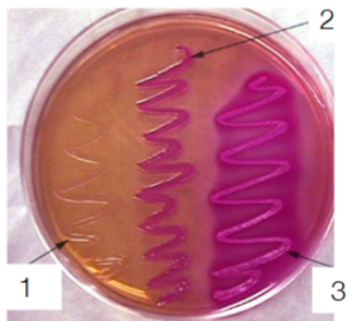
On this MacConkey agar plate, what type of microbes are in the streak labeled 1-3? What about gram positive, non-fermenters
(Lecture #1-2 Questions)
Answer: #1 = no fermentation. #2 = gram negative fermentation. #3 = strong gram negative fermentation. MacConkey inhibits gram positive
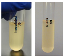
What should you do if you come into the lab, and the culture you need to use looks like this (yellow)?
A. throw it away and start over
B. proceed to analyze the inoculated culture, but throw away our media stock bottle
C. streak out the inoculated culture to separate the microbe we were working with from any contaminants
(Lecture #1-2 Questions)
A. throw it away and start over
B. proceed to analyze the inoculated culture, but throw away our media stock bottle
C. streak out the inoculated culture to separate the microbe we were working with from any contaminants
Answer: A. throw it away and start over
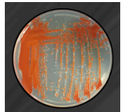
What problem in streaking technique most likely produced the plate shown?
Answer choices for all examples
A. Failure to flame loop between sections
B. Crossing back into previous section on every streak
C. Forgetting to cross into first section on second streak
D. Forgetting to cross into second section on third streak
E. Not letting the loop cool before taking sample from tube
(Lecture #1-2 Questions)
Answer: B. Crossing back into previous section on every streak
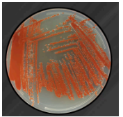
What problem in streaking technique most likely produced the plate shown?
Answer choices for all examples
A. Failure to flame loop between sections
B. Crossing back into previous section on every streak
C. Forgetting to cross into first section on second streak
D. Forgetting to cross into second section on third streak
E. Not letting the loop cool before taking sample from tube
(Lecture #1-2 Questions)
Answer: A. Failure to flame loop between sections
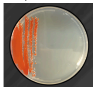
What problem in streaking technique most likely produced the plate shown?
Answer choices for all examples
A. Failure to flame loop between sections
B. Crossing back into previous section on every streak
C. Forgetting to cross into first section on second streak
D. Forgetting to cross into second section on third streak
E. Not letting the loop cool before taking sample from tube
(Lecture #1-2 Questions)
Answer: C. Forgetting to cross into first section on second streak
E is not the best answer because we would not see anything in the first streak.
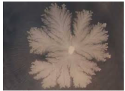
What shape is the following colony?
punctiform
rhizoid
circular
spindle
(Lecture #1-2 Questions)
Answer: rhizoid
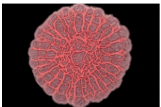
What is the margin of the following colony?
wrinkled
entire
filamentous
undulate
(Lecture #1-2 Questions)
Answer: Undulate
What kind of metabolism do the bacteria use in order to turn milk into yogurt?
Aerobic respiration
Anaerobic respiration
Lactic acid fermentation
Ethanol fermentation
(Lecture #1-2 Questions)
Answer: Lactic acid fermentation
The Cori cycle in mammals describes which process?
Aerobic respiration of lactate breakdown
Lactic acid “fermentation” in muscle cells
Ethanol “fermentation” in muscle cells
(Lecture #1-2 Questions)
Answer: Lactic acid “fermentation” in muscle cells
Although both anaerobic respiration and fermentation occur without oxygen, what is the difference? Is glycolysis a part of both? What about the electron transport chain?
(Lecture #1-2 Questions)
Both use glycolysis, but fermentation does not use an electron transport chain, while anaerobic respiration does.
Heirloom vs Commercial Yogurt
(Lecture #3 Content)
-Heirloom: undefined mix of microbes
-Commercial: defined mix of known strains
-Both required to have Streptococcus thermophilus and Lactobacillus bulgaricus
Light vs Electron Microscopy
(Lecture #3 Content)
(Study)
-Light microscopy (ex: brightfield): uses visible light, lower resolution (0.2µm), but can view living cells
-Electron microscopy: uses electron beam, ~2000× higher resolution, views very small structures like viruses, and samples must be dead and specially prepared
Transmission vs Scanning Electron Microscopy
(Lecture #3 Content)
(Study)
-Transmission Electron Microscopy (TEM): electrons pass through a thin/sectioned sample to show internal, fine structures (ex: viruses, inside cells)
-Scanning Electron Microscopy (SEM): electrons scan the surface of a thicker sample to shows 3D surface shape/texture
Cell Shapes
-Coccus, diplococci, diplococci encapsulated, streptococci, staphylococci, sarcina, and tetrad
-Coccobacillus, bacillus, diplobacillus, streptobacillus, palisades
(Lecture #3 Content)
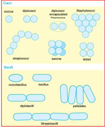
Cell Arrangement
(Lecture #3 Content)
-Diplo = two
-Strepto = chain
-Staphylo = stack/cluster = random oriented plane division
-Tetrad = 2 perpendicular planes division (vertically then horizontally)
Bacteriophage
(Lecture #4 Content)
a non-cellular virus that infect bacteria and naturally live in our bodies, especially the gut. They are generally stable over time and can mediate HGT
Two Main Viral Reproductive Cycles
(Lecture #4 Content)
-Lytic: virus injects genome → immediately produces new virions → host cell lyses
-Lysogenic: viral DNA integrates into host genome → stays dormant, then later enter lytic burst
CRISPR-Cas9:
(Lecture #4 Content)
(Study)
A gene-editing system where guide RNA directs Cas9 to cut matching DNA via double-strand break. DNA is then repaired to either knock out genes or precisely edit, allowing CRISPR to permanently change an organism’s genome. In bacteria, CRISPR acts as an adaptive immune system by storing phage DNA as spacers to recognize and destroy future phage infections.
Two methods of DNA repair:
(Lecture #4 Content)
1. Non-homologous end joining (NHEJ): broken DNA ends are joined without a template. Fast but often error-prone (indels = insertions/deletions).
2. Homology-directed repair (HDR): uses homologous DNA template to repair double-strand break, making it more precise.
Gene therapy:
(Lecture #4 Content)
Introduction of a functional gene to replace a defective one and treat disease
Challenges of Gene Therapy
(Lecture #4 Content)
-Difficult to deliver gene to enough target cells
-Viral vectors can cause immune/liver damage
-Random integration can suppress or activate oncogenes → cancer
Stem Cell Properties
(Lecture #4 Content)
-Multipotent: can become multiple cell types
-Undifferentiated: not yet specialized
-Self-renewing: divide to replace themselves and make new cells
-For multi-tissue diseases, edit stem cells so changes are passed to future cells
Hematopoietic stem cells (HSCs):
(Lecture #4 Content)
form all blood cells
Adult hemoglobin:
(Lecture #4 Content)
made of alpha and beta chain
Somatic vs Germline editing:
(Lecture #4 Content)
-Somatic: DNA changes in non-reproductive body cells → affects only the individual, not inherited
-Germline: DNA changes in eggs, sperm, or embryos → changes to all cells and passed to future generations
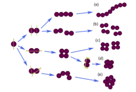
Cell Arrangement Practice Question. Use this photo to answer the following questions:
a) Which cocci are the result of division in two perpendicular planes?
b) Staphylococci are the result of division in randomly oriented planes. Which are staphylococci?
(Lecture #3-4 Questions)
a) Answer: C
Reasoning: 2 perpendicular planes division: divide vertically then horizontally = tetrad (square).
b) Answer: E
Reasoning: Division in random direction results in staphylococcі (cluster)
Are bacteriophages considered cells?
A) Yes
B) No
(Lecture #3-4 Questions)
Answer: B) No b/c bacteriophage is a non-cellular virus that infects bacteria.
True or False? The resolution of electron microscopy is better than brightfield
(Lecture #3-4 Questions)
Answer: True
To image a virus, which microscopy type would be best?
A) Phase contrast microscopy
B) Scanning electron microscopy
C) Transmission electron microscopy
(Lecture #3-4 Questions)
Answer: C) Transmission electron microscopy

What is the following cell shape?
A) Coccus
B) Bacillus
C) Streptobacilli
D) Vibrio
(Lecture #3-4 Questions)
Answer: B) Bacillus
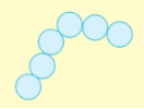
What is the following cell shape?
A) Diplococci
B) Streptobacilli
C) Streptococci
D) Staphylococci
(Lecture #3-4 Questions)
Answer: C) Streptococci
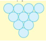
What is the following cell shape?
A) Tetrad
B) Streptococci
C) Staphylococci
D) Staphylobacilli
(Lecture #3-4 Questions)
Answer: C) Staphylococci
True or False? The CRISPR technique can permanently change the DNA genome of an organism.
(Lecture #3-4 Questions)
Answer: True
Select which of the following statements are true (choose ALL that are true)
A) CRISPR uses guide DNA to match Cas9 to target DNA
B) CRISPR-Cas9 can be used to introduce new sequence into a specific location of the genome
C) CRISPR-Cas9 can be used to cause deletions in a specific location of the genome to knock out a gene
D) Cas9 causes a single strand break in the DNA
(Lecture #3-4 Questions)
Answer: B) CRISPR-Cas9 can be used to introduce new sequence into a specific location of the genome & C) CRISPR-Cas9 can be used to cause deletions in a specific location of the genome to knock out a gene
Polymerase chain reaction (PCR):
(Lecture #5 Content)
Make many copies of a specific DNA sequence. Requires double strand DNA template, species specific forward & reverse primers, dNTPs (A, T, C, G), Mg²⁺, heat-stabled DNA polymerase (Taq)
PCR Steps:
(Lecture #5 Content)
1. Denature (~94°C): DNA strands separate
2. Anneal (45–65°C): Forward and reverse primers bind to complementary template strands
3. Extension (~72°C): Taq polymerase extends primers to synthesize new DNA strands
Taq polymerase:
(Lecture #5 Content)
a heat-stable DNA polymerase that synthesizes DNA during PCR at high temperatures.
Annealing temp rule (We might not need it):
(Lecture #5 Content)
Tm = 4(G+C) +2(A+T)°C
Ex: 5’ – ACGAAGGCAGCCTTAGCATT – 3’
—> Tm = 4(10) + 2(10) = 40 + 20 = 60°C
Why does annealing temp matter?
(Lecture #6 Content)
• Too low → primers bind non-specifically (wrong DNA amplified)
• Too high → primers don’t bind well (little/no amplification)
Positive vs Negative Control:
(Lecture #6 Content)
-Positive control: expected to give a positive result to confirm experiment works
-Negative control: expected to give no result to checks for contamination or false positives
Gel Electrophoresis:
(Lecture #6 Content)
-Purpose: separates DNA fragments by size, shape, charge density
-DNA is negatively charged so moves towards positive electrode
-Darker/thicker band = more DNA
-Smaller fragments move faster/farther; Larger moves slower/shorter
Gel Electrophoresis Components (Agarose gel, TAE Buffer, Sample/loading buffer, DNA ladder, SYBR Safe, PCR Mastermix):
(Lecture #6 Content)
(Study)
-Agarose gel: separates DNA by size → no gel = no separation. High % agarose = more solid gel is
-TAE Buffer: carries current + maintains pH → no TAE = no movement
-Sample/loading buffer: adds weight + tracking dye (bromophenol blue) → no sample buffer = sample floats, can’t visualize
-DNA ladder: size reference → no ladder = can’t estimate size
-SYBR Safe: binds DNA = visible bands → no SYBR = can’t see anything (ladder, any bands)
-PCR mastermix: amplifies DNA → no mastermix = no DNA to see
Which of the following occurs during the annealing step of a standard PCR reaction?
A) Two strands of the template strand anneal to each other
B) Both forward and reverse primers anneal to the template
C. Taq polymerase anneals to the template strand
D. The forward and reverse primers anneal to each other
(Lecture #5-6 Questions)
Answer: B) Both forward and reverse primers anneal to the template
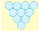
You are trying to add a scale bar to your images. If you measure the following staphylococcus cluster across and get 10 pixels, and you previously determined that using this objective, the ratio is 2.5 pixel/µm, how many µm across is the cluster?
A) 25um
B) 2.5um
C) 4um
D) 0.4um
(Lecture #5-6 Questions)
Answer: C) 4um
When picking a colony to go into the PCR reaction, if you pick a colony that is NOT S. thermophilus, do you expect a PCR product from the reaction?
A) Yes, we just can't predict the size
Yes, it should be the same no matter what kind of bacteria
No, the primers are species specific
No, this is how we are doing our negative control
(Lecture #5-6 Questions)
Answer: No, the primers are species specific

Given the DNA strand below, which of the following primer pairs would you use during PCR to successfully amplify (at least) the entire bolded gene?
A) Forward: 5’ – CTACG – 3’; Reverse: 5’ – TGACC – 3’
B) Forward: 5’ – GCCAA – 3’; Reverse: 5’ – TATAG – 3’
C) Forward: 5’ – ATGGC – 3’; Reverse: 5’ – TGACC – 3’
D) Forward: 5’ – GATGC – 3’; Reverse: 5’ – ACTGG – 3’
(Lecture #5-6 Questions)
Answer: D) Forward: 5’ – GATGC – 3’; Reverse: 5’ – ACTGG – 3’

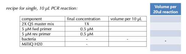
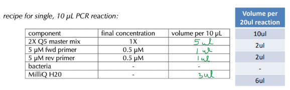
Calculate PCR reaction (C1*V1=C2*V2)
(Lecture #5-6 Questions)
Rules: C1*V1 = C2*V2
-I did it directly for 20 uL but you can do it 10 uL and multiply by 2
-For 2x Q5 Master Mix: 2X*V1 = 1X*20uL → 10 uL
-For 5 uM fwd primer: 5 uM*V1 = 0.5uM*20uL → 2 uL
-For 5 uM fwd primer: 5 uM*V1 = 0.5uM*20uL → 2 uL
-MiliQ H2O: 10 uL - 5 uL - 1 uL - 1 uL = 3 uL

Given the DNA strand below, which of the following primer pairs would you use during PCR to successfully amplify (at least) the entire bolded region?
A) Forward: 5’ – TGGCT – 3’; Reverse: 5’ – TTGGC – 3’
B) Forward: 5’ – GCTGT – 3’; Reverse: 5’ – CGGTT – 3’
C) Forward: 5’ – GAACG – 3’; Reverse: 5’ – GAACT – 3’
D) Forward: 5’ – GAACG – 3’; Reverse: 5’ – TCATA – 3
(Lecture #5-6 Questions)
Answer: D) Forward: 5’ – GAACG – 3’; Reverse: 5’ – TCATA – 3’

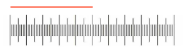
a) How long is the red line?
A) 500 um
B) 50 um
C) 50 mm
b) If the red line is 750 pixels long (measured with image J), what is the pixel/um ratio?
A) 1.5 pixels/um
B) .667 pixels/um
C) 2.5 pixels/um
c) At the same magnification, you take the following picture, and measure the tardigrade (red line) as 600 pixels. How big is the tardigrade in mm?
A) 900 um
B) 400 um
C) 300 um
(Lecture #5-6 Questions)
a) Answer: A) 500 um
Reasoning: Every 10 tick (or 2 big tick) is 100 um
b) Answer: A) 1.5 pixels/µm
Reasoning: Pixel/µm ratio = pixels ÷ actual length = 750 pixels/500 um = 1.5pixels/um
c) Answer: B) 400 µm
Reasoning: size in µm=pixels / (pixels/µm) = (600 pixels x 1 um) / (1.5 pixels) = 400 µm
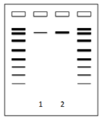
Gel Electrophoresis Example: How should the band in Lane 1 compare to the band in Lane 2 when:
Tube 1: 2 µl of DNA stock.
Tube 2: 4µl of DNA stock.
(Lecture #5-6 Questions)
Answer: Band in Lane 2 is thicker because it has twice the amount of DNA
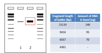
Gel Electrophoresis Example: How much DNA (in ng) is in “2” band? If you added 3µl of a DNA stock to the “2” tube, what is the concentration of the DNA stock (in ng/µl)?
(Lecture #5-6 Questions)
Answer: Band 2 thickness looks closest to ladder band with 23130 bp and 240 ng of DNA. 240 ng ÷ 3 µL = 80 ng/uL
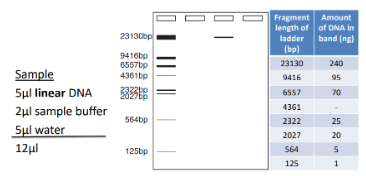
I make the following sample, and run it on the gel shown below. a) What is the length (basepairs) of the DNA fragment? b) What is the concentration (ng/µl) of DNA stock?
(Lecture #5-6 Questions)
a) Length of DNA is 23130 bp (similar distance traveled).
b) Band 2 thickness looks similar to the ladder band with 2322 bp and 25 ng amount of DNA. 25 ng by 5 uL of linear DNA → 5 ng/µl
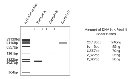
What is the approximate size (bp) and concentration (ng/ul) of Sample A if I loaded 5µl of sample in each lane?
A) 9416bp, 19ng/µl
B) 2027bp, 4ng/µl
C) 2027bp, 19ng/µl
D) 6557bp, 10ng/µl
(Lecture #5-6 Questions)
Answer: C) 2027bp, 19ng/µl
Reasoning:
-Sample A band is located on the 2027 bp of the ladder band
-19ng/uL because Sample A band thickness is similar to the band on the 9416 bp ladder. 9416 bp ladder has a DNA concentration of 95 ng. Since 5 uL was put in each lane, we must divide by 5 so 95ng/5uL = 19 ng/uL
True or False? The dye in the sample buffer binds to your DNA and lets you see where it is in the gel
(Lecture #5-6 Questions)
Answer: False b/c DNA is visualized using SYBR. Sample buffer dye tracks sample movement and does not bind DNA.
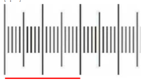
I take a picture of my micrometer at 40X (only part of it is visible, shown above), and measure the distance between the lines shown. This line is measured to have 300 pixels. What is the pixel/μm ratio for this 40X objective?
(Lecture #5-6 Questions)
-Red line = 200 um
-Ratio: 300 pixels/200um = 1.5 pixels/um
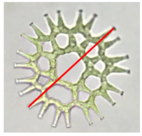
Assuming I take the following picture at 40X, and measure my area of interest to have 75 pixels. What is this length in µm? (Assume pixel/um ratio is 1.5)
(Lecture #5-6 Questions)
-pixel/μm ratio from above: 1.5 pixels/um
(75 pixels) / (1.5 um/pixel) = 50 um
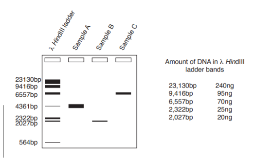
Please use the gel shown to answer the following questions and show any work if necessary.
A. What is the approximate size (in basepairs) of the Sample C fragment?
b. What is the approximate concentration of my DNA stock for Sample A if I loaded 8µl of water, 2µl of (linear) DNA sample, and 2µl of sample buffer in each lane?
(Lecture #5-6 Questions)
A) 6557 bp b/c Sample C band traveled the same length down as the ladder band at 6557 bp.
B) Sample A band thickness is most similar to the ladder band of 23130 bp with the DNA amount of 240 ng. Since we loaded with 2 uL of DNA sample: 240 ng/2 uL = 120 ng/uL
True or False?:
__________ Larger fragments of DNA would be expected to travel further down the gel than smaller fragments
__________ Syber safe binds to the DNA and allows visualization
__________ When orienting the gel, the wells should be put closest to the negative
(Lecture #5-6 Questions)
False b/c larger fragments travel less far than smaller ones
True b/c SYBR Safe binds DNA and allows visualization
True b/c wells should be near the negative electrode

Given the DNA strand below, a) please design a primer pair (each primer being 5 bases) that you would use during PCR to successfully amplify the entire given sequence.
5’ – GATTCGCCATGTACCGCGCAAGCTATTAGGTGACACTATAGAATAGCCCAGTC – 3’
3’ – CTAAGCGGTACATGGCGCGTTCGATAATCCACTGTGATATCTTATCGGGTCAG – 5’
Forward 5’ - - 3’
Reverse: 5’ - - 3’
b) How many base pairs long would the final DNA PCR fragment be using the primers shown below?
Fwd: 5’ – AAGCT – 3’
Rev: 5’ – ATAGT – 3’
(Lecture #5-6 Questions)
Answer:
a) Forward: 5’ - GATTC - 3’
Reverse: 5’ - GACTG - 3’
b) 5’ - ATTAGGTGAC - 3’ → 10 bp
3’ - TAATCCACTG – 5’ → 10 bp
Total = 20 bp
Approximate size/length of fragment in gel electrophoresis:
(Lecture #7 Content)
correlates with the ladder band of the same distance
Vertical gene transfer:
(Lecture #7 Content)
DNA passed from parent to offspring during reproduction.
Horizontal gene transfer (HGT):
(Lecture #7 Content)
DNA transfer between cells (not parents to offspring) via:
1. Conjugation: Direct cell-to-cell DNA transfer via sex pilus.
2. Transformation: Bacterium takes up free DNA from the environment and incorporates into the genome.
3. Transduction: DNA transfer via bacteriophages.
Antibiotic vs Antibiotic resistance vs Superbugs:
(Lecture #7 Content)
Antibiotic: Substance that kills or inhibits bacterial growth.
Antibiotic resistance: ability of bacteria to survive and grow despite antibiotics. Ex: spontaneous mutation, overuse of antibiotic, and horizontal transfer.
Superbugs: bacteria is resistant to multiple antibiotics.
Components needed for sequencing reaction:
(Lecture #8 Content)
(Study)
DNA Template, Primer, DNA polymerase, dNTPs, Fluorescently labeled dideoxynucleotides (ddNTPs)
Sanger Sequencing:
(Lecture #8 Content)
-Read bottom → top (5’ to 3’)
-For sequence of fragment: read bases directly bottom → top
-For template strand: take complement of sequence (A↔T, G↔C) bottom —> top
dNTP vs ddNTP:
(Lecture #8 Content)
-dNTP: normal nucleotide with 3′-OH that allows DNA elongation via phosphodiester bond
-ddNTP: lacks a 3′-OH, causing chain termination. More ddNTPs produce shorter sequencing products.
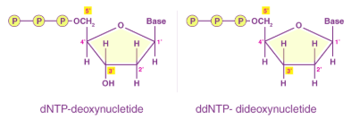
Gut Microbiome:
(Lecture #8 Content)
(Study)
Contains both bacteria (microbiome) and bacteriophages (virome), and these phage communities are generally stable over time. Bacteriophages can also mediate horizontal gene transfer through transduction.
Which of the following statements about horizontal gene transfer are correct?
A) Gene transfer only occurs between bacteria
B) Transduction is when bacteriophage infect a bacteria and produce new functional bacteriophage
C) Transformation is when a cell takes up DNA from their environment
D) None of the above are correct
(Lecture #7-8 Questions)
Answer: B) Transduction is when bacteriophage infect a bacteria and produce new functional bacteriophage
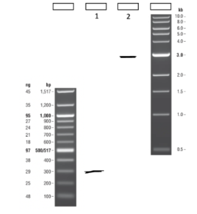
Based on the gel below, what is the approximate fragment size of Sample 1?
A) 29bp
B) 300bp
C) 3000bp
D) Cannot determine
(Lecture #7-8 Questions)
Answer: B) 300bp
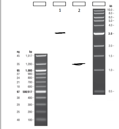
What is the approximate fragment size of a) Sample 1 & b) Sample 2?
A) 125bp
B) 1200bp
C) 3000bp
D) Cannot determine, larger than 100bp ladder
(Lecture #7-8 Questions)
Answers:
A) Sample 1: C) 3000 bp
B) Sample 2: B) 1200 bp
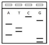
I set up an old fashioned Sanger sequencing reaction with radiolabeled ddNTPs. I run each reaction with a specific labeled nucleotide (shown on gel) and an excess of the rest of the normal dNTPs. I then run a gel with my Sanger sequencing products, shown. What is the sequence of my fragment?
A) 5’ – CGATTAGG – 3’
B) 5’ – GGATTAGC – 3’
C) 5’ – GCTAATCC – 3’
D) 5’ – CCTAATCG – 3’
(Lecture #7-8 Questions)
Answer: B) 5’ – GGATTAGC – 3’
Reasoning: Read bottom up. For sequence, read bases directly bottom → top. For template strand: take complement of sequence (A↔T, G↔C) bottom —> top
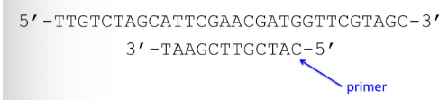
You set up the following sequencing reaction by adding DNA polymerase, dNTPs, a small amount of ddATP, and the 12bp primer shown. What sizes of products would you expect from this reaction?
A) 14, 15, 18, 21 nucleotides long
B) 19, 21 nucleotides long
C) 15, 21 nucleotides long
D) 16, 18, 20, 21 nucleotides long
E) 3, 6, 7, 12, 15, 21 nucleotides long
(Lecture #7-8 Questions)
Answer: D) 16, 18, 20, 21 nucleotides long
Reasoning: Primers are always built from 5’ to 3’ so in this scenario it will move from right to left. ddATP meant you look for “T” after the strand
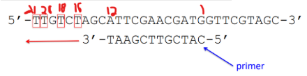

You set up a sequencing reaction by adding DNA polymerase, all four dNTPs, a small amount of ddTTP, and the DNA template and primer below. What sizes of products would you expect from this reaction?
(Lecture #7-8 Questions)
Answer: ddTTP: 13,18,23, 24
ddTTP means you look for “A” after the strand
Look for polarity, where the primer goes 5’ to 3’, so it is going right to left.


You set up the following sequencing reaction by adding DNA polymerase, dNTPs, a small amount of ddGTP, and the following template and 12bp primer. What sizes of products would you expect from this reaction?
a. 13, 17, 29 nucleotides long
b. 14, 19, 23, 26 nucleotides long
c. 16, 18, 20, 21 nucleotides long
d. 16, 21, 25, 36 nucleotides long
e. 13, 17, 20, 24, 29, 32, 35 nucleotides long
(Lecture #7-8 Questions)
Answer: d. 16, 21, 25, 36 nucleotides long
Reminders for myself: ddGTP means we are looking for C on the template. We must include the length of the primer (12 bp) then count from there. Also polarity matters.

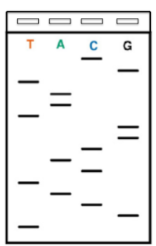
I set up an old fashioned Sanger sequencing reaction with radiolabeled ddNTPs. I run each reaction with a specific labeled nucleotide (shown on gel) and an excess of the rest of the normal dNTPs. I then run a gel with my Sanger sequencing products, shown. What is the sequence of my fragment (please include polarity)?
(Lecture #7-8 Questions)
Answer: 5’ - TGCATCACGGTAATGC - 3’
Reasoning: Read bottom up. Sequence also means reading what it says from bottom to top. Template is when you read and correlate the opposite base pair
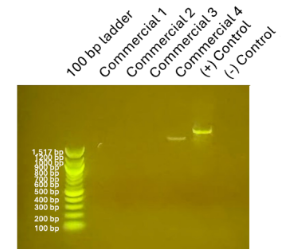
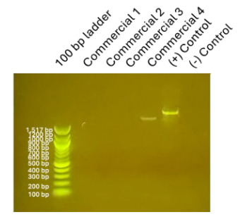
a) For the gel shown above, please select the likeliest reason for the absence of bands observed for the commercial samples 1-3.
A) Syber safe wasn’t added
B) The PCR mastermix was made incorrectly
C) Those colonies were not S. thermophilus
D) I expected the absence of those band
b) For the answers you ruled out as being incorrect: Briefly explain why you think each one is not correct.
(Lecture #7-8 Questions)
Answer a : C) Those colonies were not S. thermophilus
Answer b:
-A is incorrect b/c no SYBR Safe = can’t see anything but we can see a ladder & bands.
-B is incorrect b/c the positive control has a band, so the mastermix worked.
-D is incorrect because commercial samples contain S. thermophilus, so we expect to see bands
True or false?
________In addition to the bacteria living in our intestinal tract, we also have a natural population of bacteriophage living in our bodies
________Populations of bacteriophage in our body seems to fluctuate and change very rapidly and do not stay constant for long
________Bacteriophage are capable of mediating horizontal gene transfer
(Lecture #7-8 Questions)
Answers:
True – Our gut has a virome (bacteriophage population) in addition to bacteria.
False – Phage communities are generally stable over time, not rapidly changing.
True – Phages can transfer DNA between bacteria via transduction, carrying bacterial genes from one cell to another.
A dideoxynucleotide (ddNTP) is missing:
A) The phosphate group from the 5' carbon
B) The -OH group from the 3' carbon
C) The sugar residue
D) The base
(Lecture #7-8 Questions)
Answer: B) The -OH group from the 3' carbon
CRISPR vs BLAST analysis:
(Lecture #9 Content)
(Study)
-CRISPR: identify repeat and spacer regions to study bacterial strains and past phage exposure.
-BLAST: compares DNA sequences to databases to identify related organisms or viruses.
Repeat vs Spacer:
(Lecture #9 Content)
(Study)
-Repeats: Conserved CRISPR sequences that maintain structure/function.
-Spacers: Unique phage DNA fragments from past infections that provide immunity against matching phages
Mean vs Median vs Standard Deviation vs T-test:
(Lecture #10 Content)
-Mean: Average
-Median: Middle value
-Standard deviation: measures variation in data values
-T-test: determines if two group means are statistically different
→ Formula: t = (mean₁ − mean₂) / √(sd₁/n₁ + sd₂/n₂)
→ p < 0.05 = statistically different or p > 0.05 → not statistically different
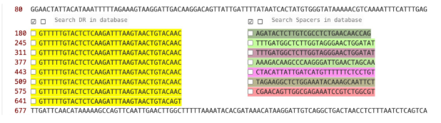
Poll 1: How many spacers are there? (photo above)
A) Eight
B) Seven
C) Six
D) Fifteen
Poll 2: How many phage is this strain immune to? (same photo above)
A) Eight
B) Seven
C) Six
D) You can’t tell based on this alone
Poll 3: Why does this spacer match so many different viruses?
A. this bacteria has been exposed to all of the viruses in the BLAST results
B. the viruses in the BLAST results are related to one another
C. the viruses in the BLAST result share a particular gene or genomic region
D. B or C are both good explanations
(Lecture #9-10 Questions)
1) Answer: B) Seven (right side of the photo)
2) Answer: D) You can’t tell based on this alone b/c we need to find out what these sequences correspond to.
3) Answer: D. B or C are both good explanations
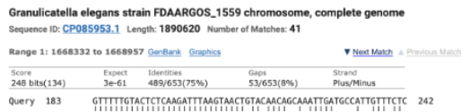
Is this match evidence of HGT of our CRISPR array from yogurt into another species
a. Yes
b. No
(Lecture #9-10 Questions)
Answer: No, because this match shows 75% identical with 8% gaps, while HGT would show ~100% identical with no gaps.
If I were to BLAST the repeat sequences identified by the program from our sequencing result, what match would I most likely expect to see?
A) Bacteriophage genome
B) Lots of different bacterial genomes
C) Streptococcus thermophilus strains genome
D) Astrophage genome
(Lecture #9-10 Questions)
Answer: C) Streptococcus thermophilus strains genome
Reasoning: CRISPR repeats are conserved bacterial sequences, so they match Streptococcus thermophilus genomes (not phage).
Simple vs Differential stain:
(Lecture #11 Content)
-Simple stain: Colors all cells the same to observe shape and arrangement.
-Differential stain: Colors different cell types differently to distinguish them. Ex: Gram staining
Gram positive vs negative bacteria
(Lecture #11 Content)
-Gram-positive: Thick peptidoglycan cell wall with no outer membrane traps crystal violet iodine, so cells remain purple.
-Gram-negative: Thin peptidoglycan cell wall with an outer membrane loses crystal violet during decolorization and is stained pink/red with safranin.
Main Steps of Gram Staining + What Happens if a Step is Missed (Crystal Violet, Iodine, Decolorizer, Counterstain (Safranin)
(Lecture #11 Content)
(Study)
1. Crystal violet:
• Purpose: stains all cells purple initially.
• If missed: Gram differentiation will not work properly.
2. Iodine (mordant):
• Purpose: forms crystal violet-iodine complex to trap dye in Gram-positive.
• If missed: Gram-positive cells may falsely appear Gram-negative.
3. Decolorizer (alcohol/acetone) — very time dependent:
• Purpose: removes crystal violet from Gram-negative while Gram-positive stay purple.
• Too long: all cells may appear Gram-negative.
• Too short: all cells may appear Gram-positive.
4. Counterstain (Safranin):
• Purpose: stains Gram-negative cells pink/red.
• If missed: Gram-negative appear colorless, so only Gram-positive cells remain visible/purple.
Biofilm
(Lecture #12 Content)
(Study)
are protective microbial communities that stick to surfaces in a sticky matrix, increase antibiotic resistance, and form best in static environments with O₂ gradients of air-liquid interface (Ex: teeth, water systems).